


There are so many cool experiments you can do with the TiFM! So excited to share this tool with everyone and eager for #Collaborations!
09.02.2026 20:55 — 👍 1 🔁 0 💬 0 📌 0
There are so many cool experiments you can do with the TiFM! So excited to share this tool with everyone and eager for #Collaborations!
09.02.2026 20:55 — 👍 1 🔁 0 💬 0 📌 0We use TiFM to measure the #MechanicalProperties of tissues using rheological principles. Here you see how our probes oscillate more in water than in tissues, with stiffer tissues resisting the probe motion most.
09.02.2026 20:49 — 👍 0 🔁 0 💬 1 📌 0Using the TiFM we can compress and/or stretch the neural folds of #ChickEmbryos
09.02.2026 20:41 — 👍 0 🔁 0 💬 1 📌 0
So excited to share that my 1st paper is published!! Read all about how to manipulate the #MechanicalProperties of tissues using our custom built TiFM! journals.biologists.com/dev/article/...
09.02.2026 20:18 — 👍 14 🔁 6 💬 3 📌 0
Fluorescent microscopy images of transgenic chicken resources maintained at the NARF, The Roslin Institute. (A) GFP chicken embryo with a ‘red’ graft placed into the limb bud with micro-surgery. The graft is about 50–100 μM. (B) Stage 32 HH limb bud from the CSF1R-eGFP embryo. Macrophages (green) are concentrated in areas of cell death, blood vessels (red) are labelled with SNA lectin. (C) ACTN line embryo spinal cord stage 16 HH. (D) Dorsal Root Ganglion of the nervous system of a Chameleon transgenic chicken embryo. Nerves going into the dorsal root ganglion are red, and nerves coming out are green. (E) Fluorescent image of the dorsal skin (periderm) from a membrane GFP chick embryo. (F) FUCHI cell cycle reporter embryo stage 23 HH. All images were provided by members of The Roslin Institute Chicken Embryology (RICE) group.
#DBfeature
Research facilities in the UK, France, and Japan maintaining transgenic lines for Avian developmental biology
By Lindsay Henderson, Yuya Okuzaki, Christophe Marcelle, Mike McGrew, and Ken-ichi Nishijima
tinyurl.com/4drw4d6a
#SpecialIssue on Avian model systems

Rainbow shading of a confocal micrograph of a developing mouse neural tube. The protein actin is stained and colored in rainbow to indicate depth in the section.
Happy #FluorescenceFriday! This is a maximum intensity projection of depth shaded actin (🌈) in a section from an E9.5 🐭 neural tube (NT). 🔬 by postdoc @christinaadaly.bsky.social 👩🔬 🧪 Image shows the floor plate and lumen of the developing NT.
#SciArt #DevBio #DevNeuro

(A) Schematic of embryonic chick skin, composed of an epithelial layer overlying a mesenchyme containing cells migrating within an extracellular matrix. During early stages of feather development dermal fibroblasts aggregate underneath epithelial thickenings, known as placodes, to form dermal condensates. (B) Skin explant dissected from an embryonic day 9 (E9) tdTomato transgenic embryo showing feather buds growing out and oriented towards the posterior. Scale bar 1 mm. (C) Dermal condensates form in sequential rows, originating from a dense mesenchymal stripe along the midline of the embryo. This stripe then separates into condensates, with subsequent rows emerging laterally in sequence. (D) In situ hybridisation detecting CTNNB1 expression in E8 chick skin. The red outline highlights the hexagonal arrangement of the 6 nearest-neighbour feather buds. (E) Haematoxylin and eosin stain of an E6.5 chick embryo section. The red dashed rectangle highlights the loose dermis, the green dashed rectangle highlights the dense dermis. Left panel scale bar = 200 μm, right panel scale bars = 100 μm.
#DBfeature 🪶
New tools bring embryonic feather bud development back to the forefront of efforts to understand vertebrate organogenesis
By Jon Riddell and Denis Headon
tinyurl.com/hzzhr5z8
#SpecialIssue on #Development and #Regeneration of ectodermal organs

#DBfeature #chick🐥
Generation of a transgenic chicken line with reporters for limb bud mesenchyme and apical ectodermal ridge cells
Yuji Atsuta, Yi-Chen Chen, Yuna Hattori, Tatsuya Takemoto, Daisuke Saito
tiom33.short.gy/DB520,53-61

Be part of the conversation on how avian models are transforming multiple fields of #research at #AvianModels25 – in person or virtually.
🎟️Register today: bit.ly/3ZBw2qS

#Science takes flight at the XII Avian Model Systems Meeting — and you can save by registering early! #AvianModels25
🐥 Secure your early bird rate today: bit.ly/3ZBw2qS
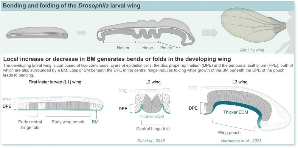
Tissue bending through localized BM growth or dissolution.
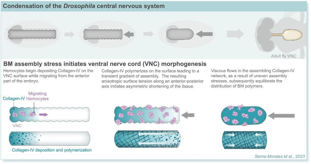
Alteration in tissue morphology by intrinsic stresses in an assembling BM network.
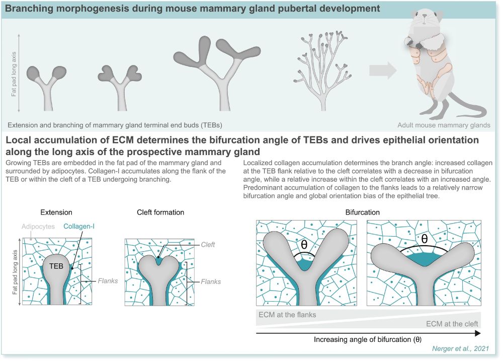
Branching morphogenesis driven by localized deposition of interstitial matrix.
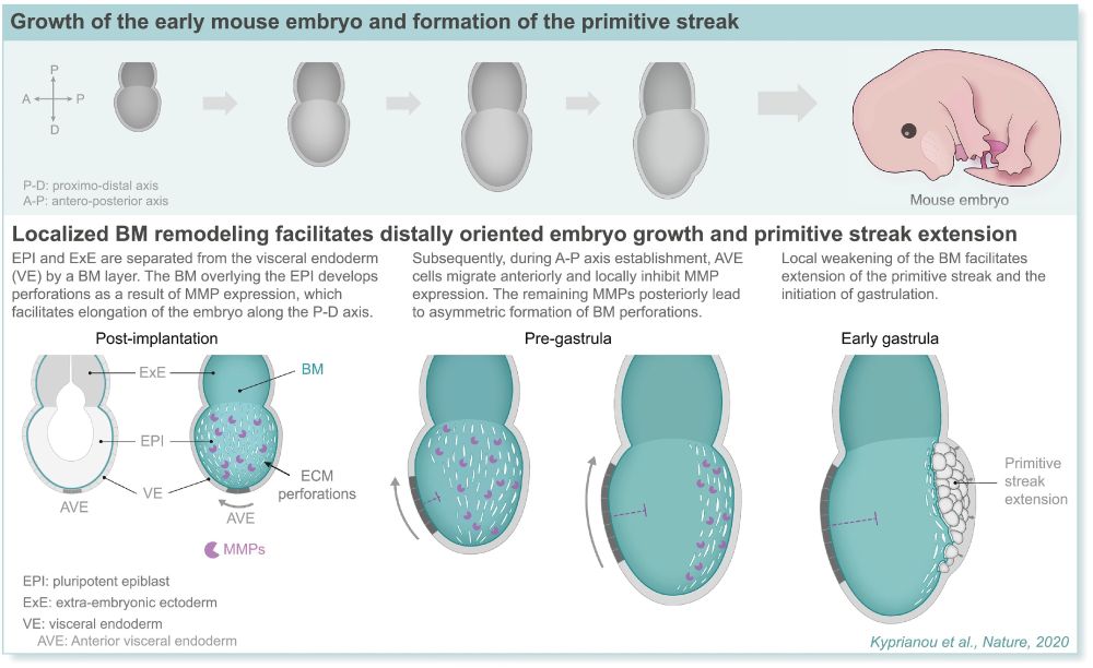
Asymmetric tissue expansion through anisotropic BM remodeling.
Maybe it's time we rethink the idea that development is a cell centric process? In this beautiful review, María-del-Carmen and @stramerlab.bsky.social discussed how the ECM underlies and influences many morphogenesis processes from wing unfolding to mammary gland development.
doi.org/10.1016/j.cd...
Excited to be giving a talk and sharing my work here! I look forward to making new friends and learning new things! #DevBio #BioPhysics
06.08.2025 16:39 — 👍 1 🔁 1 💬 0 📌 0
One of the reasons I love #DevelopmentalBiology is that it's so visually engaging. I had the pleasure of organising an image competition at the GRC last March, and share this passion. You can now check out these images on @the-node.bsky.social: thenode.biologists.com/grs-developm...
15.05.2025 08:01 — 👍 29 🔁 12 💬 0 📌 3
A heartfelt thank you to all the generous supporters of the Gordon Research Conference on Developmental Biology 2025. This event would not have been possible without your invaluable contribution.
04.04.2025 20:21 — 👍 12 🔁 1 💬 0 📌 0
It was an honour to contribute to the #DevelopmentalBiology community! Congratulations to Zainab Afzal
@xainabafzal.bsky.social and Akshada Shankar Ganesh on being elected 2027 GRS DB chairs!

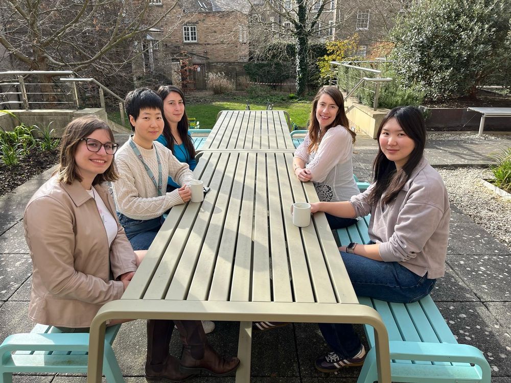
The Xiong Lab at #biologist100
A wonderful conference, great to meet so many people and share our passion for development!
@lakshmib02.bsky.social
@fengtongji.bsky.social

I'm so excited to share my work at #biologist100 Come and talk to me about how tissue stiffness regulates axis elongation! Poster 197.
24.03.2025 17:54 — 👍 7 🔁 1 💬 0 📌 0

@gurdoninstitute.bsky.social We celebrate the contributions of women in science and beyond. We recognise women who inspire, lead and push the boundaries of discovery every day.
Together we work towards a future where science thrives through diversity, inclusion and equality.
#InternationalWomensDay
And the link for tye associated GRS! www.grc.org/developmenta...
27.02.2025 09:39 — 👍 0 🔁 0 💬 0 📌 0
And this is the link for the associated GRS :)
www.grc.org/developmenta...

Only one week left to register for the #DevelopmentalBiology GRC and GRS! And thank you to all our funders for the support!
@swathiarur.bsky.social

Here is the 2025 embryo alphabet from alligator to zebrafish. Developmental biology is stunning & leads to important discoveries for human medicine.
@socdevbio.bsky.social

We’re honoured to receive funding from @biologists.bsky.social for our upcoming conference. 🙏 Thank you for championing the biology community!
🧬 Explore their journals, meetings, and grants at www.biologists.com.
📅 Don’t wait—register now and join us! 🎉 @swathiarur.bsky.social

The GRC has an exciting set of speakers! www.grc.org/developmenta...
16.01.2025 16:58 — 👍 0 🔁 0 💬 0 📌 0
Abstract submission for the #DevelopmentalBiology GRS and GRC is still open, apply now! Looking forward to sharing research and meeting new people! See the full programme in the comments. www.grc.org/developmenta... @swathiarur.bsky.social
16.01.2025 16:44 — 👍 2 🔁 1 💬 2 📌 1
And here is the link for the GRS! www.grc.org/developmenta...
Thank you for sharing!
Thank you! It was drawn by the GRS co-chair, Anastasia Repouliou
03.12.2024 21:33 — 👍 2 🔁 0 💬 0 📌 0
