

Blurry #MicroscopyImages even with a good sample? The issue is often the objective.
Check our new video to see which objective fits each imaging challenge.
#MicroscopyEducation #ZEISSMicroscopy
Blurry #MicroscopyImages even with a good sample? The issue is often the objective.
Check our new video to see which objective fits each imaging challenge.
#MicroscopyEducation #ZEISSMicroscopy
When your “bouquet” turns out to be ancient ocean life under the #microscope🌸🌊
Not flowers, but fossil coral. Silica slowly replaced calcium carbonate and formed these petal like structures over millions of years.
Thanks, Norm Barker, for this cooperation!
Happy #ValentinesDay!
On this World Cancer Day, we highlight the pioneering efforts of the Department of Urology and Pediatric Urology at University Hospital @rwth.bsky.social, Germany, to enhance prostate cancer patient care.
👉 Explore the full story: zeiss.ly/user-story-p...
#WorldCancerDay #CancerResearch
If your #microscopy images look almost right, the problem is rarely the sample.
Small objective lens setup mistakes can quietly ruin data quality, even in experienced labs.
We collected the most common brightfield pitfalls & how to fix them.
#MicroscopyCommunity #ZEISSMicroscopy

BiteSize Bio GxP-Webinar with ZEISS Microscopy on January 20, 2026
Are your microscopy workflows ready for regulated environments? Join us on January 20 for our BiteSize Bio webinar and learn about:
✅ Key GxP principles for imaging labs
✅ Reproducible, GxP-compliant imaging workflows
✅ GxP-ready microscopy implementations
Register here: zeiss.ly/by-gxp-webin...
As cell division shows us: renewal is part of progress.
2026 begins – and with it, a new cycle of discovery. Happy New Year!
📷 Image courtesy: Slide 2: Aude Nommick, Institut Jacques Monod – CNRS; Slide 3: Generated with AI
#FromCuriosityToLastingImpact
#MicroscopyMatters
#ScientificInnovation
Using advanced microscopy, Dr. Mario Hentschel of the University of Stuttgart has recreated Kandinsky's masterpiece on just 180 x 180 µm, roughly the size of a pinpoint! On Kandinsky's birthday today, join us in honoring the fusion of art and science: zeiss.ly/user-story-kandinsky
16.12.2025 12:26 — 👍 6 🔁 1 💬 0 📌 0
These 5 terms define every image you acquire – but they're rarely taught as an integrated framework. Whether you're in research, industry QC, or running a core facility, this reference guide clarifies the fundamentals.
Technical clarity leads to better imaging decisions.
Your #microscopy cheat sheet: Magnification, numerical aperture, resolution, working distance, depth of field – explained clearly, in one place.
Most of us learned these in fragments. This carousel connects the dots.
💾 Save it. Share it with your lab.
Which term should we explain next?
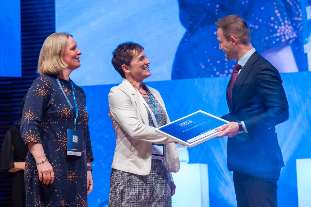
Thuringian Innovation Award 2025 for ZEISS Lightfield 4D

Thuringian Innovation Award 2025 for ZEISS Lightfield 4D

Thuringian Innovation Award 2025 for ZEISS Lightfield 4D
Join us in celebrating winning the Thuringian Innovation Award 2025 in the category "Life and Light" for ZEISS Lightfield 4D, along with the esteemed Audience Award! 🎉🏆
We are sincerely thankful. Your encouragement has been invaluable on this journey! 🙏🏼
📸 STIFT/Thomas Müller
Image Courtesy:
Dr. Jan Michels, K. Fantauzzo, Sébastien Dupichaud
On the International Day for the Elimination of Violence against Women and Girls, let’s recognize how #microscopy technologies from ZEISS empower #forensic professionals to deliver timely justice in sensitive cases ⚖️💜
25.11.2025 13:17 — 👍 0 🔁 0 💬 0 📌 0
A pink-stained forensic microscope slide with scattered cells and text stating, "In forensics microscopy, justice starts with a single cell."

Microscopy image showing stained epithelial cells and a sperm cell marked by a red box, illustrating AI-assisted sperm detection for forensic analysis.

Automated microscopy setup with computer and analysis software shown in lab, highlighting benefits of AI-powered forensic casework processing.

A purple handprint with a ribbon overlay marks International Day for the Elimination of Violence Against Women, observed on November 25. Text below highlights innovation in microscopy for justice.
Forensic labs face critical evidence backlogs 🔬⏳ZEISS Axio Imager with AI-powered software from MetaSystem cuts processing time from weeks to hours, enhancing accuracy, supporting timely evidence delivery.
👉 Explore how microscopy transforms forensics: zeiss.widen.net/s/kfzshcskjg...

ZEISS Microscopy development team of ZEISS Lightfield 4D microscope system
ZEISS Lightfield 4D has been nominated for the prestigious Thuringian Innovation Award! 🎉The jury has made their selections - now it's your turn to decide on the "Audience Award 2025". 👉 Support us with a "thumbs up" : zeiss.ly/innovationspreis2025-voting
Voting is open until Nov 21, 2025. ⌛
Driven by curiosity, our customers have been instrumental in winning over 30 Nobel Prizes through their groundbreaking research using ZEISS technology. This journey began with Robert Koch in 1905 and continues with the recent addition of Susumu Kitagawa in 2025! 🏆✨ Happy World Science Day!
10.11.2025 09:59 — 👍 1 🔁 0 💬 0 📌 0
Pharaoh Ant Head 🐜 Confocal imaging with ZEISS LSM 700 reveals stunning details.
Mouse Craniofacial Skeleton 🐭 Cartilage in blue, bone in red, captured with ZEISS Stemi 508.
Spider 🕷 Cleared with CUBIC and imaged with ZEISS Lightsheet Z1.

Close-up, color-enhanced microscope image of an ant’s head viewed from the front, showing detailed antennae and mouthparts against a black background.

A side-view microscope image of a mouse skull and upper spine, displayed in shades of blue and purple.

Fluorescent image of a spider with bright pink and green highlights on a black background, showing the spider's detailed body and leg structure.
Spooky specimens under the microscope 👻🔬
No ghosts here at #Halloween2025 – just real creatures revealing their eerie elegance through #ZEISSMicroscopy.
Details about the specimens in the comments!
Which one would you dare to look at twice? 🎃
#SpookyScience
Water squeezed into atomic-layer channels reveals entirely new physics ⚛️💧
At Manchester's National Graphene Institute, researchers fabricate angstrom-scale structures using ZEISS SEMs + RAITH ELPHY systems– enabling quantum devices, nanofluidics, & 2D photonics.
👉 Full story: zeiss.ly/2Ddevices
Facility Managers, take note! 🔬 Learn how Dr. Peter O’Toole & the University of York team excel in managing #ZEISS confocal systems.
👉 Full story: zeiss.ly/user-story-c...
#CoreImagingFacility #ZeissMicroscopy
Happy belated #WorldHeartDay! 🌍❤️
Let's celebrate heart formation & development. Understanding normal growth vs. defects is vital for #HeartHealth ⚕️
🐟💓 Check out this amazing 4D data of a beating #zebrafish heart w/ #ZEISS Lightfield 4D.
📷🔬 Toby Andrews & Rashmi Priya, @crick.ac.uk, London, UK


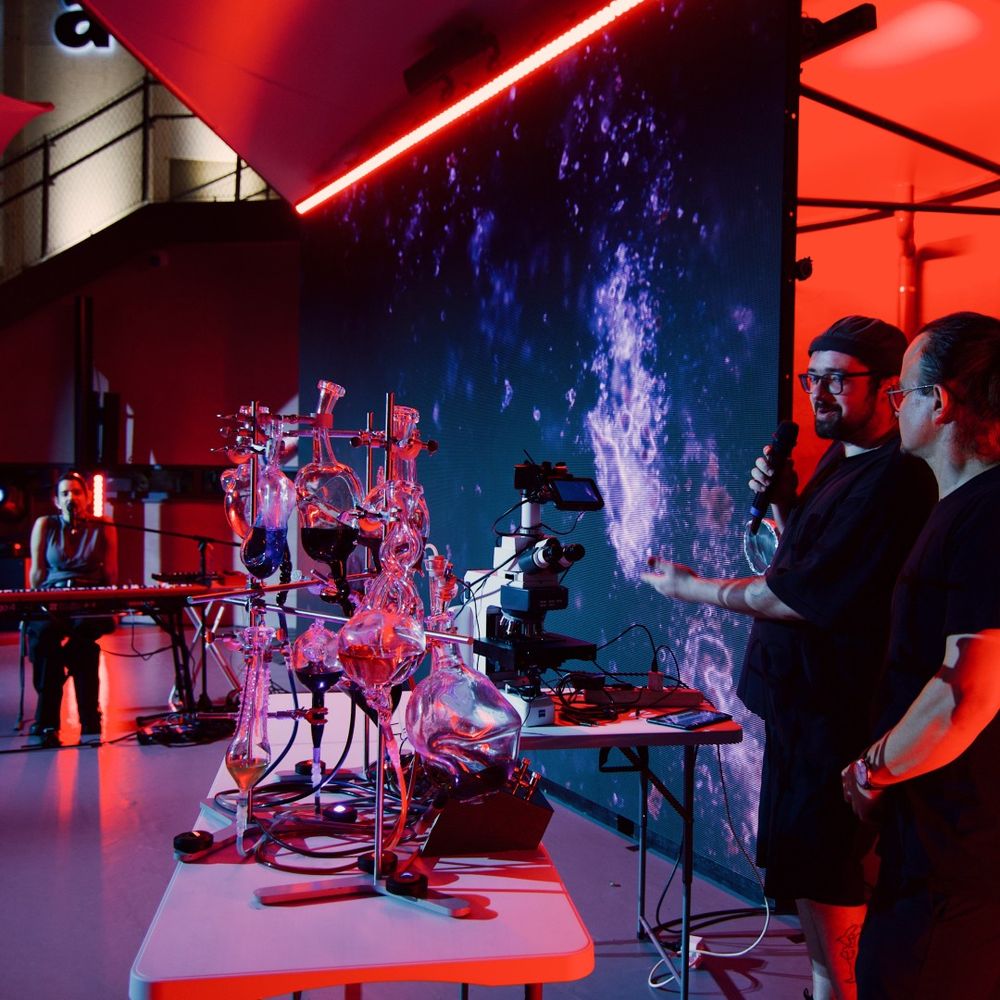
Live from Osaka! 📷🗾The fascinating performances with our collaboration partner Magnificent Matter are happening this week at the Expo 2025, blending science and art in a truly unique experience. 🎶🔬 Don’t miss your chance to witness – performances run until September 13!
12.09.2025 10:27 — 👍 1 🔁 0 💬 0 📌 0
We're thrilled to announce our participation in the World Expo 2025 in Osaka, Japan! 🎉🗾 With our collaboration partner Magnificent Matter, we are ready to blend the worlds of science and art in a performance at the German Pavilion next week.
#WorldExpo2025 #Osaka #GermanPavilion #TheInvisibleMatters
Salt & pepper like never before! 🔬
SEM images by Stefan Meichtry & Langley Anderson Photography reveal everyday beauty and discover the unseen.
Let’s uncover the hidden wonders – one image at a time.
📸 Share your ZEISS microscope images using #MyZEISSMicroscopy
What happens when a forgotten collection of amber is rediscovered at the Phyletic Museum of the Friedrich-Schiller-Universität Jena? It unveils a world trapped in time with stunning details of insects preserved for millions of years: zeiss.ly/microscopy-a...
13.08.2025 07:44 — 👍 0 🔁 0 💬 0 📌 0Join Vanessa Chappiani and Prof. Corrado Calì from the University of Turin as they delve into the fascinating world of astrocytes, star-shaped glial cells in the central nervous system, and discover how their work is shaping brain research: zeiss.ly/user-story-astrocyte-morphology
06.08.2025 08:18 — 👍 1 🔁 0 💬 0 📌 0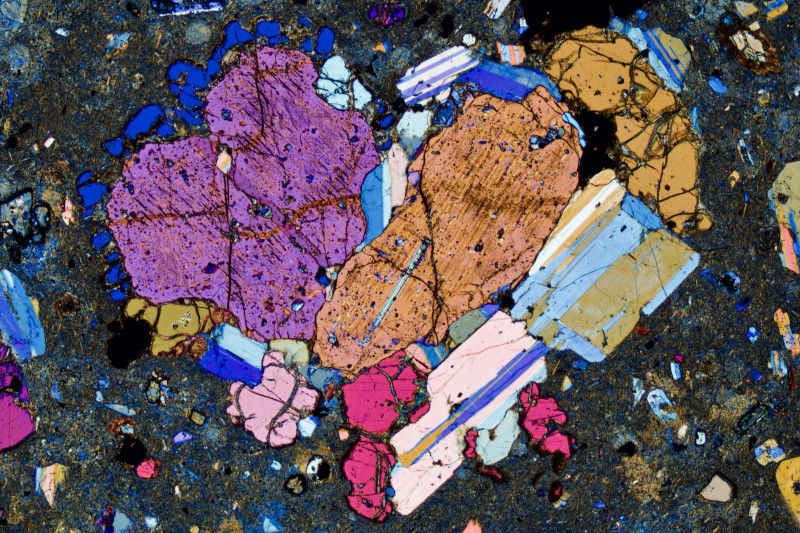
On International Friendship Day, this heart-shaped volcanic crystal symbolizes beauty & connection under pressure 🔬
Captured with ZEISS Microscopy, plagioclase and pyroxene reveal nature's bonds.
30-µm section 💎 2.5x objective | 5.4 mm view 🔬
Lipari, Italy 📍
Bernardo Cesare, Univ. of Padova
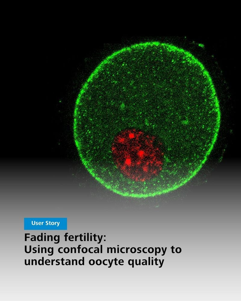
Confocal microscopy image showing an oocyte with a large green-stained area and a smaller red-stained area, illustrating research on oocyte quality. Text overlay "User Story - Fading fertility: Using confocal microscopy to understand oocyte quality"

Fluorescent microscope image of an egg cell, with the nucleus highlighted in red and the surrounding cell in green, alongside text about egg quality and fertilization research.
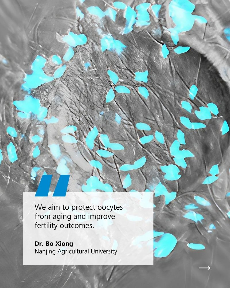
Microscopic view of tissue with blue-stained cells, overlaid with a quote box about oocyte protection from aging and fertility improvement by Dr. Bo Xiong of Nanjing Agricultural University.
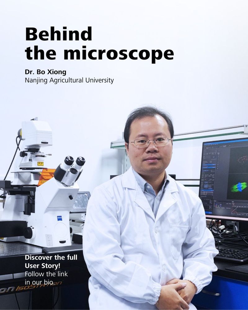
Dr. Bo Xiong in the lab at Nanjing Agricultural University with ZEISS microscope and imaging software, featured in the “Behind the Microscope” user story.
Discover #OocyteQuality with Dr. Bo Xiong from Nanjing Agricultural University!
His team studies spindle dynamics & sperm binding in aging oocytes using #ConfocalMicroscopy, revealing insights into fertility decline.
Researching similar topics? Let’s connect! 🔬
#ZeissMicroscopy #IVFScience
Celebrating 140+ years of #ART on Louise Brown's birthday, the first #IVF baby!
IVF has transformed fertility care, with ZEISS microscopy at every step.
Which milestone impacted #ART most? Share your thoughts 💬
#ZeissMicroscopy #ReproductiveMedicine
Every detail matters. Every dream counts.
From unseen beginnings to unforgettable moments – this is the journey of IVF. Microscopy makes ART possible by revealing what our eyes can’t see.
💙 Join us in making the invisible visible.
👉 Follow for more stories of hope, science, and transformation.
Happy #AIAppreciationDay! Check out how #AI is enhancing #microscopy every day 👇
16.07.2025 07:55 — 👍 2 🔁 0 💬 0 📌 0


