🔬 Join Imaging Bites seminar 28 Jan at 12:30 at @kingscollegelondon.bsky.social.
Prof. Dr. Felipe Opazo @opazo.bsky.social presents #OneStep-IF, a simplified approach to highly #multiplexed IF using same-species primaries and Smart Secondaries® premixes.
Register here: www.kcl.ac.uk/events/imagi...
21.01.2026 11:25 —
👍 6
🔁 3
💬 0
📌 0

Our article "Ångström-resolution imaging of cell-surface glycans" made it to the cover of Nature Nanotechnology! 🤩 @natnano.nature.com
www.nature.com/nnano/volume...
18.10.2025 14:32 —
👍 74
🔁 8
💬 3
📌 0

Localization of single molecules with structured illumination and structured detection - Light: Science & Applications
Light: Science & Applications - Localization of single molecules with structured illumination and structured detection
Happy to share this article with my views on @elislenders.bsky.social and @vicidominilab.bsky.social work and discussing current developments in single-molecule localization combining structured excitation and detection. So many exciting perspectives in the field!
www.nature.com/articles/s41...
30.09.2025 18:17 —
👍 23
🔁 10
💬 2
📌 0
Mirror, mirror on the wall... 🪞 I'm super proud to have contributed to our latest work on left-handed DNA-PAINT! Here we introduce 6 new sequences using the left-handed enantiomers of our speed-optimized DNA-PAINT palette, enabling out-of-the-box 12-plex imaging without extra hybridization steps. 🌈🔬
02.10.2025 11:58 —
👍 8
🔁 1
💬 0
📌 0

Our new preprint is up! This is the main postdoc work of @wiesner-t.bsky.social focusing on exocytosis along the axon shaft and its regulation by the sub membrane actin-spectrin scaffold: www.biorxiv.org/content/10.1...
Read the thread below for a summary of our findings 🧵1/11
17.09.2025 14:54 —
👍 131
🔁 48
💬 6
📌 5
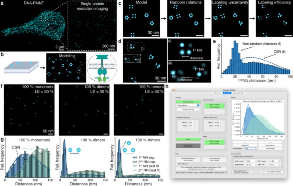
🚨 2 × PhD positions @EPFL! 🚨
Help us push the boundaries of fluorescence microscopy - DNA nanotech, custom optics & spatial omics in Lausanne 🇨🇭. Start Jan 2026. Send CV + motivation + 2 refs → fschueder@ethz.ch
#PhD #Hiring #microscopy #SuperResolution #SpatialOmics #DNAPAINT #FLASHPAINT
31.07.2025 08:56 —
👍 24
🔁 20
💬 1
📌 1

Congratulations to Ralf on your election as a new EMBO member:
❕Original press release from @embo.org : www.embo.org/press-releas...
03.07.2025 08:57 —
👍 14
🔁 2
💬 0
📌 0
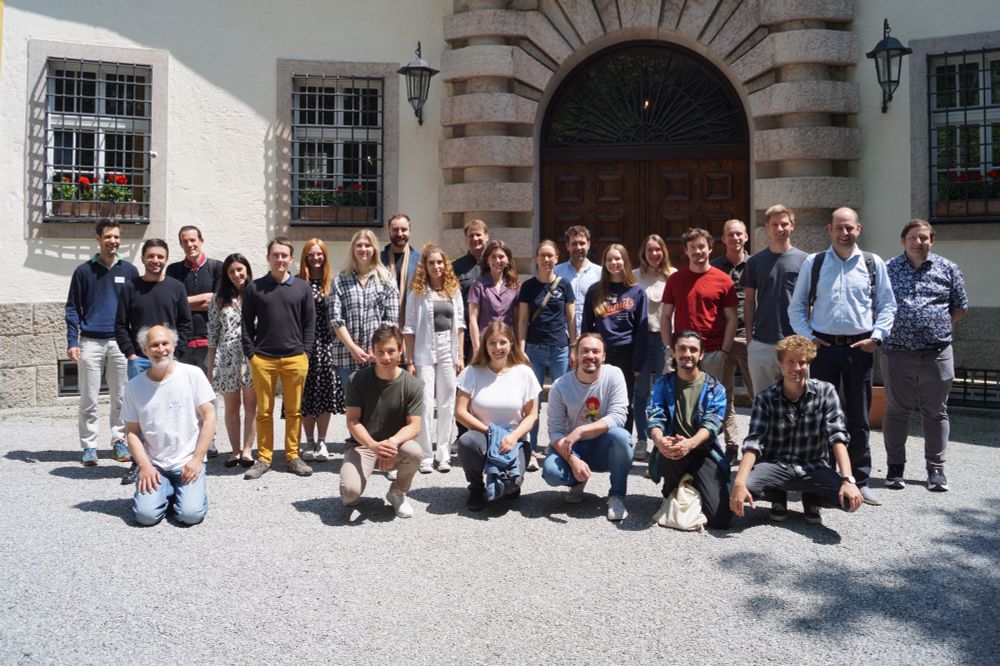
Great science, great company and stunning views at our Lab retreat on Schloss Ringberg 🏰🧬🔬. Big thanks to our guests Sabrina Simoncelli, Sebastian Kobold, Thomas Schlichthärle, @massivephotonics.bsky.social & students from the @lfmilles.bsky.social and @mlsb-borgwardt.bsky.social Labs for joining!
14.06.2025 18:31 —
👍 14
🔁 2
💬 0
📌 0
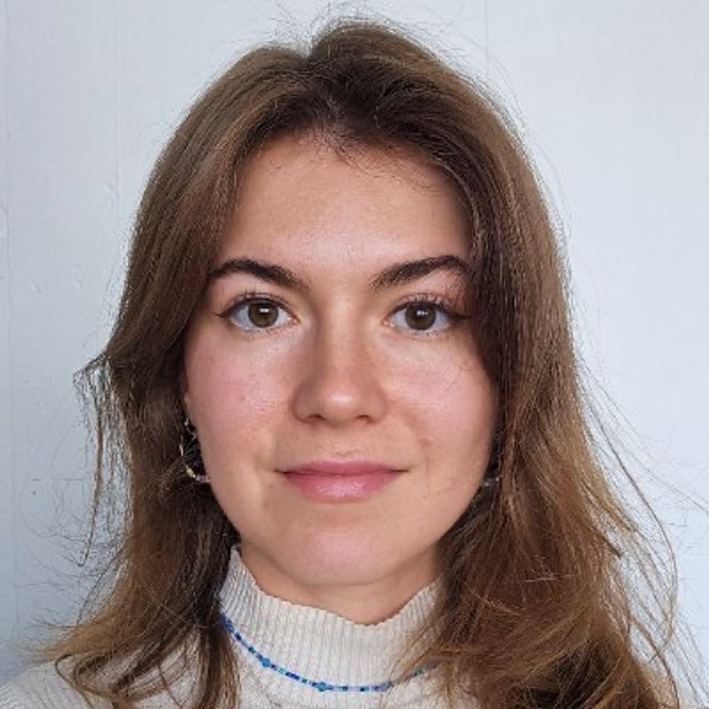
Very excited to present our latest work: SPINNA, an analysis framework and software package for single-protein resolution data! 🖥️🤩
We can directly quantify stoichiometry and oligomerization from super-res (DNA-PAINT, RESI) images!! 🧬🎨
07.05.2025 14:56 —
👍 30
🔁 11
💬 0
📌 0
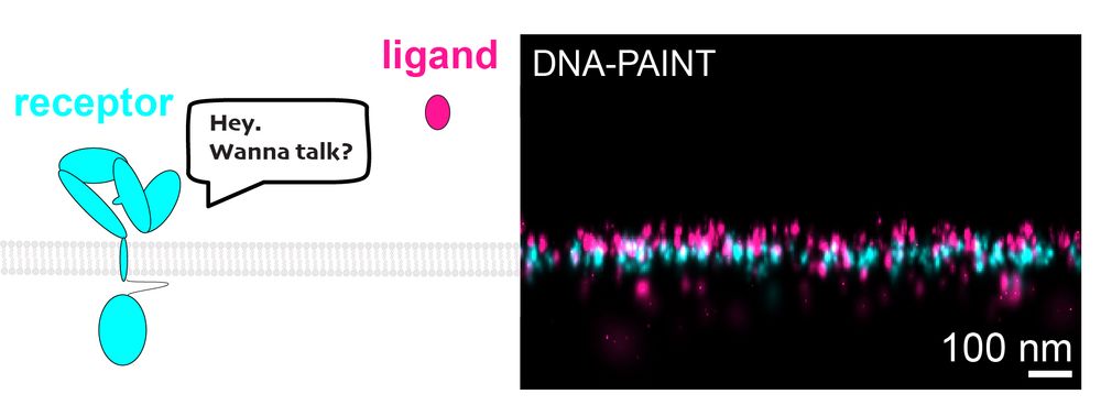
IMAGING LIGAND-RECEPTOR INTERACTIONS AT SINGLE-PROTEIN RESOLUTION WITH DNA-PAINT🔬
Ever wonder how cells "talk"? It starts when ligands bind to receptors on cell surfaces. We have cracked the challenge of imaging small ligands on cell surfaces. #DNAPAINT #celltalk 💬
doi.org/10.1002/smtd...
10.04.2025 11:40 —
👍 20
🔁 6
💬 2
📌 2
Thanks to @eduardunterauer.bsky.social, @jekristina.bsky.social, @forna.bsky.social, @opazo.bsky.social and @carstenmarr.bsky.social for putting together this great step-by-step guide on how to achieve up to 30-color DNA-PAINT spatial proteomics at sub-15 nm resolution. 🚀
07.04.2025 19:58 —
👍 6
🔁 1
💬 0
📌 0

Cell Press: STAR Protocols
STAR Protocols is an open access, peer-reviewed journal from Cell Press. We offer structured, transparent, accessible, and repeatable step-by-step experimental and computational protocols from all are...
One month ago today, I published my first paper with the @jungmannlab.bsky.social 🥳
What better way to celebrate #MicroscopyMonday than with this STAR Protocol on SUM-PAINT spatial proteomic imaging: a guide for highly multiplexed DNA-PAINT imaging in neurons. star-protocols.cell.com/protocols/4066
07.04.2025 19:58 —
👍 9
🔁 4
💬 1
📌 0
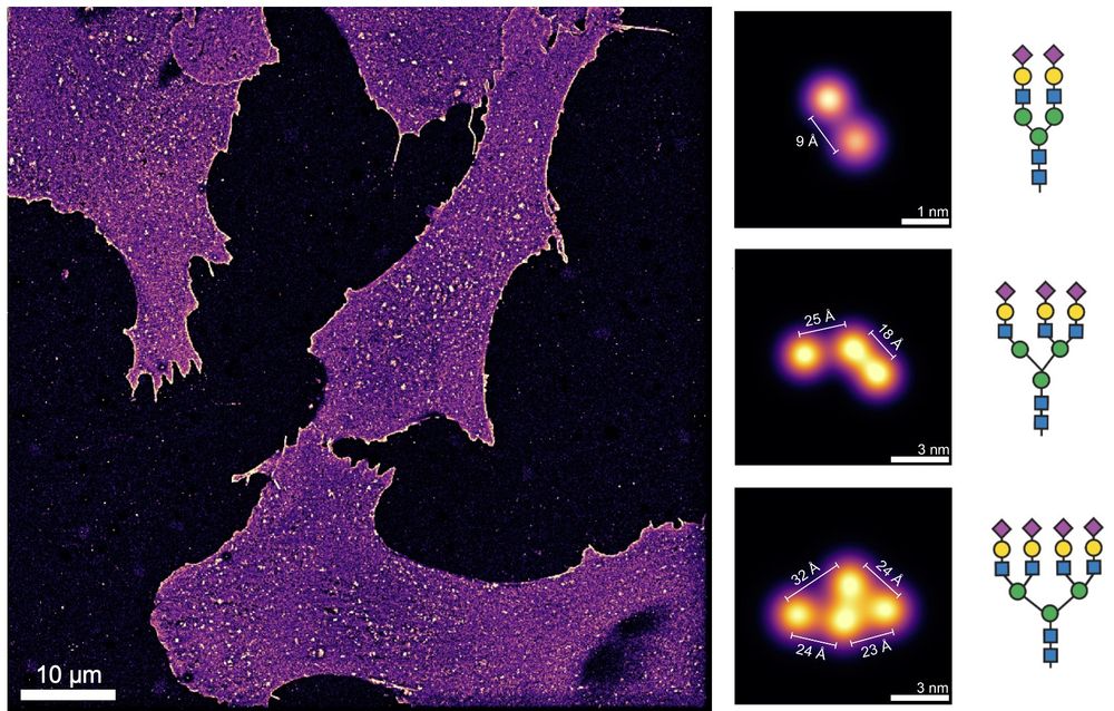
ÅNGSTRÖM-RESOLUTION IMAGING OF CELL-SURFACE GLYCANS 🧬🎨🍬
The glycocalyx, our cells' sugar coat, holds secrets in immunology, cancer, viral infections, and more. Visualizing its molecular architecture was impossible… until now. #glycotime #microscopy
www.biorxiv.org/content/10.1...
10.02.2025 08:21 —
👍 314
🔁 101
💬 15
📌 15
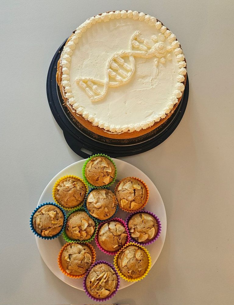
Whipped up a DNA-themed cake for the
@JungmannLab - sweet science at its finest! 🍰🔬 #PhDlife
24.01.2025 13:44 —
👍 4
🔁 0
💬 0
📌 0
Do you know what I love most about my PhD @JungmannLab? Actually seeing the things I learned from biology textbooks come to life🪄. Check out this DNA-PAINT image of the Golgi, showing both cis (GM130) and trans (TGN38) faces together with peroxisomes. Happy
#FluorescenceFriday😊
24.01.2025 13:44 —
👍 2
🔁 0
💬 0
📌 0
If you're curious about imaging more than 6 targets using DNA-PAINT, take a look at our recent publication on SUM-PAINT for high-throughput multiplexing
cell.com/cell/fulltext/…
09.08.2024 12:36 —
👍 1
🔁 0
💬 0
📌 0
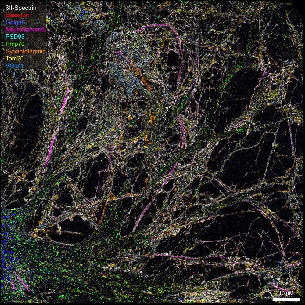
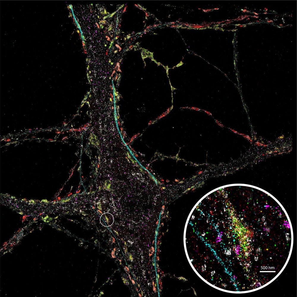
Looking at this beautifully detailed neuron, stained for 9 different proteins using SUM-PAINT, feels like stepping into one of David S. Goodsell's paintings illustrating the crowded cellular environment. #FluorescenceFriday #phdlife @JungmannLab
09.08.2024 12:36 —
👍 0
🔁 0
💬 1
📌 0
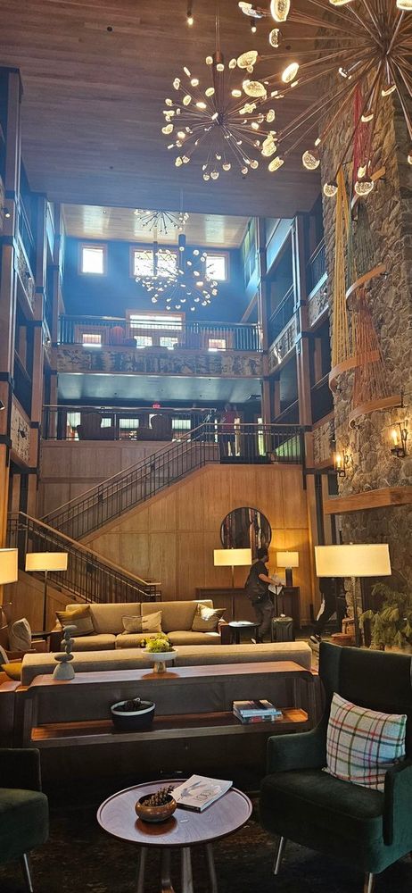
Excited to attend my first ever conference on single molecule approaches to biology by @GordonConf in beautiful Maine!
Caught a fun 'Where's Waldo' moment in the hotel lobby - can you spot Ralf Jungmann and Luciano Masullo waving at me in the background? 🔍 @JungmannLab @l_masu
15.07.2024 15:34 —
👍 0
🔁 0
💬 0
📌 0
Happy Friday! To wrap up another exciting week of learning highly multiplexed DNA-PAINT in the @JungmannLab, here's a nice #cellfie of a rat hippocampal neuron stained for synaptic vesicle markers (Vamp2, VGlut1 and synaptotagmin), clathrin, neurofilament and peroxisomes.
14.06.2024 13:20 —
👍 0
🔁 0
💬 0
📌 0
With this my MINFLUX chapter may has concluded for now, but this was just the kick-off to my journey into super-resolution microscopy as a PhD student @JungmannLab 🚀
29.05.2024 17:07 —
👍 1
🔁 0
💬 0
📌 0
Uncovering kinesin dynamics in neurites with MINFLUX
Communications Biology - A MINFLUX tracking study of a truncated kinesin-1 mutant in gently fixed primary rat hippocampal neurons revealed 8 nm substeps and a rotation of its head during the...
Here it is - fresh off the press. Tracking substeps of kinesin in mildly fixed neurons using MINFLUX 🔬.
I am proud to see my master thesis in close work together with Otto Wirth finally published!
nature.com/articles/s4200…
29.05.2024 17:07 —
👍 1
🔁 0
💬 1
📌 0