Maximilian Madern's Avatar

Maximilian Madern

@maxmadern.bsky.social

PhD student in the Tanenbaum lab, Hubrecht institute

66 Followers  |  76 Following  |  2 Posts  |  Joined: 19.01.2025
Posts Following

Posts by Maximilian Madern (@maxmadern.bsky.social)

Post image

Excited to share our new paper! We developed a method to visualize proteasomal degradation at the single–molecule level in live cells, enabling us to dissect distinct modes of substrate engagement, probe co-factor dependence, and study proteasome–ribosome collisions.
www.biorxiv.org/content/10.6...

20.01.2026 12:27 β€” πŸ‘ 48    πŸ” 15    πŸ’¬ 1    πŸ“Œ 1
In vivo kinetics of protein degradation by individual proteasomes Protein degradation by the proteasome is central to cellular homeostasis and has been studied extensively using biochemical and structural studies. Despite an in-depth understanding of core proteolytic activity, it has remained largely unresolved how individual proteasomes process substrates inside living cells where many substrate types and co-factors exist. Here, we establish a live-cell single-molecule imaging approach that enables direct visualization and quantification of protein degradation by individual proteasomes. Using this approach, we find that substrate identity, folding and protein-protein interaction have a surprisingly modest impact on processing efficiency, whereas the mode of substrate engagement greatly impacts substrate processing; degradation initiated from protein termini typically proceeds rapidly and with high processivity, whereas internal engagement constitutes a distinct processing mode that exhibits poor processivity and a specific requirement for the AAA+ family ATPase p97/VCP. Furthermore, degradation initiated from opposite termini proceeds with asymmetric rates in a sequence-dependent manner, demonstrating that directionality is an important feature of proteasomal processing in vivo. Notably, poly-glutamine substrates associated with neurodegenerative disease are efficiently degraded from one terminus but resist degradation when engaged from the opposite terminus, highlighting the importance of substrate engagement mode. Together, our results show that different modes of substrate engagement lead to different proteasomal processing outcomes in vivo and revise the prevailing view of the proteasome as a uniform degradation machine. ### Competing Interest Statement The authors have declared no competing interest.

New lab paper!! We develop a technology for real-time, single-molecule visualization of proteasomal substrate degradation in cells. We find that the site of substrate engagement by the proteasome determines decay kinetics, efficiency and co-factor requirement.

www.biorxiv.org/content/10.6...

20.01.2026 08:32 β€” πŸ‘ 47    πŸ” 15    πŸ’¬ 1    πŸ“Œ 2
Post image

Our paper is out! We delevoped a method to follow individual translating ribosomes for hours in living cells, and discovered that ribosomes are great friends and help each other in problematic situations:
www.cell.com/cell/fulltex...

03.02.2025 08:31 β€” πŸ‘ 43    πŸ” 11    πŸ’¬ 0    πŸ“Œ 0
Preview
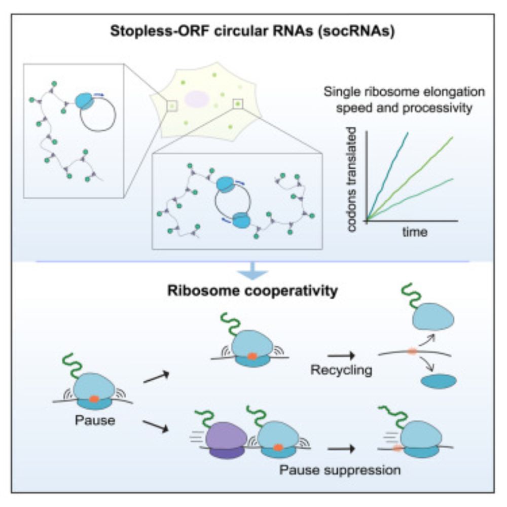
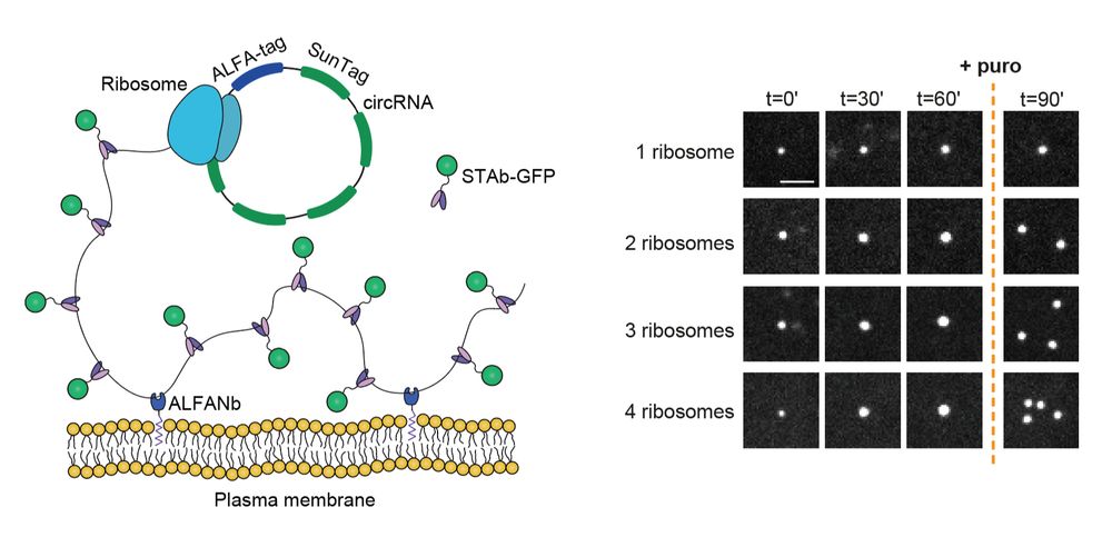
Long-term imaging of individual ribosomes reveals ribosome cooperativity in mRNA translation Ribosomes cooperate through transient collisions to ensure efficient translation.

Our paper on Stopless-ORF Circular RNAs (socRNAs) is now out in Cell. By high-res tracking and comparing translation by either single or multiple ribosomes, we find that ribosomes cooperate to overcome pausing to ensure fast and efficient translation

www.cell.com/cell/fulltex...

03.02.2025 07:38 β€” πŸ‘ 114    πŸ” 47    πŸ’¬ 4    πŸ“Œ 6
Video thumbnail

Very happy to share our preprint on visualizing the life cycle of Influenza viruses using single-molecule imaging! πŸ₯³ We developed two techniques to visualize infections of unmodified influenza viruses in live cells from endosomal release to budding of new viruses. For more details&videos see below ⬇️

21.01.2025 10:16 β€” πŸ‘ 65    πŸ” 14    πŸ’¬ 2    πŸ“Œ 1