![Fig. 5.
Detection of beet mosaic virus (BtMV)-derived A, nuclear inclusion body b double-stranded RNA (dsNIb) and B, P1 double-stranded RNA (dsP1) after mechanical application on Beta vulgaris in treated (local) and untreated (systemic) leaves at different time points (1 h after application [hpi], 2 and 7 days after application [dpi]) by RT-PCR. Mock-inoculated plants were treated only with carborundum and water. M, DNA marker (GeneRuler 100 bp DNA Ladder, Thermo Fisher Scientific).](https://cdn.bsky.app/img/feed_thumbnail/plain/did:plc:ypn4w5ifq3ag6mlwd4rd5t2j/bafkreiboq64a7oikqlbg5gk4v47yfjftuxu2rjgyfhzdjhaoxmck4p22yu@jpeg)
Fig. 5.
Detection of beet mosaic virus (BtMV)-derived A, nuclear inclusion body b double-stranded RNA (dsNIb) and B, P1 double-stranded RNA (dsP1) after mechanical application on Beta vulgaris in treated (local) and untreated (systemic) leaves at different time points (1 h after application [hpi], 2 and 7 days after application [dpi]) by RT-PCR. Mock-inoculated plants were treated only with carborundum and water. M, DNA marker (GeneRuler 100 bp DNA Ladder, Thermo Fisher Scientific).
Beet mosaic virus (BtMV), once managed via neonicotinoid seed treatments, is now being targeted with RNA-based resistance. Dennis Rahenbrock et al. report dsRNA designed from small RNA sequencing effectively reduced BtMV in sugar beet and N. benthamiana in the greenhouse: doi.org/10.1094/MPMI...
22.09.2025 15:57 — 👍 0 🔁 0 💬 0 📌 0
Deadline extended: Submit to the forthcoming MPMI focus issue by November 1, 2025! 🚨
08.09.2025 14:04 — 👍 1 🔁 2 💬 0 📌 0

Life cycle of Verticillium longisporum-infected oilseed rape. When Brassica napus plants start to senesce, microsclerotia protrude from the stem tissue, become visible, and begin to fall to the ground. Commonly, microsclerotia are observed or spotted on the stubble and leaf debris after harvest and become mixed into the soil, following the next soil management step. The microsclerotia are resistant structures and can survive in the soil for many years under adverse conditions. Upon germination, fungal hyphae enter openings or wounds in the root, and the fungus progresses into the xylem tissue. V. longisporum can remain in the vascular tissue for a long time, making it invisible during field plant inspections. Upon plant maturation, grayish stripes appear on the ripening stems by densely formed microsclerotia. They can start dropping to the ground and will be ready to initiate a new round of infections when the conditions become favorable.
New interactions review on Verticillium longisporum, the cause of stripe disease in Brassica crops. Rafiei, Dixelius & Tzelepis outline the pathogenicity factors, host immune responses, knowledge gaps, and molecular tools for studying this threat.
Read the full article here: doi.org/10.1094/MPMI...
03.09.2025 18:12 — 👍 4 🔁 0 💬 0 📌 0

Fig. 1.
Visual symbiotic phenotypes. Strains were inoculated onto alfalfa plants grown under nitrogen-deficient conditions. Representative pictures were taken of A, plant growth and B, their representative nodules after 28 days of growth. Strains used are as follows: wild type (wt), Rm1021; tktA::Tn5, SRmD397; stkR1, SRmD410. Scale bar in Panel B represents 1 mm.
In Sinorhizobium meliloti, loss of the carbon metabolism blocks symbiosis with alfalfa. Kaur et al. demonstrate that disrupting the key metabolic enzyme transketolase (tktA) in S. meliloti completely impairs its ability to engage in nitrogen-fixing symbiosis with alfalfa: doi.org/10.1094/MPMI...
29.08.2025 16:05 — 👍 1 🔁 0 💬 0 📌 0

Representative images showing the outcome of cell death assays for MLA7L902S in Nicotiana benthamiana. Agrobacterium strains carrying Mla13, Mla7, or Mla7L902S were co-infiltrated with Agrobacterium strains carrying AVRA13-V2, AVRA13-1, or AVRA7-2 into N. benthamiana leaves. Images were taken 5 days after infiltration. Cell death is indicated by yellow to dark spots on the leaves. The numbers indicate the numbers of leaves showing cell death phenotypes as a proportion of the total number of leaves infiltrated.
Crops often lack diverse resistance genes, making them vulnerable to rapidly evolving pathogens. Xiaoxiao Zhang et al. used DNA shuffling on barley Mla7 and Mla13 genes to create MLA variants, some gaining the ability to recognize the powdery mildew effector AVRA13: doi.org/10.1094/MPMI...
19.08.2025 17:53 — 👍 1 🔁 0 💬 0 📌 0
Deadline approaching—submit to the forthcoming focus issue by September 1!
08.08.2025 17:42 — 👍 1 🔁 4 💬 0 📌 0

Representative images of callose deposition. EV, empty vector.
Editor’s Pick! The cassava pathogen Xanthomonas phaseoli pv. manihotis (Xpm) causes severe disease, but its effector functions are not well understood. Diana Gómez De La Cruz et al. show that the Xpm effector XopAE suppresses plant defenses. Learn more: doi.org/10.1094/MPMI...
28.07.2025 13:42 — 👍 4 🔁 2 💬 1 📌 0
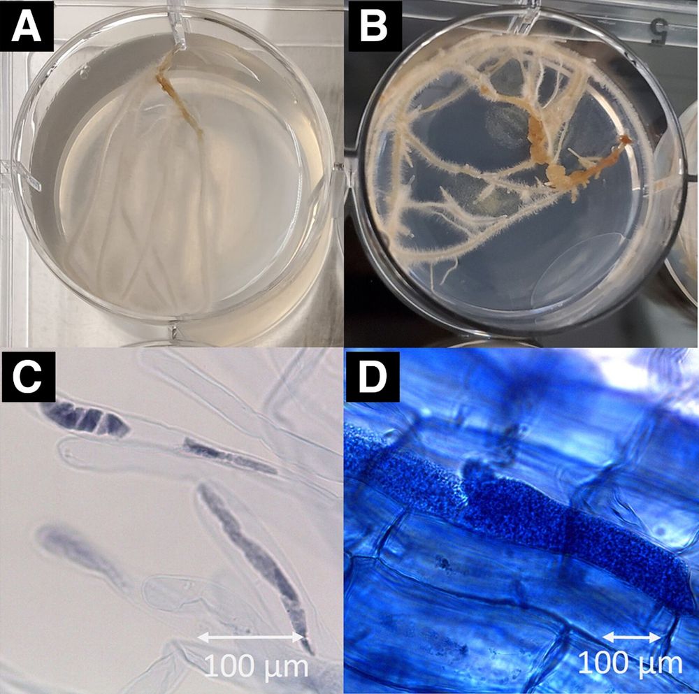
Infection of Spongospora subterranea f. sp. subterranea (Sss) in a hairy root culture system. A, Healthy potato hairy roots 21 days after induction with Rhizobium rhizogenes. B, Formation of gall-like structures on hairy roots. C, Sss sporangia stained with trypan blue observed in root hairs 4 weeks post-inoculation. D, Sss sporangia stained with trypan blue observed in the cortex of hairy roots 8 weeks post-inoculation.
A breakthrough for potatoes: Samodya K. Jayasinghe et al. identify salicylic acid as a key defense hormone against Spongospora subterranea f. sp. subterranea, a persistent soilborne protist threatening yields around the world. 🥔
Read the press release to learn more: www.apsnet.org/about/newsro...
23.07.2025 20:02 — 👍 4 🔁 3 💬 1 📌 0

A Viral Brake on Bloom: BYDV-GAV Delays Flowering via VOZ Degradation | Molecular Plant-Microbe Interactions®
Our commentary on how a virus infection delays flowering published in MPMI titled "A Viral Brake on Bloom: BYDV-GAV Delays Flowering via VOZ Degradation" | Molecular Plant-Microbe Interactions® apsjournals.apsnet.org/doi/10.1094/...
@mpmijournal.bsky.social
#plantvirus
#BYDV
#virusinfection
29.06.2025 17:44 — 👍 2 🔁 1 💬 0 📌 0
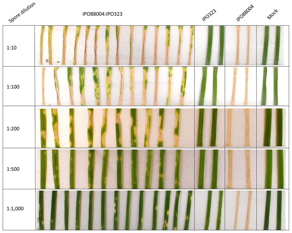
Co-inoculation of Zymoseptoria tritici strains IPO323 and IPO88004. Co-inoculation experiments were performed with the avirulent strain IPO323 and the virulent strain IPO88004. Disease severity was evaluated across a series of virulent-to-avirulent ratios ranging from 1:10 to 1:1,000. Disease symptoms were recorded 21 days postinfection (dpi).
Haider Ali, Megan C. McDonald, and Graeme J. Kettles developed a rapid forward genetics methodology to identify genes that enable Zymoseptoria tritici to gain virulence on previously resistant wheat varieties. Learn more: doi.org/10.1094/MPMI...
12.06.2025 21:46 — 👍 6 🔁 5 💬 0 📌 0

Disease symptoms for bacterial blight (BB)-susceptible and -resistant rice varieties based on Kauffman's leaf-clipping assay.
Eliza P. I. Loo et al. discuss gaps in information acquisition and transfer between the field and laboratory and indicate that collaboration among stakeholders, scientists, farmers, and policymakers could enhance the development of pathogen-resistant crops. doi.org/10.1094/MPMI...
19.05.2025 19:30 — 👍 1 🔁 0 💬 1 📌 0

Call for Papers! Symbiotic and Pathogenic Interactions in the Rhizosphere—a Molecular Plant-Microbe Interactions Focus Issue
New call for papers alert 🚨🌱🦠 Submit to the "Symbiotic and Pathogenic Interactions in the Rhizosphere" focus issue by September 1, 2025!
Focus Issue Guest Editors: Tarek Hewezi, Hari Krishnan, Kevin Garcia, and Mina Ohtsu
Learn more: apsjournals.apsnet.org/mpmirhizosph...
08.05.2025 21:13 — 👍 8 🔁 7 💬 1 📌 2

B, Sporulating lesion on a rice leaf. C, Sporulating lesions on a wheat leaf. D, Rice panicle neck infections block all grain filling above the infection points (red arrows). E, Wheat spike blast with individual directly infected spikelets. F, Wheat rachis infections kill all spikelets above the infection points. Removed spikelets show stem infection points (red arrows) with gray sporulation. G, Finger millet neck infection (red arrow) kills all fingers on the seed head. H, Leaf lesions on perennial ryegrass showing white lesion centers after spore release.
Just published! Barbara Valent honors H. H. Flor by exploring how rice blast fungus evades plant resistance via AVR gene changes—driving boom–bust cycles—and warns of rising wheat and ryegrass blast threats from similar shifts and crossbreeding. Read the article: doi.org/10.1094/MPMI...
25.04.2025 16:15 — 👍 8 🔁 8 💬 0 📌 0
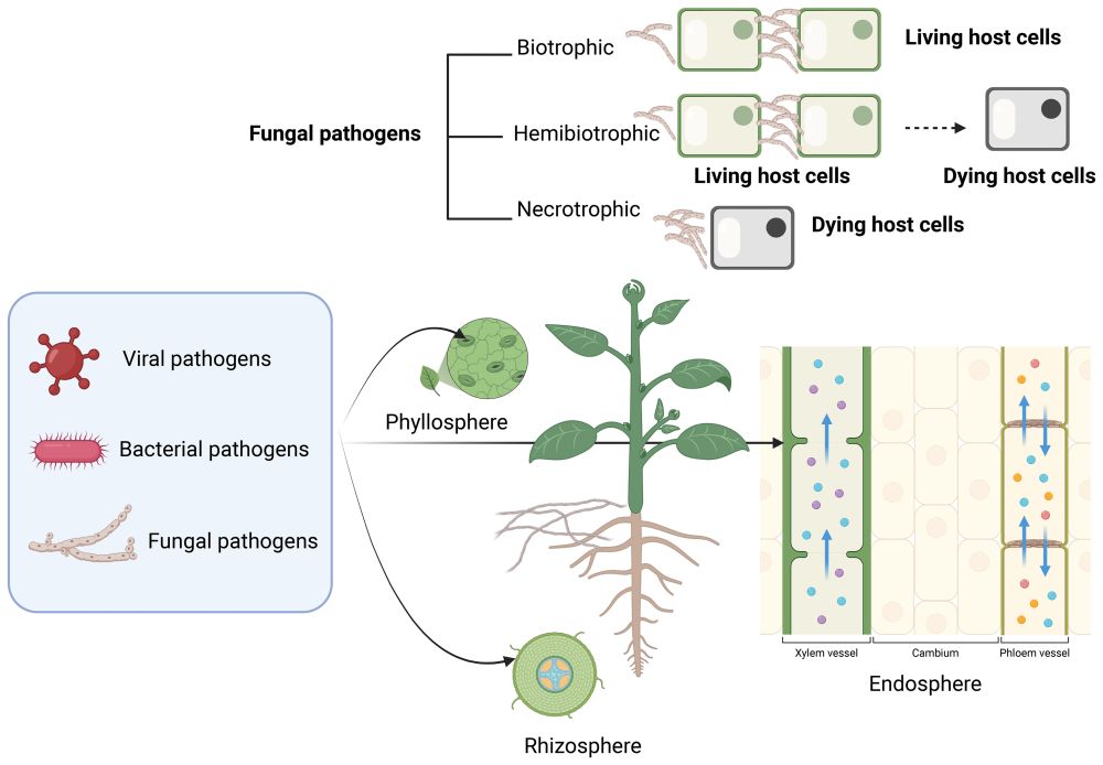
Types of phytopathogens (viruses, bacteria, and fungi) invade plant organs. Pathogens can invade the aboveground organs, the phyllosphere (leaves, stems, branches, and fruits), the roots or rhizosphere, the vascular system (phloem and xylem), or the endosphere. Phytopathogens can be biotrophic feeding on living host cells, necrotrophic feeding on dying host cells, and hemibiotrophic with an initial biotrophic phase and a later necrotrophic stage.
Alissar Cheaib and Nabil Killiny review how pathogens disrupt photosynthesis, from tissue destruction to chlorosis, and how plants adapt. Understanding these interactions is essential for #sustainableagriculture and ecosystem health. ☀️🌱 Learn more: doi.org/10.1094/MPMI... @alissarcheaib.bsky.social
14.04.2025 19:15 — 👍 4 🔁 3 💬 1 📌 0

Localization of tomato spotted wilt virus (TSWV) proteins with infected and healthy plant cells. Left: TSWV proteins expressed in agroinfiltrated plant cells. Column 1, green fluorescent protein (GFP) tagged to TSWV protein; column 2, unfused red fluorescent protein (RFP); and column 3, overlay of columns 1 and 2. Rows are as follows: I to III, TSWV-N:GFP; IV to VI, TSWV-NSs:GFP; VII to IX, TSWV-GFP:NSm; X to XII, TSWV-GN:GFP; and XIII to XV, TSWV-GC:GFP. Right: TSWV proteins expressed in TSWV-infected plant cells. Column 1, GFP tagged to TSWV protein; column 2, unfused RFP; and column 3, overlay of columns 1 and 2. Rows are as follows: XVI to XVIII, TSWV-N:GFP; XIX to XXI, TSWV-NSs:GFP; XXII to XXIV, TSWV-GFP:NSm; XXV to XXVII, TSWV-GN:GFP; and XXVIII to XXX, TSWV-GC:GFP. Scale bar is equal to 20 μm.
New research by K. M. Martin et al. expands our knowledge of tomato spotted wilt virus infection and elaborates on the intricate relationships between viral proteins, cellular dynamics, and host responses.
Learn more: doi.org/10.1094/MPMI...
03.04.2025 19:06 — 👍 6 🔁 2 💬 0 📌 0

Differentially expressed long noncoding RNAs (lncRNAs) contain putative mimic binding sites for microRNAs (miRNAs). Schematic representations showing base-pairing between A, TCONS_00160362 and miR47; B, TCONS_00188971 and miR156e-5p; C, TCONS_00041366 and miR403-5p; D, TCONS_00084313 and miR184; and E, TCONS_00146883 and miR698.
Selin Ozdemir et al. investigated the differential regulation and function of lncRNAs during two different stages of tomato infection by the root-knot #nematode Meloidogyne incognita. 🍅🪱 Learn more: apsjournals.apsnet.org/doi/10.1094/...
01.04.2025 19:20 — 👍 4 🔁 3 💬 0 📌 0
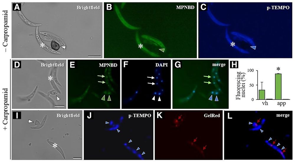
Colletotrichum graminicola conidia/appressoria were fluorescence-labeled for Fe2+/Fe3+ with/without carpropamid. MPNBD/p-TEMPO stained Fe2+/Fe3+ (A-C). MPNBD/DAPI showed Fe3+ in nuclei (D-G). p-TEMPO/GelRed stained Fe2+, not in nuclei (I-L). Quantification of Fe2+/Fe3+ in hyphae/appressoria (H). Asterisk: conidium, arrowhead: appressorium. Scale: 10μm.
Iron's redox states (Fe2+/Fe3+) are critical in fungal pathogenesis, influencing ROS production during plant-pathogen interactions. Aliyeva-Schnorr et al. developed p-TEMPO and MPNBD dyes to visualize Fe2+/Fe3+ distribution in #Colletotrichumgraminicola hyphae. Learn more: doi.org/10.1094/MPMI...
31.03.2025 16:52 — 👍 3 🔁 2 💬 0 📌 1

New research by Joydeep Chakraborty et al. elucidates the tomato PP2C-immunity associated candidate 6 (Pic6) as a negative regulator of MAPK signaling and its pivotal role in modulating tomato immunity against bacterial pathogens. 🍅🦠
Learn more: doi.org/10.1094/MPMI...
26.03.2025 05:22 — 👍 3 🔁 1 💬 0 📌 0

Tan et al. developed efficient base editing vectors, achieving high A-to-G conversion rates in Arabidopsis thaliana and successfully targeting multiple genes, including NLRs, to modify protein function, demonstrating the potential of base editing 🧬
Learn more: doi.org/10.1094/MPMI...
25.03.2025 16:19 — 👍 5 🔁 2 💬 0 📌 0

Two ‘Candidatus Liberibacter asiaticus’ (Las) virulence proteins inhibited bacterial infection-triggered callose deposition when expressed in Nicotiana benthamiana.
Editor's Pick! Amelia Lovelace et al. examined the transcriptomic landscape of ‘Candidatus Liberibacter asiaticus’ in citrus seed coat vasculatures, revealing unique gene expression patterns vs. previous midrib studies and new insights into citrus #huanglongbing. 🍊🦠 doi.org/10.1094/MPMI...
21.03.2025 18:00 — 👍 3 🔁 1 💬 0 📌 0
Our first bimonthly issue (Jan-Feb) of 2025 is now online!! 👉 apsjournals.apsnet.org/toc/mpmi/cur...
03.03.2025 15:12 — 👍 2 🔁 2 💬 0 📌 0

2025 Early Career Showcase
......2025 Early Career Showcase...Registration NOW OPEN!Join the 2025 Early Career Showcase: A Global Gathering for Advancing Plant-Microbe Research Explore new breakthroughs in plant...
I just got a sneak preview of talks for Part I of the IS-MPMI Early Career Showcase next week. You won't want to miss any. Breakout discussions too. Register now! www.ismpmi.org/Events/2025-... @ismpmi.bsky.social @mpmijournal.bsky.social #plantimmunity #plantdiseaseresistance #effectorbiology
12.02.2025 19:01 — 👍 6 🔁 5 💬 0 📌 0

Support emerging researchers when you donate to the Mishkind Travel Fund!
Support #EarlyCareer members attending the upcoming #2025ISMPMI Congress in Cologne, Germany! Your donations to the #MichaelMishkindTravelFund will assist and support the next generation researchers in the field of plant-microbe interactions. Thank you! my.ismpmi.org/ISMPMI/Donat... #donate
27.01.2025 20:29 — 👍 0 🔁 3 💬 1 📌 0
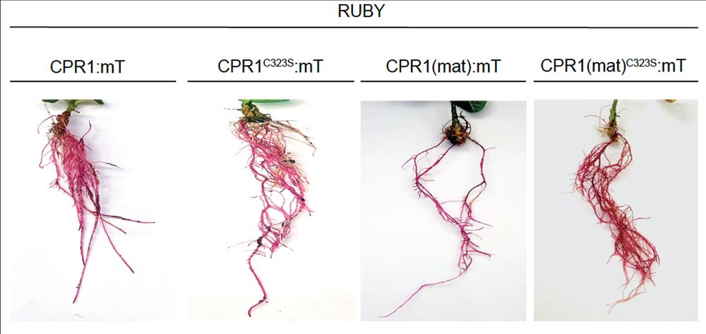
New Discovery Paves the Way for More Resistant Soybeans to Combat $1.5 Billion Crop Loss from Nematode Infection (original article by Alexandra Margets et al. @acmargets.bsky.social @inneslab.bsky.social doi.org/10.1094/MPMI...): www.eurekalert.org/news-release...
28.01.2025 15:34 — 👍 0 🔁 0 💬 0 📌 0
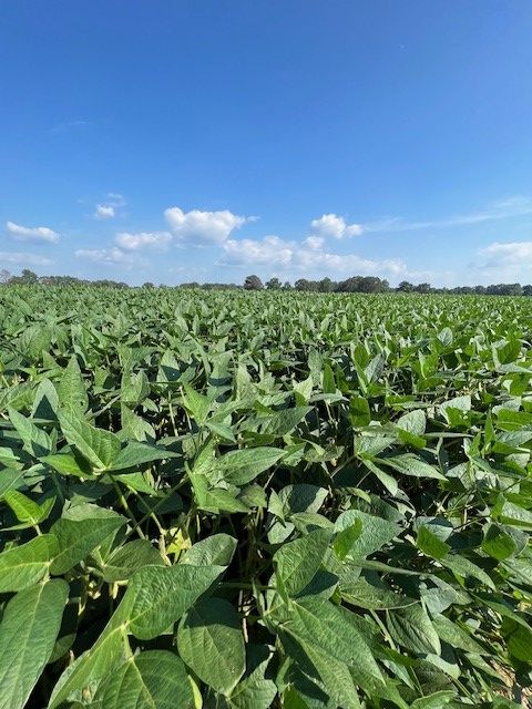
Rooting for Resistance: How Soybeans Tackle Nematode Invaders Is No Secret Anymore (original article by Mst Shamira Sultana et al. doi.org/10.1094/MPMI...): www.eurekalert.org/news-release...
28.01.2025 15:33 — 👍 2 🔁 0 💬 0 📌 0

Harnessing Nature to Defend Soybean Roots: Using Novel Cry Protein to Control the Number-One Soybean Pest (original article by R. Howard Berg et al. doi.org/10.1094/MPMI...): www.eurekalert.org/news-release...
28.01.2025 15:33 — 👍 1 🔁 0 💬 0 📌 0

MPMI recently published three prominent papers on breakthrough research in soybean cyst nematode management. See the thread below to read the press releases about the novel research 🫛🪱:
www.eurekalert.org/news-release...
www.eurekalert.org/news-release... www.eurekalert.org/news-release...
28.01.2025 15:32 — 👍 2 🔁 3 💬 3 📌 0

![Fig. 5.
Detection of beet mosaic virus (BtMV)-derived A, nuclear inclusion body b double-stranded RNA (dsNIb) and B, P1 double-stranded RNA (dsP1) after mechanical application on Beta vulgaris in treated (local) and untreated (systemic) leaves at different time points (1 h after application [hpi], 2 and 7 days after application [dpi]) by RT-PCR. Mock-inoculated plants were treated only with carborundum and water. M, DNA marker (GeneRuler 100 bp DNA Ladder, Thermo Fisher Scientific).](https://cdn.bsky.app/img/feed_thumbnail/plain/did:plc:ypn4w5ifq3ag6mlwd4rd5t2j/bafkreiboq64a7oikqlbg5gk4v47yfjftuxu2rjgyfhzdjhaoxmck4p22yu@jpeg)


