Last but not least: this achievement was only possible due to the amazing dedication and insight by @dijo-mj.bsky.social, @linussison.bsky.social, @nazlicanyurekli.bsky.social, and Sheston Culpepper!
02.10.2025 11:05 — 👍 3 🔁 0 💬 0 📌 0
This methodology adds a fundamentally new perspective to our understanding of cell biology. We’re looking forward to ideas and feedback – please get in touch!
02.10.2025 11:05 — 👍 2 🔁 0 💬 1 📌 0

Have you ever wondered where different sialylation states of the EGF receptor are located on the cell surface? Here you go:
02.10.2025 11:05 — 👍 1 🔁 0 💬 1 📌 0

This enables us to map glycoforms of individual proteins as well as their organization on the cell membrane in the native state.
02.10.2025 11:05 — 👍 2 🔁 0 💬 1 📌 0

Here, we demonstrate the first spatial mapping of the cell-surface glycoproteome at true molecular resolution. We target proteins of interest with antibody-nanobody constructs and address glycosylation with either lectins or metabolic oligosaccharide engineering.
02.10.2025 11:05 — 👍 1 🔁 0 💬 1 📌 0
Various methods like mass spectrometry have been used to study the cell-surface glycoproteome with great success, however, no technique could analyze the spatial organization of proteins and their glycosylation patterns at molecular resolution.
02.10.2025 11:05 — 👍 1 🔁 0 💬 1 📌 0

Every cell in the human body is surrounded by the glycocalyx, the "sugar coat" of the cell. A key component of the glycocalyx are glycosylated proteins. Indeed, virtually all cell-surface proteins are glycosylated.
02.10.2025 11:05 — 👍 1 🔁 0 💬 1 📌 0
Maybe I’m biased: but well deserved:) 🙌
01.09.2025 21:03 — 👍 2 🔁 0 💬 0 📌 0

Attended #SMLMS2025 last week in Bonn, Germany. Many thanks to the organizers @uendesfelder.bsky.social @heilemannlab.bsky.social and Prof. Kubitscheck for introducing #youngSMLMS. That was really mindful of the young researchers including me:). Had a great time in Bonn discussing science.
01.09.2025 17:22 — 👍 7 🔁 2 💬 1 📌 0
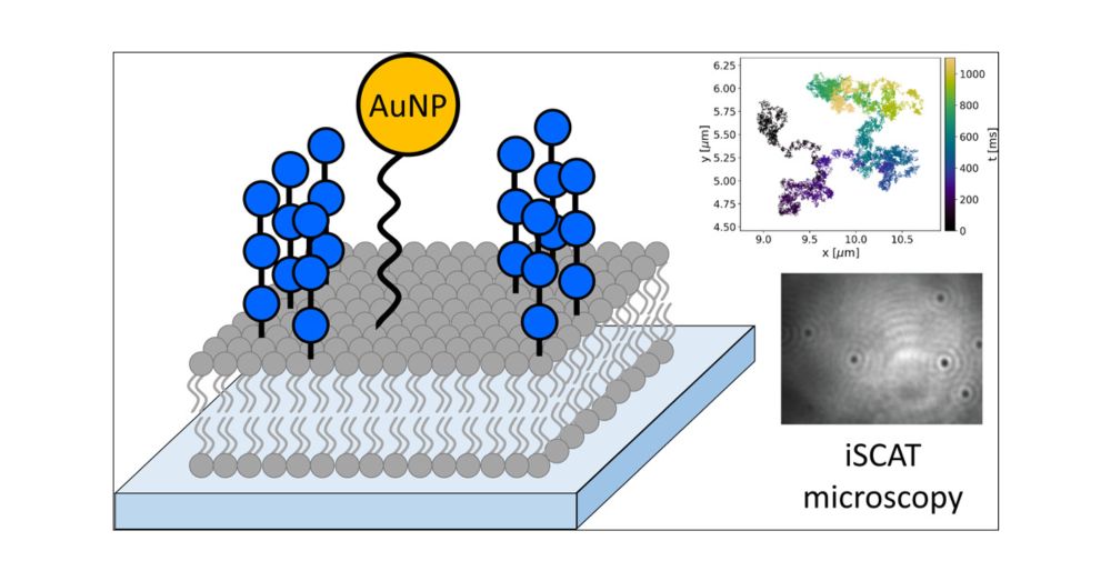
Bottom-up Investigation of Spatiotemporal Glycocalyx Dynamics with Interferometric Scattering Microscopy
Over recent decades, the glycocalyx, an extracellular organelle composed of a multitude of glycolipids, glycoproteins, proteoglycans, and glycoRNA, has gained considerable interest in cellular biology. While research in this field has revealed its tremendous importance in ever more aspects of physiological and pathological cellular processes, many of the principles that govern the role of the glycocalyx in these processes on a molecular level are still unknown. In order to unravel the fundamental laws underlying glycocalyx function, new technologies are required that enable the distinction between individual subprocesses within the intricate environment of the glycocalyx. Here, we establish an experimental platform to investigate the dynamics of the glycocalyx at the nanometer and microsecond length and time scales in a bottom-up fashion. We synthesized defined oligosaccharides and installed them on supported lipid bilayers. This way, synthetic glycolipids were assembled to glycocalyx model systems with tunable properties. By investigating these tunable model systems with interferometric scattering (iSCAT) microscopy, we gain access to the required spatiotemporal resolution. We found a strong correlation between the molecular structure of several investigated model glycans and global dynamics of the system. Our findings are corroborated by atomistic molecular dynamics simulations and coarse-grained Brownian dynamics simulations. Our results provide the first direct experimental evidence on the relationship between glycan structure, organization, and dynamics, offering a robust and versatile basis for a quantitative understanding of glycocalyx biology and physics at the molecular level.
🔔
Excited to share our latest work on the dynamics of synthetic glycolipids in model membranes! 🤩
@biomemphys.bsky.social!
Bottom-up Investigation of Spatiotemporal Glycocalyx Dynamics with Interferometric Scattering Microscopy | Journal of the American Chemical Society pubs.acs.org/doi/full/10....
28.08.2025 11:47 — 👍 3 🔁 2 💬 1 📌 0
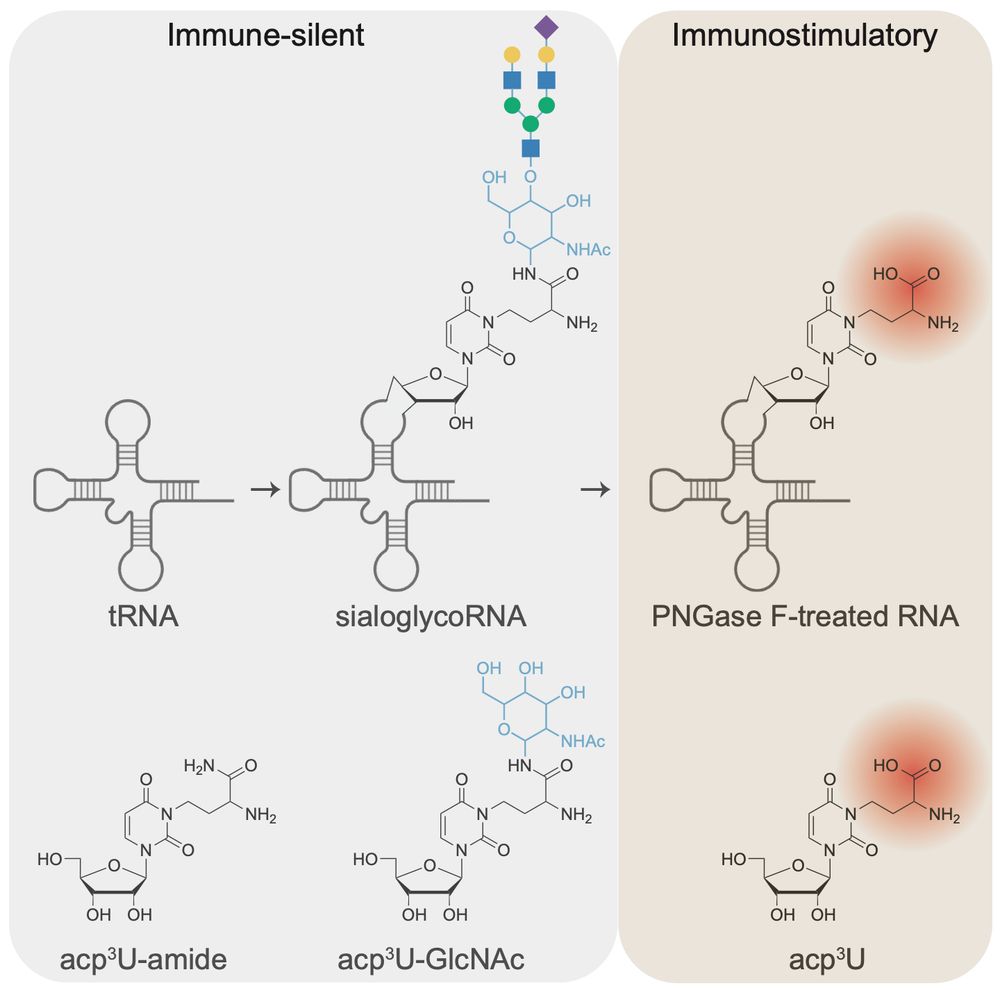
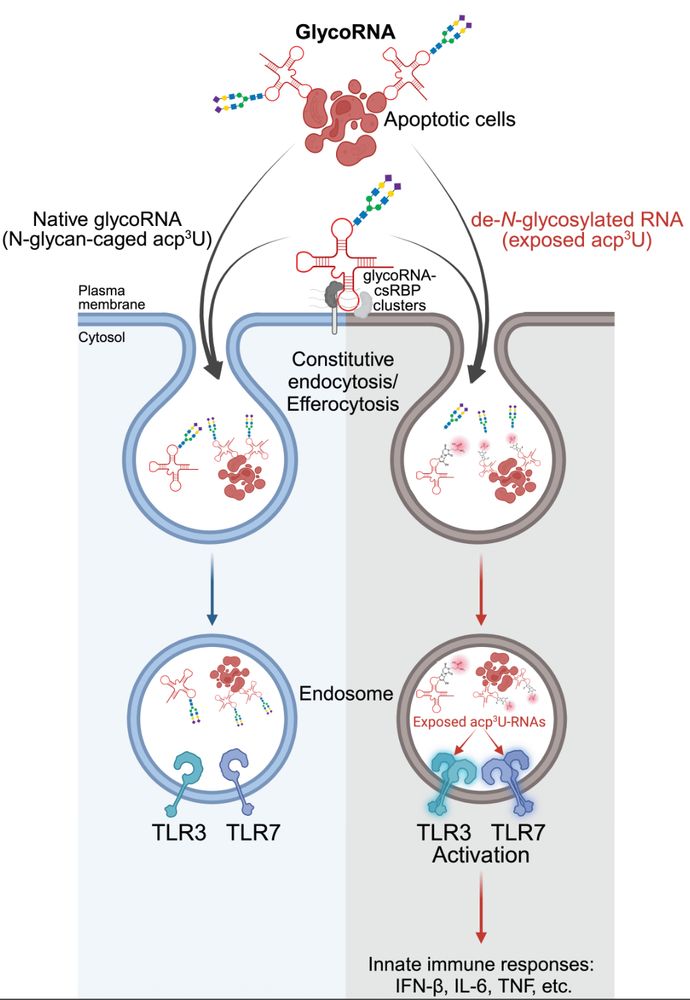
RNA N-glycosylation enables immune evasion and homeostatic efferocytosis by chemically caging acp3U. Excited to report this work lead by Vinnie @vinnieviruses.bsky.social and in collaboration with @vijayrathinam.bsky.social in @nature.com www.nature.com/articles/s41...
06.08.2025 15:36 — 👍 108 🔁 46 💬 7 📌 2
A more personal impression on our recent work on molecular resolution microscopy of the glycocalyx - check it out!
#glycotime
06.08.2025 11:38 — 👍 4 🔁 0 💬 0 📌 0
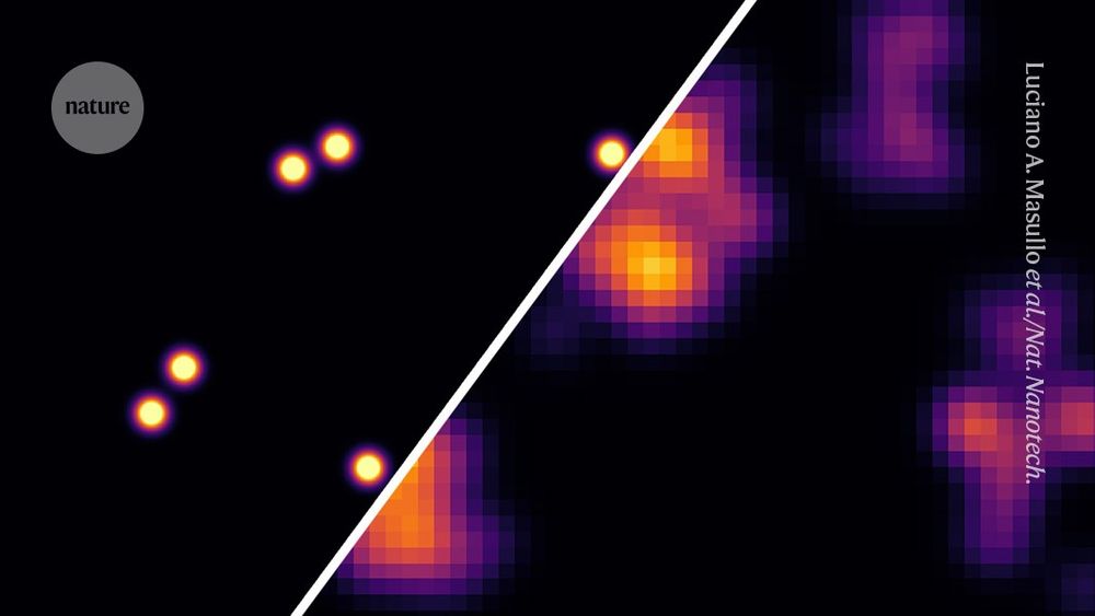
Cell's sugar coating mapped at below-nanometre resolution
Super-resolution technique works with off-the-shelf optical microscopes.
Scientists have mapped individual sugar molecules on the surface of cells at a resolution once thought to be impossible for light microscopes
go.nature.com/4l2wVjS
30.07.2025 07:57 — 👍 65 🔁 15 💬 0 📌 3
Geeking out is the biggest compliment:D
29.07.2025 21:34 — 👍 0 🔁 0 💬 0 📌 0
Very happy to see this one out - the first molecular-resolution imaging of glycocalyx components in their native environment! Great collaboration with @jungmannlab.bsky.social - congrats to first authors @karimalmahayni.bsky.social and @lumasullo.bsky.social and to the whole team!
28.07.2025 12:38 — 👍 10 🔁 5 💬 0 📌 0

Glycan Atlassing: Nanoscale analysis of glycocalyx architecture enables functional tracing of cell state
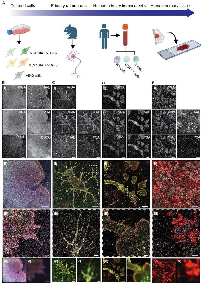
The glycocalyx is a complex layer of glycosylated biomolecules surrounding all cells in the human body. It is involved in the regulation of critical cellular processes such as immune response modulation, cell adhesion, and host-pathogen interactions. Despite these insights, the functional relationship between glycocalyx architecture and cellular state has remained elusive so far, mainly attributable to the structural diversity of glycocalyx constituents and their nanoscale organization. Here, we show that DNA-tagged lectin labeling and metabolic oligosaccharide engineering enables multiplexed super-resolution microscopy of glycocalyx constituents, yielding an atlas of glycocalyx architecture with nanometer resolution. Quantitative analysis of the obtained nanoscale map of glycocalyx constituents facilitates the extraction of characteristic spatial relationships that accurately report on cellular state. We demonstrate the capacity of our approach, which we term Glycan Atlassing, across cell and tissue types, ranging from cultured cell lines to primary immune cells, neurons, and primary patient tissue. Glycan Atlassing establishes a powerful strategy for investigating glycocalyx remodeling in development and disease, potentially enabling the development of new glycocalyx-centered targets in diagnosis and therapy. ### Competing Interest Statement The authors have declared no competing interest. Else Kröner-Fresenius-StiftungElse Kröner-Fresenius-Stiftung, https://ror.org/03zcxha54, 2020_EKEA.91 Deutsche ForschungsgemeinschaftDeutsche Forschungsgemeinschaft, https://ror.org/018mejw64, 529257351, 460333672, 270949263 Wilhelm Sander StiftungWilhelm Sander Stiftung, https://ror.org/02q83sc19, 2023.025.1 European Research CouncilEuropean Research Council, , 101118729 Alexander von Humboldt FoundationAlexander von Humboldt Foundation, https://ror.org/012kf4317,
Check out this latest
#glycotime achievement from Leonhard Moeckl’s lab: single sugar superresolution imaging to create a high res glycan atlas of cell surfaces!
www.biorxiv.org/content/10.1...
02.05.2025 14:11 — 👍 38 🔁 7 💬 0 📌 1
Huge shoutout to @dijo-mj.bsky.social and @nazlicanyurekli.bsky.social who spearheaded this study! Also, thanks to our amazing collaborators Sarah Fritsche, Reem Hashem, Oana-Maria Thoma, Imen Larafa, Tina Boric, Chloe Bielawski, @karimalmahayni.bsky.social, Kristian Franze, and Maximilan Waldner!
02.05.2025 08:55 — 👍 2 🔁 1 💬 0 📌 0
Glycan Atlassing links glycocalyx structure to cell function, and we are looking forward to applying this strategy to exciting questions in fundamental and clinical glycoscience!
02.05.2025 08:55 — 👍 2 🔁 0 💬 0 📌 0

Strikingly, we found that glycan patterns communicate cell state via the glycocalyx. We can make these visible: Below, each dot is one cell, and each color is a different stage of cancer progression. Just looking at the glycocalyx, we can see which cell is at which stage in the oncogenic cascade.
02.05.2025 08:54 — 👍 2 🔁 0 💬 0 📌 0

These images look pretty, but can we do more with them? Turns out, we can! We developed an analysis pipeline to understand how the different glycan species talk to each other. It turns out: They are organized in highly specific ways on the cell surface.
02.05.2025 08:54 — 👍 2 🔁 0 💬 0 📌 0

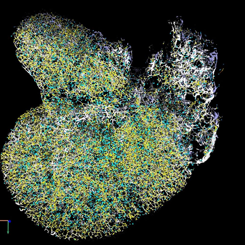
With this, we obtained an “atlas of glycans” on a range of sample types, from cultured cells over primary immune cells to neurons and patient tissue. Each dot in the image below is one glycan (sub-)unit, resolved at the nanometer scale.
02.05.2025 08:54 — 👍 2 🔁 0 💬 0 📌 0

We tackled this problem by labeling different glycan units within the glycocalyx using lectins. These lectins were tagged with DNA barcodes, which we used for multiplexed DNA-PAINT super-resolution microscopy.
02.05.2025 08:54 — 👍 2 🔁 0 💬 0 📌 0
To understand how the glycocalyx state and the cell state are interconnected, we must structural glycocalyx architecture. Unfortunately, this is not so easy: The glycocalyx is organized at the nanometer length scale and contains numerous different building units.
02.05.2025 08:53 — 👍 3 🔁 0 💬 0 📌 0

All cells in the human body are covered by the glycocalyx, a complex and dense layer glycosylated species. The glycocalyx is centrally involved in various processes in health and disease, for example, in cancer progression and immune system regulation.
02.05.2025 08:53 — 👍 2 🔁 0 💬 0 📌 0
GLYCAN ATLASSING: NANOSCALE ANALYSIS OF GLYCOCALYX ARCHITECTURE ENABLES FUNCTIONAL TRACING OF CELL STATE 🍭🔬🧬
Don’t judge a book by its cover - judge a cell by its cover! We can now trace how cell state is encoded in glycocalyx state. #glycotime #microscopy
www.biorxiv.org/content/10.1...
02.05.2025 08:52 — 👍 22 🔁 11 💬 8 📌 2
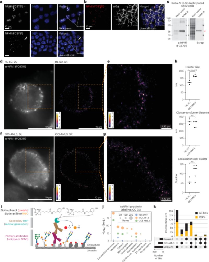
A wonderful study, published today in Nature Biotech.: NPM1 is a marker of AML and can be targeted for efficient therapy! @karimalmahayni.bsky.social created the first SR reconstructions of NPM1 on the cell surface. Great collaborative work, spearheaded by @raflynn5.bsky.social and Kostas Tzelepis!
23.04.2025 14:14 — 👍 4 🔁 3 💬 0 📌 0
Postdoc|Flynn lab| @BCHStemCell @HSCRB @SamratLabMohali @IiserMohali @iitdelhi Everything about RNA|Single-molecule biophysics|Super-resolution imaging 🔬.
Single Molecule Localization and Super resolution microscopy enthusiast @labtinnefeld.bsky.social , LMU Munich
Incoming assistant professor | Postdoc in Kai Johnsson Lab at MPIMR Heidelberg | Ph.D in Xing Chen Lab at PKU | Chemical biology, Expansion🔬, Molecular labeling
scholar.google.co.kr/citations?user…
Chemical biologist @ Max Planck Institut for Medical Research in Heidelberg
Technologist, scientist. Co-founder of Convergent Research.
Ph.D. Student, Nobel laureate Carolyn Bertozzi and Longzhi Tan Labs, Stanford University🌲, Departments of Chemical and Systems Biology|Neurobiology|Sarafan ChEM-H, Passionate about Chemical Biology🧪, Neuroscience 🧠and Artificial Intelligence🤖
Playing with #singlemolecules, bending light, and algorithms to see really small things. Led by Matthew Lew, Associate Professor @WashUESE @WashUengineers @WashU #superresolution #microscopy #SMLM #computationalimaging
Photonics researcher at ARCNL.
Interested in ultrafast light matter interaction.
📍🇳🇱
Neuroscientist at KISN at NTNU, Tronheim, Norway
Nobel prize in Medicine or Physiology, 2014 together with Edvard I. Moser and John O’Keefe for the discoveries of cells that constitute a positioning system in the brain
Democrat and Humanist by Nature with strong personal believes💖
Outdoor swimming jock🏊
Head Optical Imaging Competence Centre Erlangen OICE
Hooray, I awake from yesterday... I live by that!🖖
CERC in Glycomics, Director of Glycomics Institute of Alberta(www.glyco-alberta.ca), Professor of Chemistry, Univ. of Alberta. Work on #glycotime with lectin microarrays and discovering new aspects of #miRNA. Opinions are mine (she/her). www.glycocode.org
she/her; I use computers to study chemistry! Junior Professor in the Institute of Physical Chemistry at Albert-Ludwigs Universität Freiburg!
Husband, Father and grandfather, Datahound, Dog lover, Fan of Celtic music, Former NIGMS director, Former EiC of Science magazine, Stand Up for Science advisor, Pittsburgh, PA
NIH Dashboard: https://jeremymberg.github.io/jeremyberg.github.io/index.html
Researcher | glycobiology | blood groups | enviroment | photography digital and analog | Poland
Physicist and engineer, into multiple scale models of proteins/RNA/membranes, polymers, scientific computing, more --> http://tinyurl.com/goCompModeling
Light Microscopy Facility @mdibiolab.bsky.social
https://lmf.mdibl.org/
Postdoc at Bertozzi lab, ChEM-H, Stanford University.
Alumni of Chudasama and Walker labs, UCL.
Chemical biology, bioconjugation, antibody-drug conjugates, chemical generation of bispecific antibodies.
Scientist. Chemist. Nanocar racer. Research Prof. and Deputy Director @imdeananociencia.bsky.social. Nanotubes, 2D materials, rotaxanes, and supramolecular chemistry...mostly