
@mmestas.bsky.social @laurengroner.bsky.social
Just a perceptual teaser for your entertainment and edification:
@mmestas.bsky.social @laurengroner.bsky.social
Just a perceptual teaser for your entertainment and edification:

Finally, the time has come.
I'm just going through Rule #13 and really enjoying it.
Highly recommended for any radiologist (not to say mandatory).
@pmccoubrie.bsky.social


Lipoid pneumonia: Aspiration/inhalation of animal, vegetable, or mineral oils. Low CT attenuation dependent consolidation (-10 to -150 HU). Examine all lung lesions w/ mediastinal & bone windows - look for fat (80%).
Tx - avoid source of aspiration
#radiology
#radsky
@danielvargasmd.bsky.social


Nice example of increased systemic arterial flow into the left pulmonay artery
Hypertrophied left bronchial artery in a patient with LUL lung cancer
See more here: pubs.rsna.org/doi/abs/10.1...
@howardm19.bsky.social you will like this one

Large pleural lipoma 🫁
- rare benign neoplasm
- asymptomatic
- can appear more opacified than fat on CXR
- two types: hour-glass (with extrathoracic extension) & intrathoracic
#radiology
@laurengroner.bsky.social
@thexraydoctor.org
@pulmpeeps.bsky.social
@chestdb.com
@mmestas.bsky.social

Common findings associated with interstitial lung disease 🫁
#radiology
#radsky
@pulmpeeps.bsky.social
@laurengroner.bsky.social
@mmestas.bsky.social
@thexraydoctor.org


Great case.
A bit old, but one of the best articles about lung adenocarcinoma in my opinion
doi.org/10.1259/bjr/...
And two slides from our recent RSNA exhibit

"But, that right focal consolidation and contralateral tiny nodules bother me though… *lepidic/muc adeno alert*"
Indeed!
Surgical Lung Biopsy: Diffuse adenocarcinoma with areas of lepidic growth.
A relevant citation below:
Another morning arriving at Hospital del Mar 🤩
16.12.2024 09:30 — 👍 3 🔁 0 💬 0 📌 0
A clinical-imaging diagnosis of ILD was made in this patient.
How would you describe/report the (representative) CT images ?
I'll post a follow-up CT series later.
@mmestas.bsky.social
@tlhm-md.bsky.social
@danielvargas.bsky.social
@laurengroner.bsky.social
#radsky
#chestradsky
GGO with asymmetric and basilar distribution in a patient with emphysema and mild bronchiectasis
ILD isn’t my first choice. Might be Inflammtory in the appropiate clinical setting
But, that right focal consolidation and contralateral tiny nodules bother me though… *lepidic/muc adeno alert*


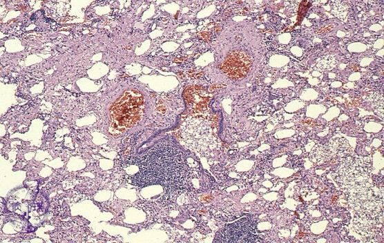
#Chestrad - Pulmonary alveolar microlithiasis 🫁
- idiopathic, F>M
- middle/lower lung predilection
- Look for 'crazy paving', calcified interlobular septa (virtually pathognomonic), small subpleural cysts/emphysema
- black pleura sign
- Tx: lung tx
#radiology
#radsky
@danielvargasmd.bsky.social

"Barely looking at the images purely to get on to the next case isn’t why we became doctors"
@pmccoubrie.bsky.social on @auntminnie.bsky.social
www.auntminnie.com/practice-man...
Proud of my team and colleagues for our educational exhibits that earned Certificates of Merit at #RSNA24
✅ Gaps in the Heart: ASDs Revisited
✅ Radiologic Approach to Persistent Consolidations
@rsnasky.bsky.social @mmestas.bsky.social
Great teamwork! 💪🏼💪🏼
08.12.2024 21:28 — 👍 1 🔁 0 💬 0 📌 0


Elastofibroma dorsi (bilateral): asymptomatic benign soft-tissue tumor (mean age: 65 - this pt was 62).
MC location - infrascapular regions, deep to serratus anterior & latissimus dorsi musculature. Mild-moderate FDG uptake on PET
#radiology
#radsky
@laurengroner.bsky.social
@mmestas.bsky.social


Non-Athersclerotic Vascular Disease by Drs Broncano, Ghoshhajra, @katehanneman.bsky.social , Robb
✅Remember diff patterns of involvement (TKA vs GCA)
✅Beading 📿 in FMD
✅Arch sidedness is determined by bronchus crossed
✅TAA in the young or unusual think of HTAD
#RSNA24 #cvrad #radsky #radiology


Immotile Cilia Syndrome (aka - Kartaganer): congenital, a/w situs inversus, impaired fertility
CXR: hyperinflation, airway thickening/bronchiectasis
CT: mid & lower lung bronchiectasis
#radiology #radsky
@laurengroner.bsky.social
@chestradiologist.bsky.social
@danielvargasmd.bsky.social


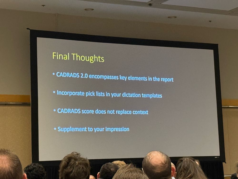

ICYMI 🔑 in CAD #yescct session by Drs Vliegenthart, Ghoshhajra, Agarwal, and Schoepf
✅ Qualify CAC in plain chest CT
✅ Use multiple views, especially in LM dx
✅ CADRADs score does not replace the impression, give context
✅ FFR-CT is helpful and seeing ⬆️ use
#RSNA24 #radsky #radiology
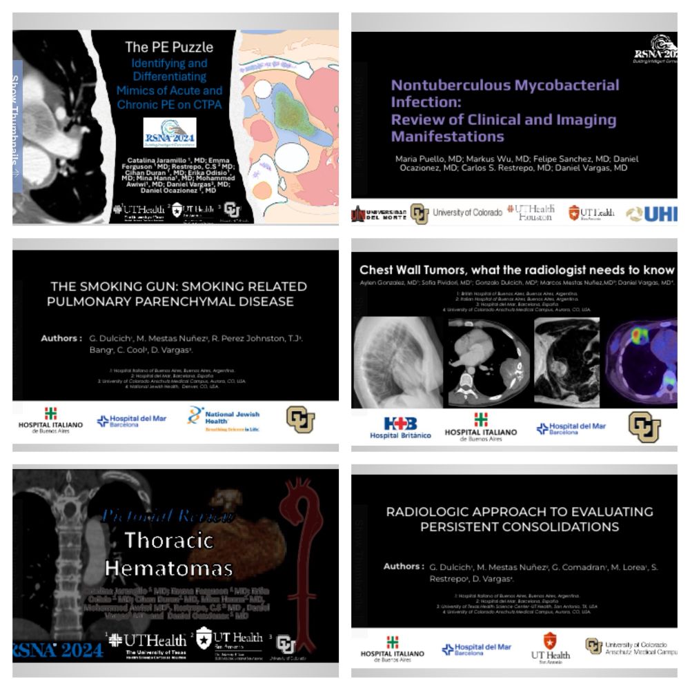
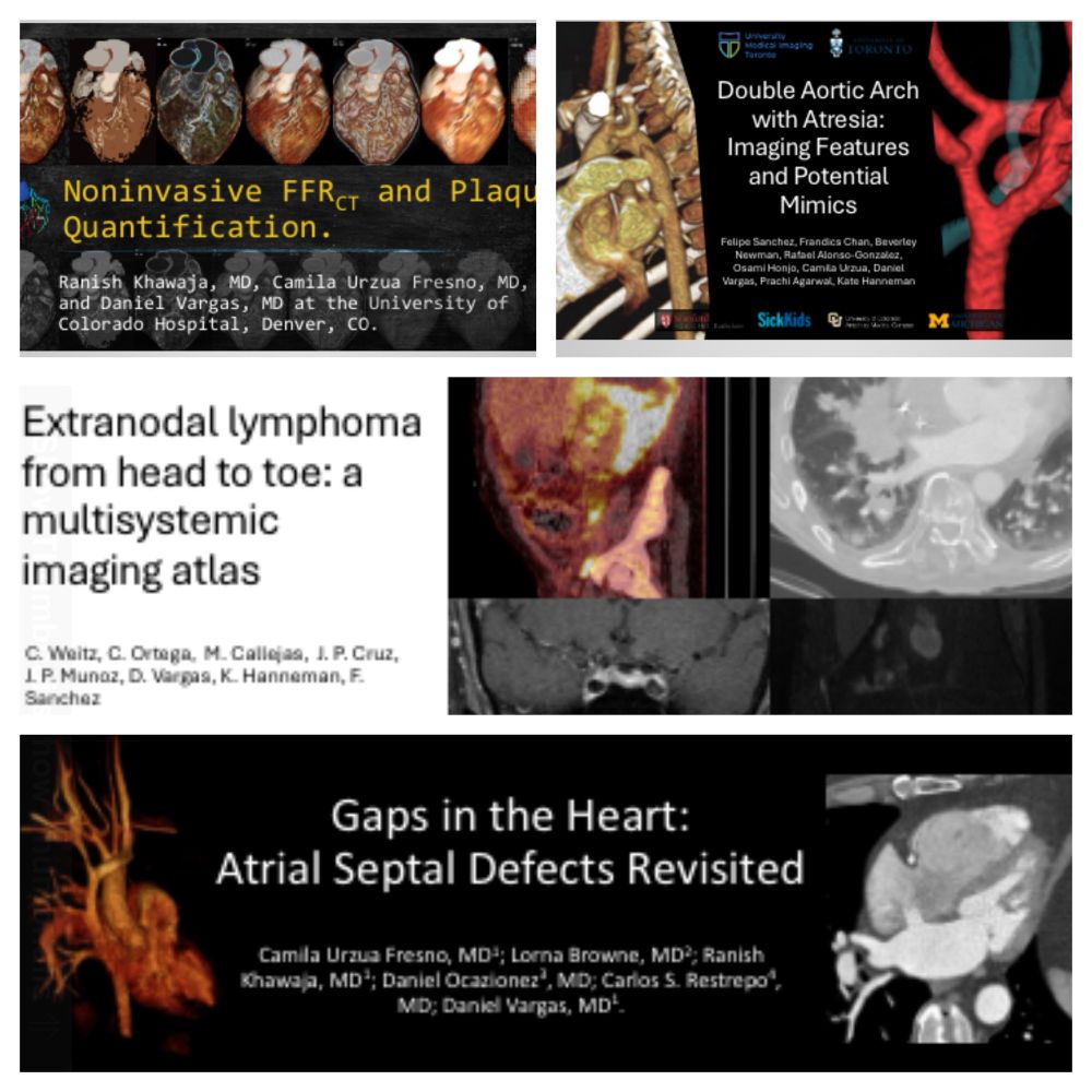
Another fantastically productive year with wonderful educational exhibits presented at #RSNA24
Amazing team of colleagues and trainees!
@katehanneman.bsky.social @mmestas.bsky.social @rsnasky.bsky.social


#RSNA2024 starts today!
If you are there, check out our 3 exhibits on:
- Smoking Related Pulmonary Parenchymal Disease;
- Persistent Pulmonary Consolidations;
- Chest Wall Tumors
Hope you like them!
#radiology #Radsky

Emphysema: centrilobular, panlobular and paraseptal 🫁 Cr. @radiopaedia-org.bsky.social #radsky #chestrad
@laurengroner.bsky.social
@lauraheyneman.bsky.social
@stefantigges.bsky.social
@mmestas.bsky.social
@avrahamcoopermd.bsky.social
