
🚨 Postdoc position 🚨
I’m moving back to Switzerland this summer to launch my lab at the University of Zurich and am recruiting a Postdoctoral Fellow (start: late 2026).
Details & application:
jobs.uzh.ch/job-vacancie....
Please share! 😊
@nicolas3474.bsky.social
SNSF Postdoctoral fellow at Cambridge Uni Perkinsids ⁽ᵃⁿᵈ ᵃᵖⁱᶜᵒᵐᵖˡᵉˣᵃⁿ⁾ cell biology 🦠🔬 🇫🇷Montpellier➡️🇨🇭Geneva➡️🇬🇧Cambridge https://scholar.google.com/citations?user=pddz8toAAAAJ&hl=fr&oi=ao
🚨 Postdoc position 🚨
I’m moving back to Switzerland this summer to launch my lab at the University of Zurich and am recruiting a Postdoctoral Fellow (start: late 2026).
Details & application:
jobs.uzh.ch/job-vacancie....
Please share! 😊
FYI were starting weird structure/evolution/biology stuff at Glasgow University!
If you're interested let me know! Tell your friends!
CRYO www.jobs.gla.ac.uk/job/research...
BIOINFO www.jobs.gla.ac.uk/job/research...
TECHNICIAN www.jobs.gla.ac.uk/job/technici...
#science #hiring #evolution

A super detailed protocol + video on Cryo-ExM - Cryo-Expansion Microscopy, led by the labs of our former postdocs @marinelap.bsky.social & @ebertiaux.bsky.social.
Clear, practical, and very useful for anyone doing nanoscale imaging 🚀 app.jove.com/t/68595/expa...

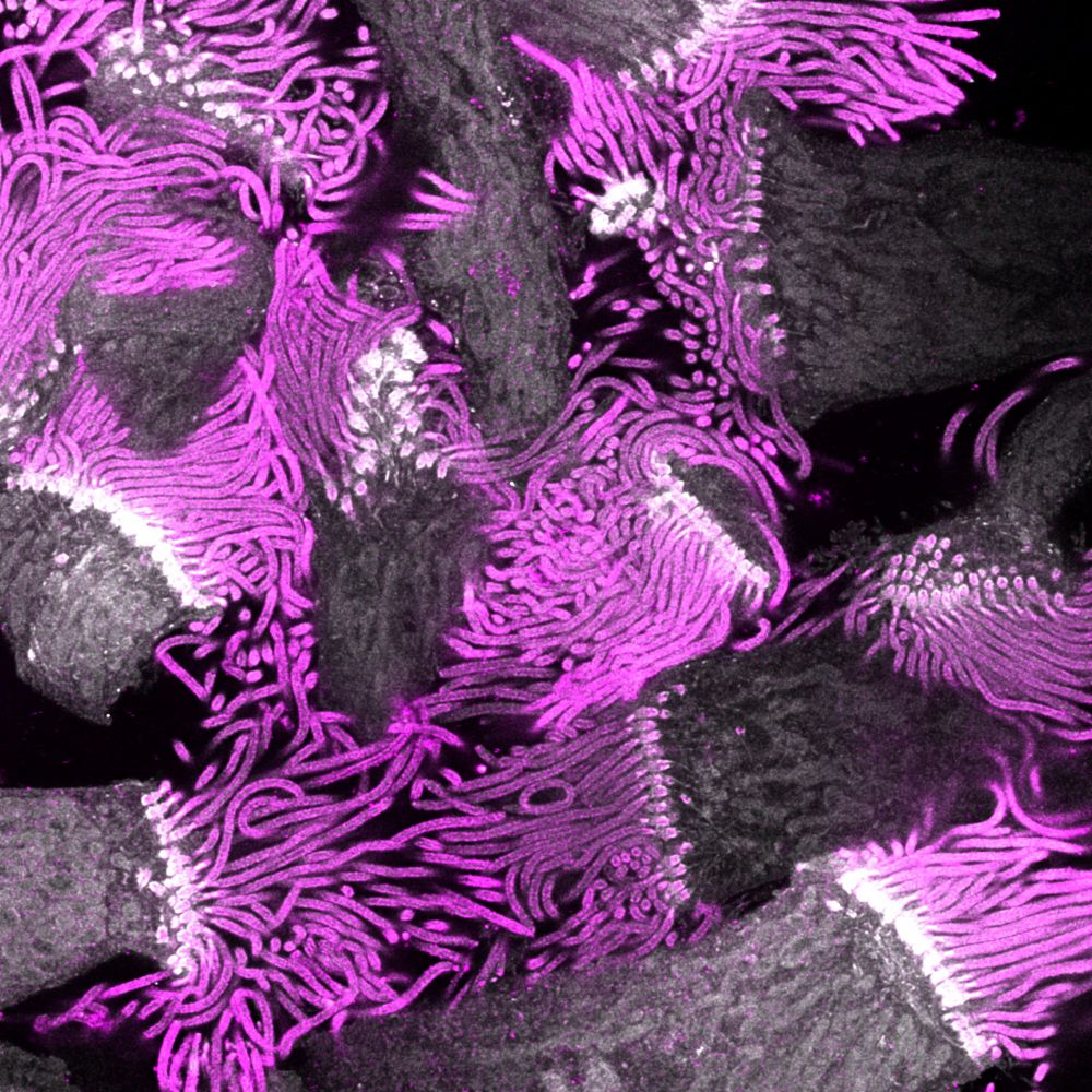
C2CD3 localizes to a ring structure observed in the lumen of the distal centriole by in situ cryo-electron tomography. Top: Confocal image of an expanded mouse photoreceptor cell immunolabeled for tubulin (magenta) and C2CD3 (green). DC, daughter centriole; BB, basal body; TZ, transition zone; CC, connecting cilium. Scale bar: 500 nm. Middle: In situ cryo-tomogram slices taken along the longitudinal axis of the centriole and connecting cilium. White arrowheads indicate microtubule triplets, black arrowheads indicate microtubule doublets, orange arrowheads mark the luminal ring structure, and yellow highlights the membrane. Scale bar: 50 nm. Bottom: Model of the architecture of the distal region of the human centriole.
How does C2CD3 contribute to distal appendage formation in centrioles? @centriolelab.bsky.social show that C2CD3 acts as a central architectural organizer of the distal #centriole, bridging the luminal distal ring complex & peripheral appendage sites @plosbiology.org 🧪 plos.io/496XZuH
10.12.2025 09:10 — 👍 23 🔁 10 💬 0 📌 4Great study 🤩 Congrats 🎉
29.11.2025 12:43 — 👍 1 🔁 0 💬 0 📌 0New preprint! Really excited to share our spatial proteomic study on Plasmodium falciparum schizonts . We used hyperLOPIT to map thousands of proteins—from apical invasion organelles to the infected red blood cell surface.
28.11.2025 10:39 — 👍 18 🔁 3 💬 1 📌 0
Excited to release the Herculean efforts of @scottchisholm.bsky.social &Co. defining the subcellular #hyperLOPIT spatial proteome of #Plasmodium schizonts. Proteomes defining 24 subcellular niches, including exported compartments in the blood cell, are identified.
www.biorxiv.org/content/10.1...
When Toxoplasma's conoid is literally unhinged 😅
26.11.2025 18:33 — 👍 5 🔁 0 💬 0 📌 0
Top: Model RNG2 as a molecular tether for conoid apical anchorage. Illustration of the consequences of RNG2 depletion on the apical complex structure of Toxoplasma gondii tachyzoites. Bottom: Toxoplasma gondii extracellular tachyzoite imaged through Ultrastructure-Expansion Microscopy and stained for tubulin (magenta ) and RNG2-mAID-HA (green). The original image has been modified for aesthetic purposes Credit : the data was produced by Romuald Haase and the image was designed by Albert Tell i Puig
The conoid is a structure in #Apicomplexans that extrudes during egress, gliding motility & invasion. This study shows that the coiled-coil protein RNG2 tethers the conoid to the apical polar ring in #Toxoplasma and is essential for invasion @plosbiology.org 🧪 plos.io/489blpq
26.11.2025 17:40 — 👍 9 🔁 3 💬 0 📌 1Félicitations 🥳
18.11.2025 13:08 — 👍 0 🔁 0 💬 1 📌 0In French - Virginie on Swiss radio! 🎙️ She talks about our latest collab with @dudinlab.bsky.social and @gautamdey.bsky.social #UExM #ProtistsOnSky
03.11.2025 17:13 — 👍 30 🔁 5 💬 0 📌 0
Who-cysts? OOCYSTS! That's right, it's almost time for ❄️Coccidia UK 2025❄️
Your one-stop shop (/conference) for all like-minded sporulating apicomplexan parasites! This year we're in London @crick.ac.uk, looking forward to welcoming parasitologists from across the UK. Register-
tinyurl.com/4ny5bwu2


Les Embiez harbour at sunrise

View from the highest point of the island
Last week I attended my first #EMBO #ParaFrap Host-Parasite Interaction meeting in Les Embiez 🇫🇷
I had an amazing time packed with great science and people 👨🔬👩🔬
Thank you to the organizers for the chance to talk about my work on Perkinsus 🦠🦪 and thank you to everyone for the support and kind words! 🥹
Thanks a lot for the kind words and support! 😃
06.10.2025 22:12 — 👍 1 🔁 0 💬 0 📌 0
Presentation on Perkinsus cell biology in the main auditorium at the MBL Woods Hole

MBL - Woods Hole

MBL - Woods Hole

Jetlag-fueled sunrise are the best!
Had an incredible time at the MPM 🇺🇸! It was an honor to talk about my work on Perkinsus, especially at the Marine Biological Laboratory, a perfect venue for a marine parasite 🌊🦠 So great to see such a diverse range of parasites and topics this year! Thanks to everyone for the fantastic discussions!
19.09.2025 13:53 — 👍 10 🔁 0 💬 0 📌 0
🚨 New paper out! 🚨
🧬 Parasite sex as a discovery tool — new insights into Cryptosporidium virulence!
Huge thanks to my co-authors, collaborators & mentors 🙌
#Parasites #Cryptosporidium #Genetics #InfectiousDiseases #ForwardGenetics
www.sciencedirect.com/science/arti...
Transfection of the free-living alga Chromera velia enables direct comparisons with its parasitic apicomplexan relative, Toxoplasma gondii https://www.biorxiv.org/content/10.1101/2025.08.26.672290v1
29.08.2025 22:30 — 👍 3 🔁 2 💬 0 📌 0Finally out! Did you ever wonder how a phylum with 10 core genes can make a conserved organelle? Read the paper! It's all about intrinsically disordered proteins! | PNAS www.pnas.org/doi/10.1073/...
27.08.2025 18:07 — 👍 9 🔁 6 💬 1 📌 0
Now available in a polished format! An important step toward accelerated functional genomics for the Cryptosporidium parasite. Brought to you by the incomparable @lucy-watson.bsky.social
www.nature.com/articles/s41...
Want to acquire #ExM images like this and help us understand the true extent of cytoskeletal diversity across the tree of life? This position might be for you!
embl.wd103.myworkdayjobs.com/en-US/EMBL/j...
With @dudinlab.bsky.social
@embl.org @biology-unige.bsky.social @moorefound.bsky.social
Join our crazy beautiful project of ExM Atlas !
One more position to fill, this time for a Research Technician at @embl.org in @gautamdey.bsky.social lab.
Come have fun with us !.
🚨 New EMBO Practical Course!
Ultrastructure Expansion Microscopy: From Cells to Tissue 🔬
📅 20–24 Apr 2026 | 📍 EMBL Heidelberg
Co-organised with @banterlegroup.bsky.social @gautamdey.bsky.social
💡 Register your interest to get notified when registration opens:
🔗 www.embl.org/about/info/c...
The anticipation is over, the MPM registration and abstract submission is open!!
05.06.2025 17:00 — 👍 10 🔁 4 💬 0 📌 0
Check out our dispatch on apicomplexans that infect corals!
www.sciencedirect.com/science/arti...
These parasite-like partners have lost photosynthesis but retained chlorophyll... are they friend or foe?
@currentbiology.bsky.social #protistsonsky #symbiosky #microsky #coralreefs

Cryptosporidium modifies intestinal microvilli through an
exported virulence factor!! Phenomenal work from the newly DR’ed Elena Rodrigues
www.biorxiv.org/content/10.1...
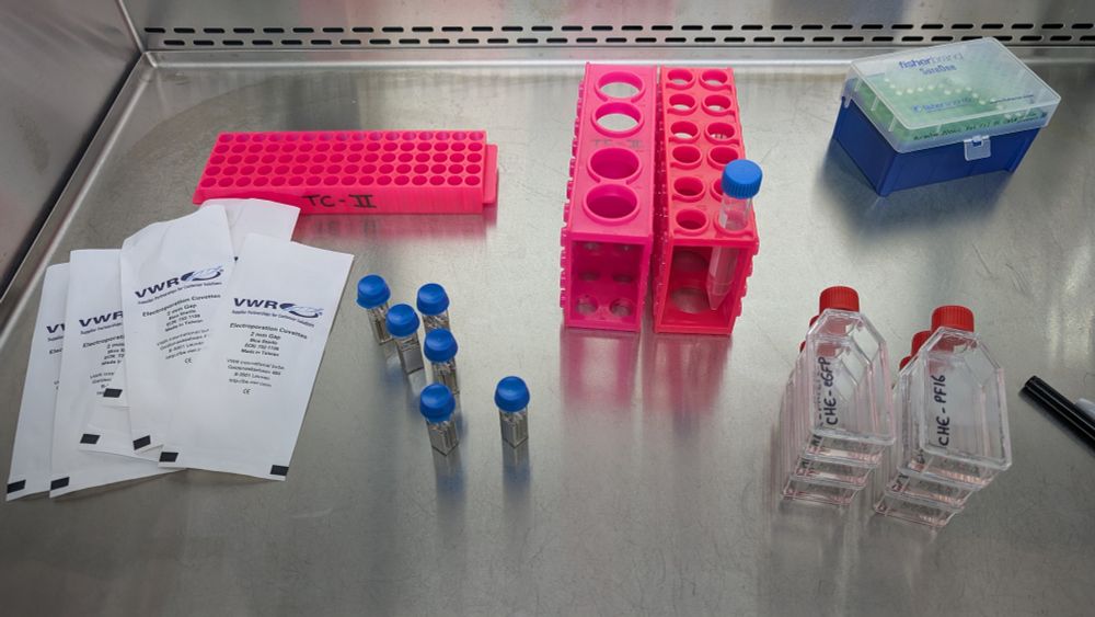
It's transfection time for Perkinsus marinus and chesapeaki ⚡🦠🧬⚡ Hoping to have some fluorescent parasites in the coming days 🔬 So exciting!! #ProtistOnSky
24.01.2025 18:17 — 👍 5 🔁 0 💬 0 📌 0Very nice 🤩
09.01.2025 23:18 — 👍 0 🔁 0 💬 0 📌 0Thanks Omaya! I'm planning to do U-ExM very soon on those cells! 😁🔬
04.01.2025 13:53 — 👍 2 🔁 0 💬 1 📌 0Dynamic remodeling of centrioles and the microtubule cytoskeleton in the lifecycle of chytrid fungi https://www.biorxiv.org/content/10.1101/2025.01.03.631223v1
04.01.2025 12:31 — 👍 10 🔁 7 💬 0 📌 1I haven't observed enough cells to confidently conclude about synchronicity just yet 😅. Here's a video of a sporangium where the first division appears asymmetrical. I'm unsure if this is physiological tho... It might just be cell division going wrong !
03.01.2025 19:14 — 👍 3 🔁 0 💬 1 📌 0
