I am looking forward to present our recent works on volumetric light-sheet FLIM (www.nature.com/articles/s42...) and two-photon multiscale imaging & analysis (elifesciences.org/reviewed-pre...) of organoids in the Euro-BioImaging #VirtualPub on March 27th!
04.03.2026 09:24 —
👍 2
🔁 1
💬 0
📌 0
I’d like to thank @hfspo.bsky.social for continuous support with a long-term fellowship, providing the freedom to embark on such exciting projects.
24.02.2026 22:11 —
👍 1
🔁 0
💬 0
📌 0
This was a fantastic collaboration with @mundhegayatri.bsky.social, Claudio Collinet and @thomaslecuit.bsky.social at @ibdm.bsky.social during my postdoc time in Marseille. The paper is available in Nat. Comms., featured in the collection “Organizers and self-organization in developmental biology”.
24.02.2026 22:11 —
👍 1
🔁 0
💬 1
📌 0
To turn spatial data into a 3D object, check our protocol (open source, napari plug-in). @guignardlab.bsky.social @adrianobolondi.bsky.social @ibdm.bsky.social
For example, if you have spatial RNA-seq as sections, you can align them automatically to analyse directly in 3D.
bio-protocol.org/e5607
06.02.2026 14:13 —
👍 18
🔁 9
💬 1
📌 0
For good reasons. Some of my favourite SPIM acronyms were born back then!
16.12.2025 16:34 —
👍 0
🔁 0
💬 1
📌 0
Oh, I am very sorry. I thought we had cited and listed all previous SPIM-FLIM publications. We missed yours somehow (by no means intentionally of course!). Your implementation was frequency domain based then?
15.12.2025 14:38 —
👍 0
🔁 0
💬 1
📌 0
Thank you! This is fully volumetric time lapse FLIM data over a ca. 50 um depth range (1 plane every 2 um). In the end we only analysed Flipper-TR lifetimes in one 2d section per cell, but in principle one could analyse individual cells or junctions and their dynamics in 3D.
15.12.2025 13:25 —
👍 1
🔁 0
💬 0
📌 0
Thank you Amir!
15.12.2025 11:58 —
👍 0
🔁 0
💬 0
📌 0
Thank you Joachim! I think there are many cool applications that hopefully we and others will explore in the future. Could be also promising in combination with OPM-like systems.
15.12.2025 11:26 —
👍 1
🔁 0
💬 1
📌 0
Lastly, I want to thank the Human Frontier Science Program @hfspo.bsky.social for continuous support with a long-term postdoctoral fellowship.
15.12.2025 10:23 —
👍 2
🔁 0
💬 1
📌 0
I want to thank my postdoc mentor Pierre-François Lenne @pflenne.bsky.social for giving me the freedom to embark on this exciting project.
15.12.2025 10:23 —
👍 0
🔁 0
💬 1
📌 0
This project was a great collaboration with Johan Hummert and colleagues at @picoquant.bsky.social kindly providing equipment, PI Imaging Technologies in Lausanne where the detector was developed, co-workers in Marseille and in Bordeaux and Singapore that originally developed the soSPIM microscope.
15.12.2025 10:23 —
👍 0
🔁 0
💬 1
📌 0

Finally, by sub-sampling our data, we show that sub-100ms acquisition times can still provide robust lifetime estimates.
15.12.2025 10:23 —
👍 1
🔁 0
💬 1
📌 0

We also achieved time-lapse 3D imaging of membrane tension in live organoids. This revealed illumination-induced lifetime changes of Flipper-TR requiring further investigation. Strikingly though, we could detect subtle changes in individual cells and junctions tracked over time with 1s frame times.
15.12.2025 10:23 —
👍 0
🔁 0
💬 1
📌 0

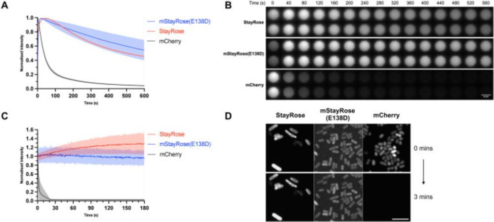
This allowed us to perform 3D FLIM and volumetric lifetime-multiplexed imaging on dense multicellular aggregates (embryonic organoids).
15.12.2025 10:23 —
👍 0
🔁 0
💬 1
📌 0

We benchmarked soSPIM-FLIM with both static and scanning light-sheet illumination, making our implementation transferable to many light-sheet configurations.
We achieved excellent quantitative agreement to confocal FLIM at orders of magnitude shorter acquisition times, down to 100ms per image.
15.12.2025 10:23 —
👍 0
🔁 0
💬 1
📌 0

New paper out! Combining single-objective light-sheet microscopy and time-resolved SPAD array detection, we massively accelerate fluorescence lifetime imaging (FLIM) compared to confocal FLIM, making FLIM applicable to 3D specimen such as organoids and embryos.
www.nature.com/articles/s42...
15.12.2025 10:23 —
👍 36
🔁 13
💬 6
📌 2
Thanks! You are very welcome to visit! It has BIMSB-like stairs, just wider, and microscopes will be on an upper floor and not in the basement :)
12.12.2025 13:04 —
👍 1
🔁 0
💬 0
📌 0
Thank you, Helge! You are welcome to visit once the doors are opened.
12.12.2025 10:32 —
👍 1
🔁 0
💬 0
📌 0
My research activities will continue and exciting projects are ahead.
12.12.2025 09:29 —
👍 1
🔁 0
💬 0
📌 0
Personally, I seriously doubted a future in academia and its compatibility with family life after just missing out on a DFG Emmy Noether Grant at the final Interview stage in June, when our twins were just 5 months old. The long-term perspective of the new position brought back a lot of motivation.
12.12.2025 09:29 —
👍 1
🔁 0
💬 1
📌 0
I look forward to contribute my expertise in quantitative optical microscopy and biophysics to the interdisciplinary scope of Si-M to make an impact for the biomedicine of the future.
12.12.2025 09:29 —
👍 2
🔁 0
💬 1
📌 0

Very happy to share that starting in January, I will take on a new position as microscopic imaging facility leader and researcher at Der Simulierte Mensch (Si-M), a new joint institute by Charité and TUBerlin, which aims at advancing human model systems (e.g. organoids) for biomedical research.
12.12.2025 09:29 —
👍 12
🔁 1
💬 3
📌 0
Of course. Too tempting 😄 Looking forward to see more!
22.10.2025 19:41 —
👍 1
🔁 0
💬 0
📌 0
Very nice, congratulations! I am sure you did try FCS on it!?
22.10.2025 18:26 —
👍 1
🔁 0
💬 1
📌 0

It has been great two days of scientific exchange at @picoquant.bsky.social 30th Single Molecule Workshop in Berlin #WS30. Looking forward to present a poster on in vivo applications of FCS tomorrow and a talk on fast, volumetric light-sheet FLIM in organoids on Friday!
24.09.2025 22:02 —
👍 8
🔁 1
💬 0
📌 0
Latest paper elifesciences.org/articles/107... closes an important cycle in our efforts to study regeneration: week-long recordings allow us to observe the behaviour of cells during the entire course of regeneration in a crustacean leg – bright objects in movie are fluorescent nuclei of cells. 1/6
08.08.2025 17:39 —
👍 142
🔁 50
💬 2
📌 3