

A new MRI protocol can scan the human brain and its venous network in under 7 minutes at 0.35 mm iso. resolution, marking it as a potential tool for diagnosing cerebrovascular diseases and monitoring neurodegeneration.
scim.ag/3LkRfRY
A new MRI protocol can scan the human brain and its venous network in under 7 minutes at 0.35 mm iso. resolution, marking it as a potential tool for diagnosing cerebrovascular diseases and monitoring neurodegeneration.
scim.ag/3LkRfRY

It's the second paper in the same day with mesoscale/UHF/vascular imaging in the title (check out also the new paper of @ofgulban.bsky.social and Dimo Ivanov in Science Advances www.science.org/doi/10.1126/...). A sign of things to come
10.01.2026 14:28 — 👍 2 🔁 0 💬 0 📌 0with Pilou Bazin, Emma Brouwer, @jorgefmejias.bsky.social, @thijsdebuck.bsky.social, Anneke Alkemade, Wietske van der Zwaag and Matthan Caan
10.01.2026 14:28 — 👍 0 🔁 0 💬 1 📌 0We observe a convergent interlobular gradient in cortical thickness and vascular-density. This heterogeneity seems driven by the granular layer cytoarchitecture when comparing to 3D histology. Still work to be done though to further improve SNR and segmentation fidelity.
10.01.2026 14:25 — 👍 0 🔁 0 💬 1 📌 0We show that we can derive measures like cortical thickness aligned to the biological ground-truth —bringing cerebellar imaging closer to the level of detail already available in the cerebrum.
10.01.2026 14:25 — 👍 1 🔁 0 💬 0 📌 0Until recently, getting a reliable in-vivo approximation of the human cerebellar cortex was out of reach.
10.01.2026 14:24 — 👍 0 🔁 0 💬 1 📌 0
🎓🧠 It's finally out in @pnas.org 📄
doi.org/10.1073/pnas...
We reconstruct the cerebellar cortex and vasculature in-vivo by combining motion-corrected, pTX-enabled #7T MRI with a new segmentation approach.
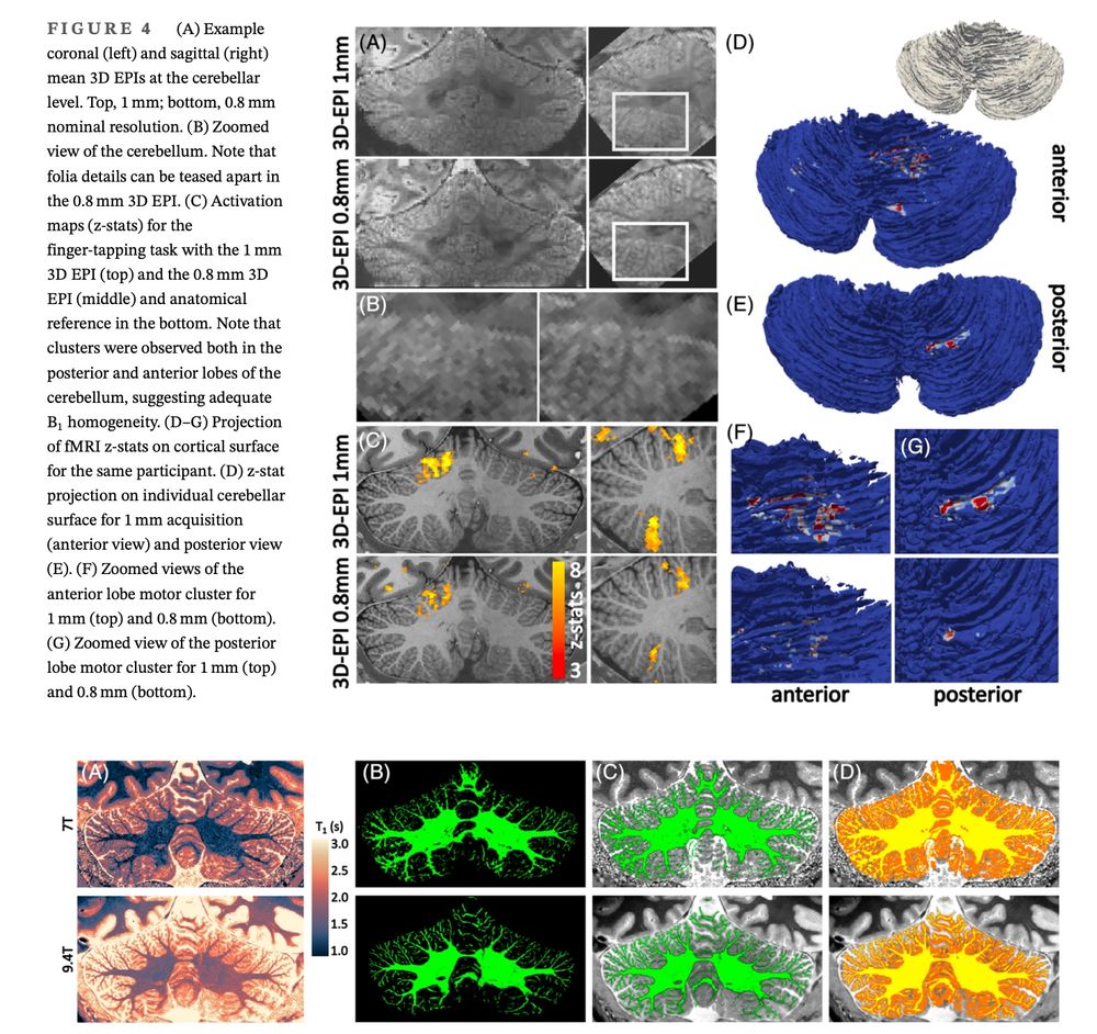
New high resolution fMRI paper describing the capabilities and challenges of 9.4T fMRI in the cerebellum.
By van der Zwaag et al., (@highonfield.bsky.social, @nikospriovoulos.bsky.social)
doi.org/10.1002/mrm....
and yet one more: routine cerebellar neuroimaging at 9.4T—no individual B1+ calibration, robust BOLD. A glimpse of what’s to come at even higher fields. in MRM onlinelibrary.wiley.com/doi/10.1002/... with Wietske van der Zwaag, @highonfield.bsky.social, Desmond Tse 🎓🧠 #fMRI #cerebellum #9.4T
30.06.2025 08:34 — 👍 3 🔁 1 💬 1 📌 0
New paper from the amazing Emma Brouwer: humble, group-optimized B1+ shims at 7T can boost or destroy signal where it matters—no subject-specific calibration needed. in MRM doi.org/10.1002/mrm.... @spinozacentre.bsky.social🎓🧠 #MRI #fMRI #Neuroimaging #7T
25.06.2025 08:02 — 👍 11 🔁 2 💬 0 📌 0