✨ We are pleased to announce the conclusion of the final session of the 2025 @cambridgeflyclub.bsky.social talks series and are now preparing for next year’s sessions, which will be announced soon (check out our upcoming posts for more details).
10.11.2025 19:25 — 👍 4 🔁 1 💬 1 📌 0

Fwd: Fwd: “I hope this email finds you well”
07.10.2025 08:37 — 👍 281 🔁 50 💬 7 📌 8
Membrane protein export in developing axons, via axonal ERES and Golgi bypass #ER_Literature
09.09.2025 16:40 — 👍 6 🔁 2 💬 0 📌 0

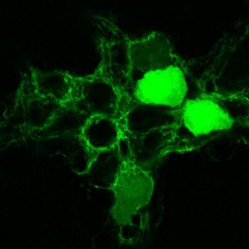
fluorescence imaging of drosophila nerves
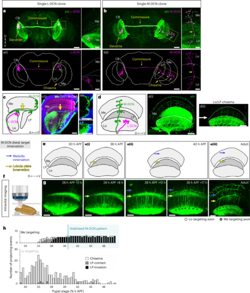
A new genetic toolkit lets researchers target most neuronal lineages in the #Drosophila ventral nerve cord across development and into adulthood, opening the door to mapping structure, chemistry, and behaviour.
buff.ly/zHQK9dC
05.07.2025 16:28 — 👍 16 🔁 2 💬 0 📌 0
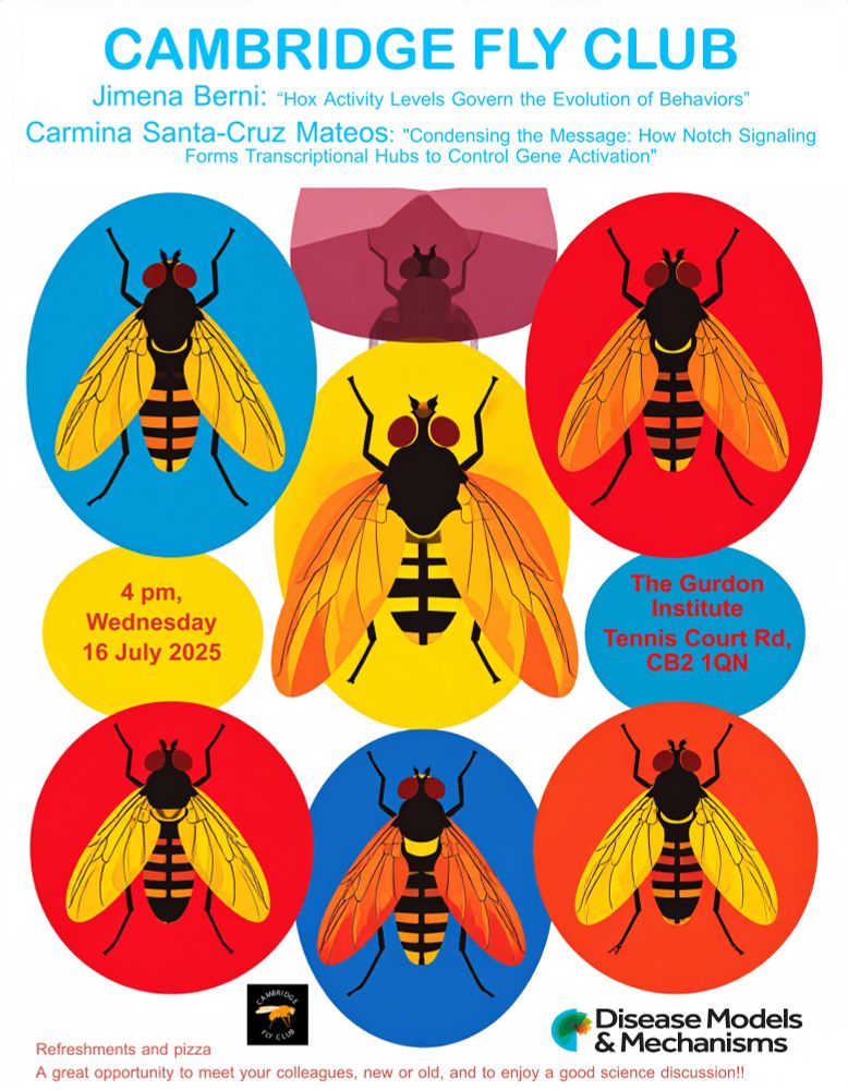
Carmina Santa-Cruz Mateos from PDN, University of Cambridge: "Condensing the Message: How Notch Signaling Forms Transcriptional Hubs to Control Gene Activation"
Jimena Berni from Medical Research Building, University of Sussex – “Hox Activity Levels Govern the Evolution of Behaviors”
Expect refreshments and pizza, this is a great chance to meet colleagues, exchange ideas, and build collaborations. Spread the word, everyone is welcome!
🗓️#CambridgeFlyClub 2nd meeting this year on July 16th (Weds) 4pm at @gurdoninstitute.bsky.social. Our speakers are Carmina Santa-Cruz Mateos from Sarah Bray's lab @pdncambridge.bsky.social and Jimena Berni from University of Sussex.
It's not the Dmel on our poster, guess what species is it?
13.06.2025 09:26 — 👍 1 🔁 5 💬 0 📌 1
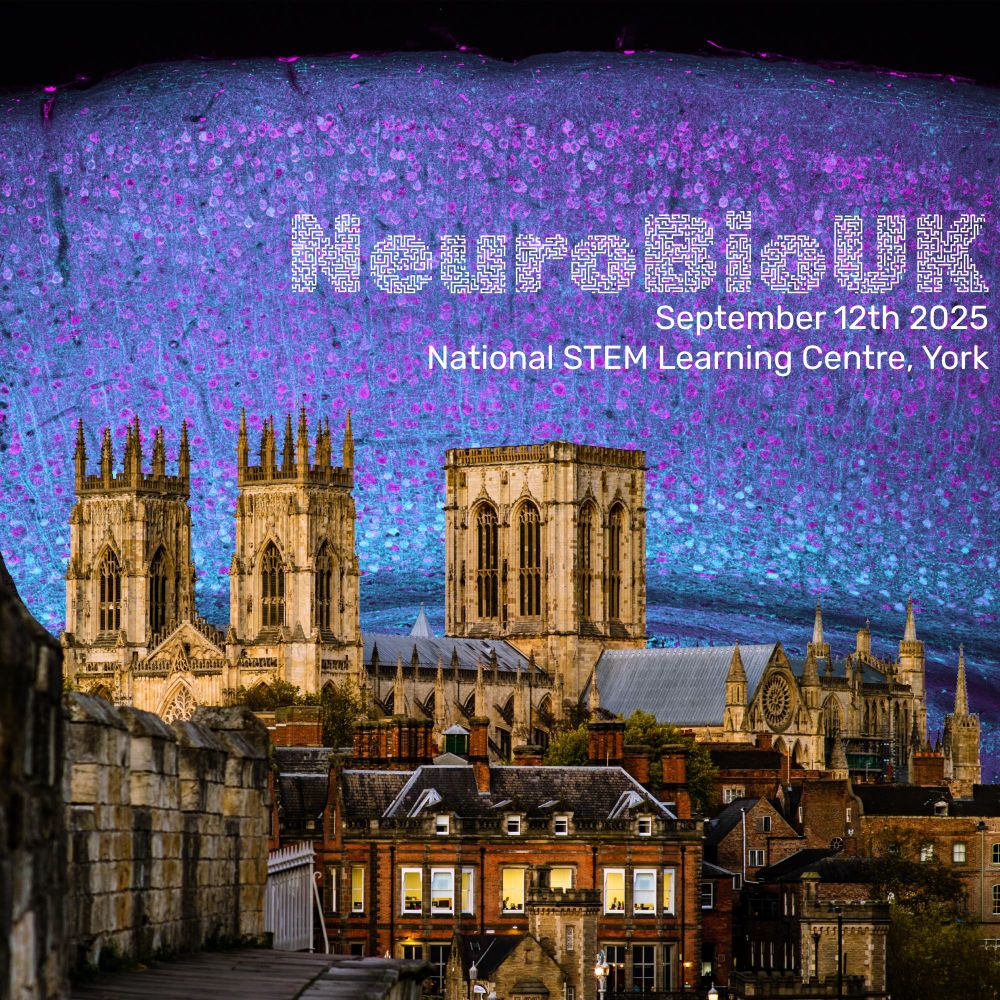
An image montage advertising NeuroBioUK - 12th of September 2025 in York. The image is York cathedral superimposed over a fluorescent cortex mouse section coloured to look a bit like a dawn sky.
Registration is now open for NeuroBioUK 2025!
This year we're looking forward to hearing from our plenary speaker @cathyabbott.bsky.social. As ever, all other talks chosen from submitted abstracts so get submitting - we can't wait to see you there!
neurobiouk.sites.sheffield.ac.uk/registration
06.06.2025 15:17 — 👍 23 🔁 21 💬 0 📌 9
We currently have a call for support that has gone out to European labs, to support FlyBase-UK. We are asking our colleagues from labs in the US and other countries to wait for a similar call to them that will go out in the near future, to support the US sites. We thank you for your patience.
03.06.2025 21:17 — 👍 83 🔁 115 💬 0 📌 12
This is so bad :(
23.05.2025 20:29 — 👍 1 🔁 0 💬 0 📌 0
Welcome to the new serie of Cambridge Fly Club meetings. We organise several events every year to foster collaboration and maintain a positive dynamic between Drosophila groups in Cambridge.
23.05.2025 14:43 — 👍 7 🔁 3 💬 1 📌 1
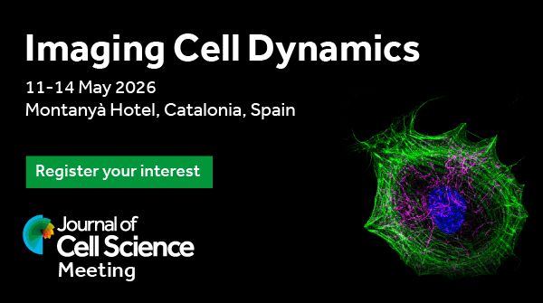
Journal of Cell Science Meeting 2026
Imaging Cell Dynamics
Date: 11 – 14 May 2026
Venue: Montanyà Hotel, Catalonia, Spain
Register your interest
After the success of our 2023 Imaging Cell Dynamics conference, we’ll be hosting a second #JCSImaging meeting in 2026, organised by @franbottanelli.bsky.social, @guijacquemet.bsky.social, @drmichaelway.bsky.social & Giulia Zanetti.
To register your interest: www.biologists.com/meetings/jcs...
19.05.2025 13:01 — 👍 74 🔁 40 💬 0 📌 8
Following the annual tradition, the student symposium of MRC LMB is happening on July 10th and 11th this year.
This year we are collaborating with Institut Pasteur!
Details 👇
22.05.2025 11:31 — 👍 0 🔁 2 💬 0 📌 0
Congratulations Maheva!
07.05.2025 14:15 — 👍 1 🔁 0 💬 1 📌 0
Mouse egg cells stash endosomes, lysosomes, and autophagy regulators in giant structures called ELYSAs. After fertilisation, they break down, timing the embryo’s cleanup and recycling systems for early development.
buff.ly/aM5oMxt
20.04.2025 13:59 — 👍 12 🔁 4 💬 0 📌 0
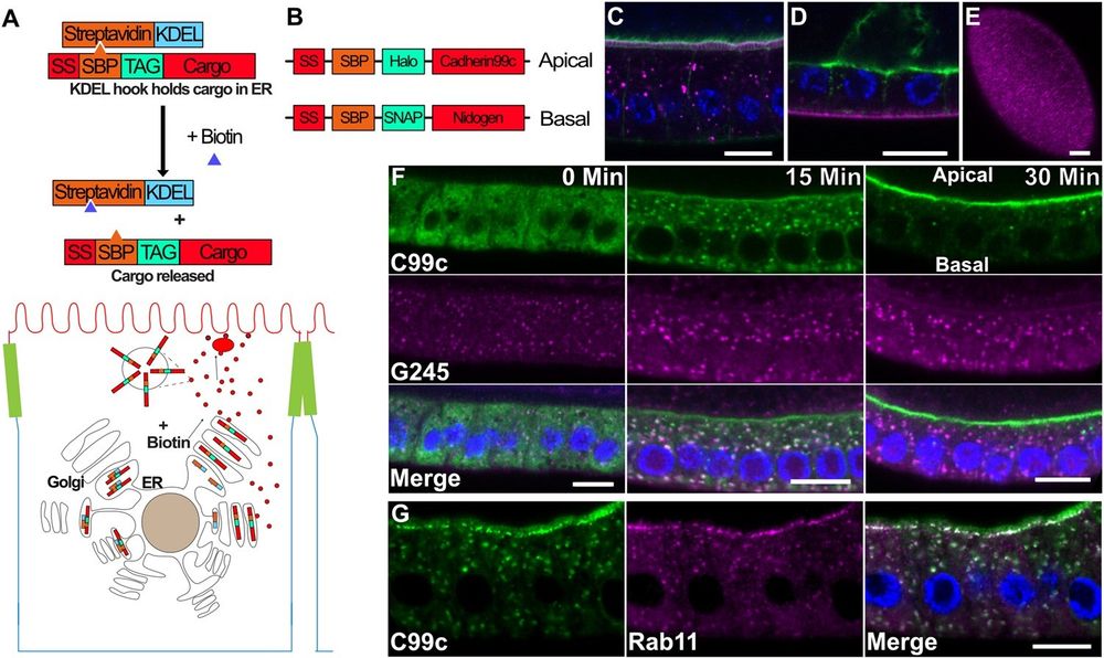
A) Diagram of the RUSH system. The Halo-tagged cargo is retained in the ER by the binding of the Streptavidin binding peptide (SBP) to Streptavidin (StrepA), which is fused to the ER retention signal, KDEL. The cargo is synchronously released upon the addition of biotin, which outcompetes SBP for binding to Streptavidin.B) Diagram of the tagged cargo constructs used in this study. C) Steady state expression of UAS-SBP-Halo-Cadherin99c (magenta) under the control of traffic jam-Gal4, showing its localization to the apical microvilli. Phalloidin staining of F-actin (green) labels the apical microvilli in the follicle cells and the oocyte. Scale bar 10 µm. D) Steady state expression of UAS-SBP-SNAP-Ndg (magenta) under the control of traffic jam-Gal4, showing its localization to the basement membrane. Actin is shown in green. Scale bar 10 µm. E) En face view of SBP-SNAP-Ndg in the basement membrane. Scale bar 10 µm. F) Time course of SBP-Halo-Cadherin99C trafficking in fixed samples. G) 25 min after release from the ER, Cad99c (green) localizes to subapical puncta that are labeled by Rab11 (magenta). Scale bar 10 µm. In all figures with a cross-section of the follicle cells, apical is toward the top of the image and basal toward the bottom.
How are specific cargos targeted to apical & basolateral domains within #EpithelialCells? This study uses a novel #vesicle tracking software "MSP-tracker" to show that the secretory pathway in #Drosophila follicle cells is unexpectedly spatially organized @plosbiology.org 🧪 plos.io/3EfJsBp
14.04.2025 11:58 — 👍 35 🔁 14 💬 0 📌 1

Amazing microscopy from Bewersdorf/Rothman groups shows golgins in a striking four-layered structure at the Golgi rim. Biochemical reconstitution shows long filaments. Does a Golgi stack form due to golgin filament properties rather than dedicated stacking proteins?
www.biorxiv.org/content/10.1...
29.03.2025 21:02 — 👍 50 🔁 20 💬 0 📌 0
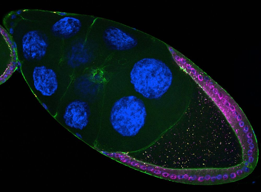
Drosophila follicle showing retrotransposons (pink & yellow) expressed in somatic cells infecting the oocyte
1/ Transposable elements are often called "jumping genes" because they mobilize within genomes. 🧬
But did you know they can also jump 𝘣𝘦𝘵𝘸𝘦𝘦𝘯 cells? 🤯
Our new study reveals how retrotransposons invade the germline directly from somatic cells.
www.biorxiv.org/content/10.1...
A short thread 🧵👇
17.03.2025 11:56 — 👍 545 🔁 259 💬 11 📌 33
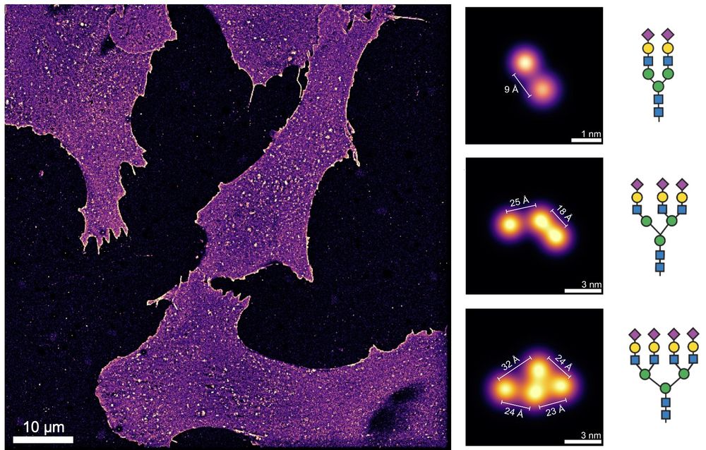
ÅNGSTRÖM-RESOLUTION IMAGING OF CELL-SURFACE GLYCANS 🧬🎨🍬
The glycocalyx, our cells' sugar coat, holds secrets in immunology, cancer, viral infections, and more. Visualizing its molecular architecture was impossible… until now. #glycotime #microscopy
www.biorxiv.org/content/10.1...
10.02.2025 08:21 — 👍 315 🔁 102 💬 15 📌 15
"Imagine a DAPI-like stain, but for the extracellular matrix." That's basically how this work was pitched to me by Kayvon and Antonio a year or so ago. Now the final product really delivers. Read about their versatile label for ECM in living tissues here: www.nature.com/articles/s41...
06.02.2025 16:02 — 👍 354 🔁 104 💬 9 📌 7

Or what about the #Drosophila computer game? Loads of stuff that I almost forgot about: scratch.mit.edu/projects/744...
15.01.2025 16:01 — 👍 10 🔁 4 💬 0 📌 1

Sensory neurons (Class I, purple) are shown in the context of the Drosophila larval body wall. Epidermal cells that sit above the neurons are shown in green. The image was acquired by Pankajam Thyagarajan in the Melissa Rolls lab.
A previous study in @plosbiology.org found that Wnt receptors are housed in early endosomes and required for dendritic #microtubule nucleation. This Update Article shows that microtubule dynamics in #dendrites can be influenced non-autonomously by surrounding epithelial cells 🧪 plos.io/3DK0M0J
07.01.2025 14:51 — 👍 22 🔁 7 💬 2 📌 1
Postdoc in the Baum lab at the MRC-LMB, studying the diversity and behaviour of Asgard archaea
Synaptic Membrane Remodeling @neurocure.bsky.social @fmp-berlin.de with @volkerhaucke-lab.bsky.social #MolecularNeuroscience and #MembraneBiophysics. #FMPGreenInitiative. Before at @mpi-nat.bsky.social with #ReinhardJahn
Developmental & cell biologist @IBDM and @Centuri in Marseille. Studying muscle making and maintenance in Drosophila and in culture.
Passionate cyclist.
Asst. Prof. at UNIGE
Evolutionary cell biology & multicellular developmental diversity of protists.
www.dudinlab.com
#Ichthyosporea, #Multicellularity, #Evolution, #Embryo, #Development, #Protist, #UExM, #Cytoskeleton, #Actin, #Expansion #Microscopy
the Node is a community site for and by developmental and stem cell biologists, covering news, meetings, and research. Hosted by Development @dev-journal.bsky.social @biologists.bsky.social. #DevBio #StemCell
https://thenode.biologists.com
Life scientist.
Head of EMBO Membership & Elections and Courses & Workshops @embo.org #EMBOevents
previously: Editor at Molecular Systems Biology, EMBO Press
Views my own
Professor at #EPFL - #RNAVelocity inventor - Laboratory of Brain Development and Biological Data Science. Single-cell and spatial biologist studying brain development and lipids. #ERCStg investigator
Cell biology PhD candidate & Golgi enthusiast with a fondness for live cell microscopy 🧫🔬
Nature lover, hobby baker, optimist ☀️
ucd.ie/hcs
A fan of ER and derivatives thereof, I’m a microscopic cell biologist.
PI at University of Namur in Belgium
Akita University, Graduate School of Medicine
Cell Biology, ER exit site, Secretion, Collagen
Professor at the Medical University of Innsbruck, Austria. Interested in Endoplasmic Reticulum, Mechanobiology, Proteostasis, Organelles, Cell migration, Pseudoenzymes
The European Drosophila Society bsky account is maintained by N.Tapon, S.J. Araújo, I.Grunwald Kadow, N.Brown, M.Milan. Logo: MC Diaz de la Loza, fly picture: N.Gompel
https://europeandrosophilasociety.org/
Neurobiology Lab studying development, maintenance, and aging of the Enteric Nervous System.
Home of 'enfant terrible' of enteric neurobiology
https://www.enteric-neuron.com
Molecular biologist. Group leader at MRC LMB Cambridge UK. Fellow at Clare Hall college. #CryoEM #RNAbiology #DNArepair
Views/opinions are my own.
https://www2.mrc-lmb.cam.ac.uk/group-leaders/n-to-s/lori-passmore/
Recent PI and Wellcome Trust fellow at the University of Sheffield. Combining microscopy with membrane reconstitution and cell biology. Lover of Phosphoinositides and rock climbing
LGBTQ+ Cell Biologists
Join us as we celebrate diversity and inclusivity in science! #CellLGBTQ
Scientist / Writer / Creative. Molecular Brain Mapping at the MRC-LMB. He/They. All views my own.
Located @Crick.ac.uk in London, we investigate how diet and other factors in the environment affect our metabolism and health
https://www.crick.ac.uk/research/labs/alex-gould