Expansion microscopy improves resolution, but integration with SMLM remains challenging.
In our new preprint, we present a simplified strategy using spontaneously blinking dyes, validated against well-defined reference structures including NPCs and PicoRulers!
➡️ www.biorxiv.org/content/10.6...
24.02.2026 12:09 —
👍 11
🔁 3
💬 1
📌 0
Fantastic look into sub-cellular patterning and functional organization of ciliary arrays in Paramecium! This is a great platform for decoding the mechanistic details that organize these sub-cellular layouts as well-- congrats @guille-rochelle.bsky.social and @daphnelaan.bsky.social !!
23.02.2026 15:42 —
👍 8
🔁 2
💬 0
📌 0

🚨NEW EPISODE🚨
How do we map the #brain at molecular resolution?
@sventruckenbrodt.bsky.social Dr. Sven Truckenbrodt explains #connectomics, #expansion microscopy, neuronal barcoding, and why scaling brain mapping is still a major challenge.
🔗Link to the video: youtu.be/a4qpFpjnOX4
23.02.2026 17:03 —
👍 8
🔁 3
💬 0
📌 0

New research provides insight into how division orientation is controlled to ensure robust tissue architecture during plant growth.
Learn more in this week’s issue of #ScienceAdvances: https://scim.ag/3Okfjp7
20.02.2026 22:27 —
👍 27
🔁 9
💬 0
📌 2
Thanks to its imaging development service and close collaboration with @bic-bordeaux.bsky.social, the LBM is overcoming technological barriers and making cutting-edge imaging accessible in the field of plant biology.
© Magali Grison
@france-bioimaging.bsky.social
www.biomemb.cnrs.fr/service-micr...
09.02.2026 09:36 —
👍 14
🔁 9
💬 0
📌 0

Using micropatterning & thousands of hours on the microscope
@manuelthery.bsky.social and the #CytoMorphoLab reimagined the Musée D’Orsay. Accompanied by an orchestra and poet, their images transported us into the cellular universe. I remembered why I am a cell biologist. All of this is in us. Wow!
25.01.2026 11:03 —
👍 103
🔁 19
💬 3
📌 3

Just leaving from @manuelthery.bsky.social exhibition @museeorsay.bsky.social with stars of actin and microtubules in my head 🤩 😍
24.01.2026 21:41 —
👍 48
🔁 4
💬 0
📌 0

Check our last review on Expansion Microscopy and how it will change the game for plant biologist: authors.elsevier.com/a/1mPIl4tPF3...
@Magali Grison and @emmanuellebayer.bsky.social
15.01.2026 13:35 —
👍 24
🔁 14
💬 0
📌 0
If you’re looking to stain DNA in ExM and in particular U-ExM, Spirochrome offers a broad range of dyes that work really well 👍
08.01.2026 17:38 —
👍 6
🔁 2
💬 0
📌 0

Antibody-free NHS-ester labeling #protocol enabling dense, uniform visualization of cytoskeletal structures. Optimized for #expansionmicroscopy, it minimizes linkage error, improves coverage, and provides an internal #nanoscale ruler for accurate #expansion-factor correction.
bio-protocol.org/e5539
23.12.2025 18:35 —
👍 3
🔁 1
💬 0
📌 0

C2CD3 localizes to a ring structure observed in the lumen of the distal centriole by in situ cryo-electron tomography. Top: Confocal image of an expanded mouse photoreceptor cell immunolabeled for tubulin (magenta) and C2CD3 (green). DC, daughter centriole; BB, basal body; TZ, transition zone; CC, connecting cilium. Scale bar: 500 nm. Middle: In situ cryo-tomogram slices taken along the longitudinal axis of the centriole and connecting cilium. White arrowheads indicate microtubule triplets, black arrowheads indicate microtubule doublets, orange arrowheads mark the luminal ring structure, and yellow highlights the membrane. Scale bar: 50 nm. Bottom: Model of the architecture of the distal region of the human centriole.
How does C2CD3 contribute to distal appendage formation in centrioles? @ebertiaux.bsky.social @centriolelab.bsky.social show that C2CD3 architecturally organizes the distal #centriole, bridging the luminal distal ring complex & peripheral appendage sites @plosbiology.org 🧪 plos.io/496XZuH
10.12.2025 14:02 —
👍 9
🔁 4
💬 0
📌 0
As a Christmas present, our Jove paper with @ebertiaux.bsky.social is out and available for everyone ! Check this out & thanks @centriolelab.bsky.social for the nice highlight
25.12.2025 15:03 —
👍 26
🔁 9
💬 0
📌 0

A super detailed protocol + video on Cryo-ExM - Cryo-Expansion Microscopy, led by the labs of our former postdocs @marinelap.bsky.social & @ebertiaux.bsky.social.
Clear, practical, and very useful for anyone doing nanoscale imaging 🚀 app.jove.com/t/68595/expa...
25.12.2025 13:41 —
👍 126
🔁 32
💬 0
📌 1
✨ Blinking #nanobodies that work for single-molecule localization 🔬
Our new preprint shows that the self-blinking dye JF635b restores robust, buffer-free blinking in #nanobodies, enabling reliable #dSTORM, #MINFLUX, and more, without chemical-switching buffers. Opening new possibilities for #ExM!
22.12.2025 10:45 —
👍 76
🔁 19
💬 3
📌 2
Exciting day for the lab: our 1rst paper is officially out in @currentbiology.bsky.social 🥳 Wonderful collaboration wt @gautamdey.bsky.social showing how Cryo-ExM achieves consistent immunostaining in diverse diatoms, from the lab and the natural environment 1/n
#ProtistsOnSky
tinyurl.com/2zxaund7
31.10.2025 15:27 —
👍 166
🔁 55
💬 1
📌 2
Thank you again for this great article @jcellsci.bsky.social
#Artificial_intelligence #Biological_imaging #Microscopy #Nanoscopy #Quantitative_imaging #Mifobio2025
@cnrs.fr @cnrsbiologie.bsky.social @cnrsingenierie.bsky.social
15.10.2025 17:37 —
👍 7
🔁 4
💬 0
📌 0

#Mifobio2025
@gdrimabio.bsky.social
11.10.2025 09:49 —
👍 12
🔁 2
💬 2
📌 0


First workshop of the week! Self-blinking dyes of Luke Lavis, with Luke himself, Eva Pinto, Sandrine Lévêque-Fort and the Abbelight @abbelight.bsky.social team.
#Mifobio2025
(I apologize for the horrible photos taken in an imaging school)
11.10.2025 13:22 —
👍 10
🔁 3
💬 0
📌 0

A Trypanosoma brucei cell visualized by confocal microscopy after ultrastructure expansion. The cell was immunolabelled for the BILBO1 protein (yellow) and stained with fluorescent NHS-ester (grey). The flagellar pocket is highlighted in pink. Credit: Marie Zelená
The flagellar pocket collar (FPC) is a cytoskeletal structure essential for nutrient uptake & immune evasion in #Trypanosome. @mbonhivers.bsky.social &co use U-ExM to provide novel insights into FPC biogenesis, and reveal 2 unknown cytoskeletal structures @plosbiology.org 🧪 plos.io/4n3bWi6
10.10.2025 16:14 —
👍 37
🔁 8
💬 0
📌 4

#Mifobio2025
Focus on the module 4 :
👉 Multi-scale imaging of biological systems
We are delighted to welcome David LABONTE
@imperialcollegeldn.bsky.social @jungeakademie.bsky.social
Lecture : “photogrammetry, synthetic data, and high-throughput automatic data extraction in insects”
#GDRimabio
05.10.2025 12:11 —
👍 9
🔁 4
💬 1
📌 1

#Mifobio2025
Focus on the module 4 :
👉 Multi-scale imaging of biological systems
We are delighted to welcome Anne BEGHIN @mbisg.bsky.social
Lecture - “Breaking scale: Image Analysis–Based AI to Streamline multilevels 3D quantitative Imaging and Tackle the vEM Challenge”
#GDRimabio
05.10.2025 12:01 —
👍 9
🔁 6
💬 1
📌 1

#Mifobio2025
Focus on the module 4 :
👉 Multi-scale imaging of biological systems
We are delighted to welcome @merlinlange.bsky.social
Institut de la Vision Paris
Lecture : “Building a Multimodal Atlas of Vertebrate Development”
👉 imabio-cnrs.fr/mifobio-2025/
#GDRImaBio #microscopy
05.10.2025 11:48 —
👍 9
🔁 4
💬 1
📌 1

#Mifobio2025
Focus on the module 4 :
👉 Multi-scale imaging of biological systems
We are delighted to welcome Professor Timo BETZ
@uni-goettingen.de
Lecture - “Flow, Force & Form : Multimodal 3-D Microscopy for Living-Tissue Mechanics
👉 imabio-cnrs.fr/mifobio-2025/
#GDRImaBio #microscopy
05.10.2025 11:35 —
👍 8
🔁 5
💬 1
📌 1

#Mifobio2025
Focus on the module 5:
👉 Physical measurements, control and handling -Mechanobiology
We are delighted to welcome Daria BONAZZI
@pasteur.fr @ijmonod.bsky.social
Lecture : “How Mechanical Forces Shape Bacterial Infections?”
👉 imabio-cnrs.fr
#GDRimabio #microscopy #biological_imaging
05.10.2025 15:45 —
👍 14
🔁 9
💬 2
📌 1

#Mifobio2025
Focus on the module 5:
👉 Physical measurements, control and handling -Mechanobiology
We are delighted to welcome Professor @ivatolic.bsky.social
Ruđer Bošković Institute
Lecture : “Mechanobiology of the Mitotic Spindle”
👉 imabio-cnrs.fr
#GDRimabio #microscopy #biological_imaging
05.10.2025 15:58 —
👍 18
🔁 8
💬 2
📌 1

#Mifobio2025
Focus on the module 5:
👉 Physical measurements, control and handling -Mechanobiology
We are delighted to welcome Professor @salaita.bsky.social @salaitalab.bsky.social
Lecture - “May the Force Be Measured : mechano probes reveal the molecular forces generated by cell
#GDRimabio
05.10.2025 16:59 —
👍 10
🔁 7
💬 2
📌 1

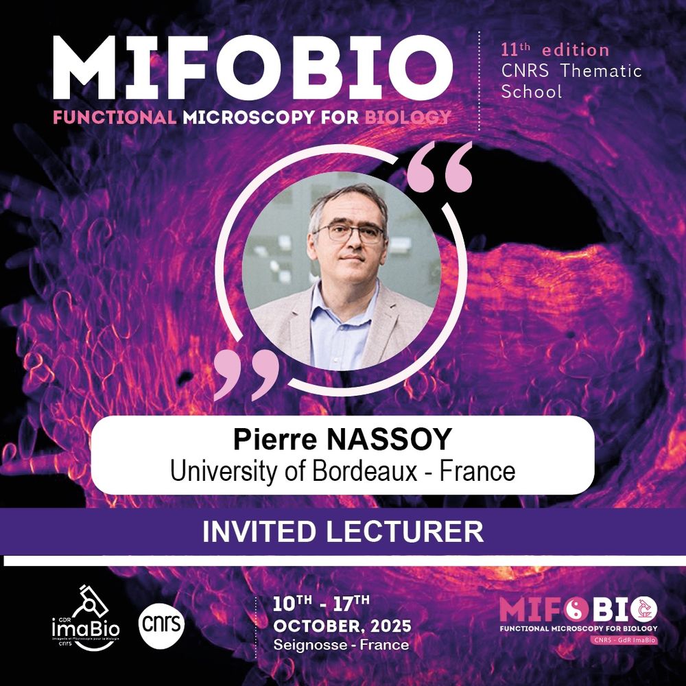
#Mifobio2025
Focus on the module 5:
👉 Physical measurements, control and handling -Mechanobiology
We are delighted to welcome Pierre NASSOY
@univbordeaux.bsky.social
Lecture : “formation, morphometrics and mechanics of epiblast models”
👉 imabio-cnrs.fr
#GDRimabio #TreeFrog_Therapeutics
05.10.2025 18:44 —
👍 9
🔁 3
💬 1
📌 2


