

Go on our website to explore it 👉 france-bioimaging.org
12.02.2026 15:34 — 👍 2 🔁 1 💬 0 📌 0
Go on our website to explore it 👉 france-bioimaging.org
12.02.2026 15:34 — 👍 2 🔁 1 💬 0 📌 0


The new FBI website is out!
We are pleased to present our redesigned website which has been built to simplify our users' journey whether from academia / industry
We have developed new features such as:
➡️Interactive map
➡️Services page to help you identify the technologies best suited to your needs

[PUBLI] A recent collaboration between the Irset Institute & the H2P2 platform (FBI Bretagne-Loire node) has introduced a new imaging protocol to explore the immune microenvironment of HCC using multiplex immunofluorescence.
Read more in our article 👉 france-bioimaging.org/announcement...
emploi.cnrs.fr/Offres/MOBIN... offre de mobilité pour les collègues ingénieurs du CNRS, préparation d'échantillons (transparisation, expansion microscopy...) et plus 🔬, à Strasbourg ! Merci de relayer !
03.12.2025 17:59 — 👍 0 🔁 0 💬 0 📌 0Wow 😍 What labelling?
28.11.2025 17:25 — 👍 0 🔁 0 💬 1 📌 0
[PUBLI] Improving DNA-PAINT with a fluorogenic DNA probe
A team led by Yves Mely (LBP/Strasbourg University) in collab with Alain Burger's team (ICN) developed DRET-PAINT, a new approach that enhances super-resolution microscopy!
🔗Read our article: france-bioimaging.org/announcement...

Very glad to be in Beijing at PKU for the Sino-French exchange on biomedical imaging infrastructures. Many thanks to @france-bioimaging.bsky.social, Laurent Bourdieu, Liangyi Chen and many others for the organization!
03.11.2025 01:20 — 👍 6 🔁 3 💬 0 📌 1
for #FluorescenceFriday : is your ExM sample too large to be imaged with high NA objectives? VIPS it! Very clever way to tackle one of the biggest challenge in large volumetric ExM www.science.org/doi/10.1126/...
17.10.2025 12:29 — 👍 20 🔁 6 💬 0 📌 1
Great first poster session at #Mifobio2025 — thanks for the inspiring discussions! 🔬
All week long, come (re)discover our poster and explore #OpenCID, making #data #storage, #management & #sharing simpler.
See you soon!
@gdrimabio.bsky.social
@imag-ic.bsky.social

📰 #Mifobio dans la presse
👉 france3-regions.franceinfo.fr/nouvelle-aqu...
Merci @france3-naquitaine.bsky.social pour cet article sur #Mifobio2025
@cnrs.fr @cnrsingenierie.bsky.social @institutfresnel.bsky.social @healthcare.nikon.com @leicamicrosystems.bsky.social @abbelight.bsky.social

🔥 It's Time ! #Mifobio2025
👉 imabio-cnrs.fr
#GDRImabio
@cnrs.fr
@cnrsingenierie.bsky.social
@cnrsbiologie.bsky.social
Have a look at this image from the lab, taken by @katkajerabkova.bsky.social, and vote for it as it is competing for the Grand Public Prize of the @sbcf.bsky.social
25.07.2025 15:11 — 👍 25 🔁 5 💬 1 📌 1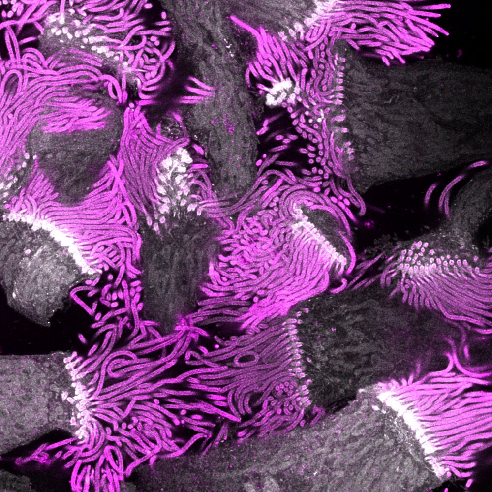
🚨 New EMBO Practical Course!
Ultrastructure Expansion Microscopy: From Cells to Tissue 🔬
📅 20–24 Apr 2026 | 📍 EMBL Heidelberg
Co-organised with @banterlegroup.bsky.social @gautamdey.bsky.social
💡 Register your interest to get notified when registration opens:
🔗 www.embl.org/about/info/c...
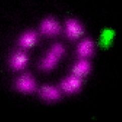
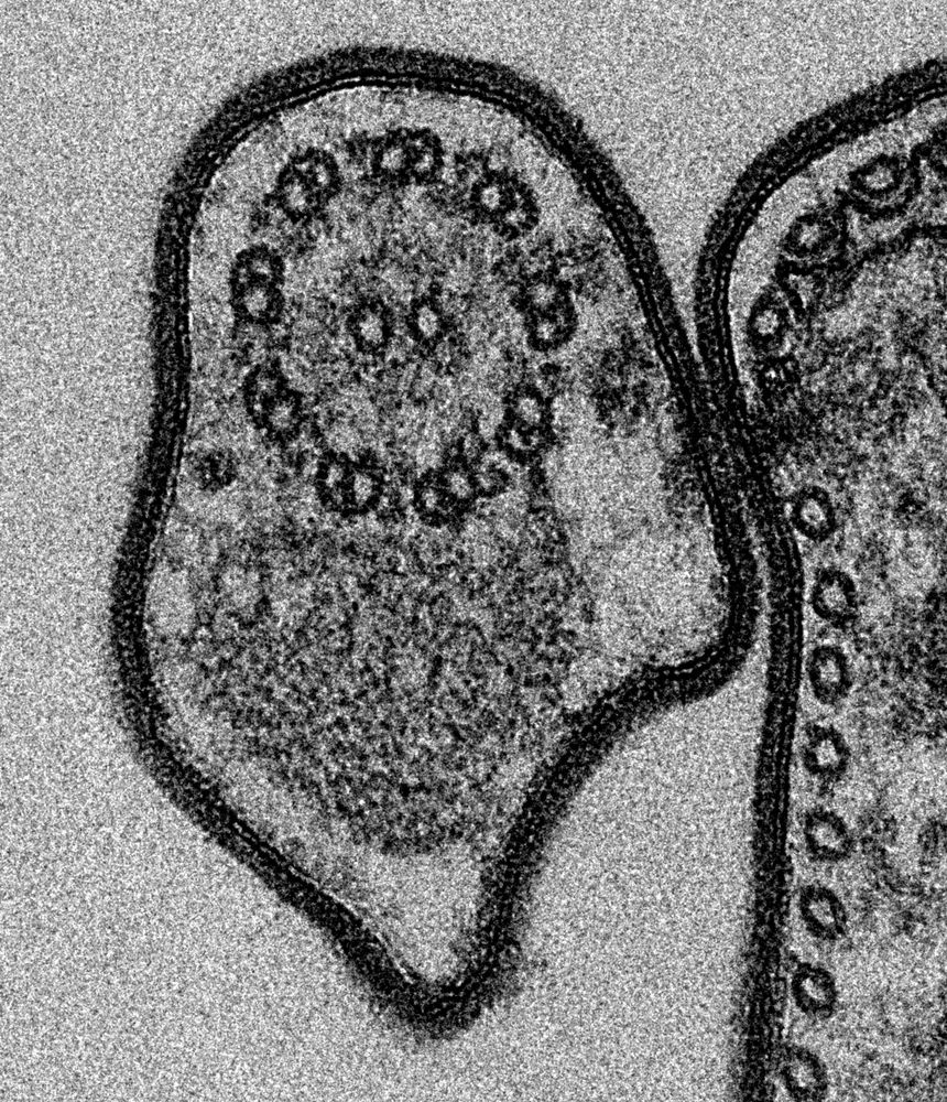
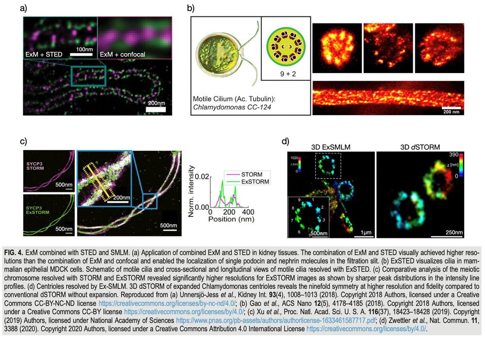
The nucleus is fantastic, but can it beat #cilia and #flagella 😉 here stained for tubulin (magenta) with an intraflagellar transport train (green) after #expansion microscopy and #STED or shown by classic TEM. JCS @jcellsci.bsky.social found the solution and is featuring both! 😍
17.07.2025 09:02 — 👍 33 🔁 7 💬 3 📌 0@cnrsecologie.bsky.social regrette profondément l'adoption de cette loi à la vision court-termiste & ses conséquences graves sur l’environnement, qui méprise santé & bien-être de la population & le rôle des espèces sauvages dans la prod. agricole. La communauté scientifique n'a pas été entendue.
09.07.2025 08:38 — 👍 1625 🔁 1121 💬 36 📌 83🚨 #ExM meets #eSRRF 🔬🏄🌊 the full protocol now in @natprot.nature.com: doi.org/10.1038/s415...
03.07.2025 08:05 — 👍 31 🔁 12 💬 1 📌 2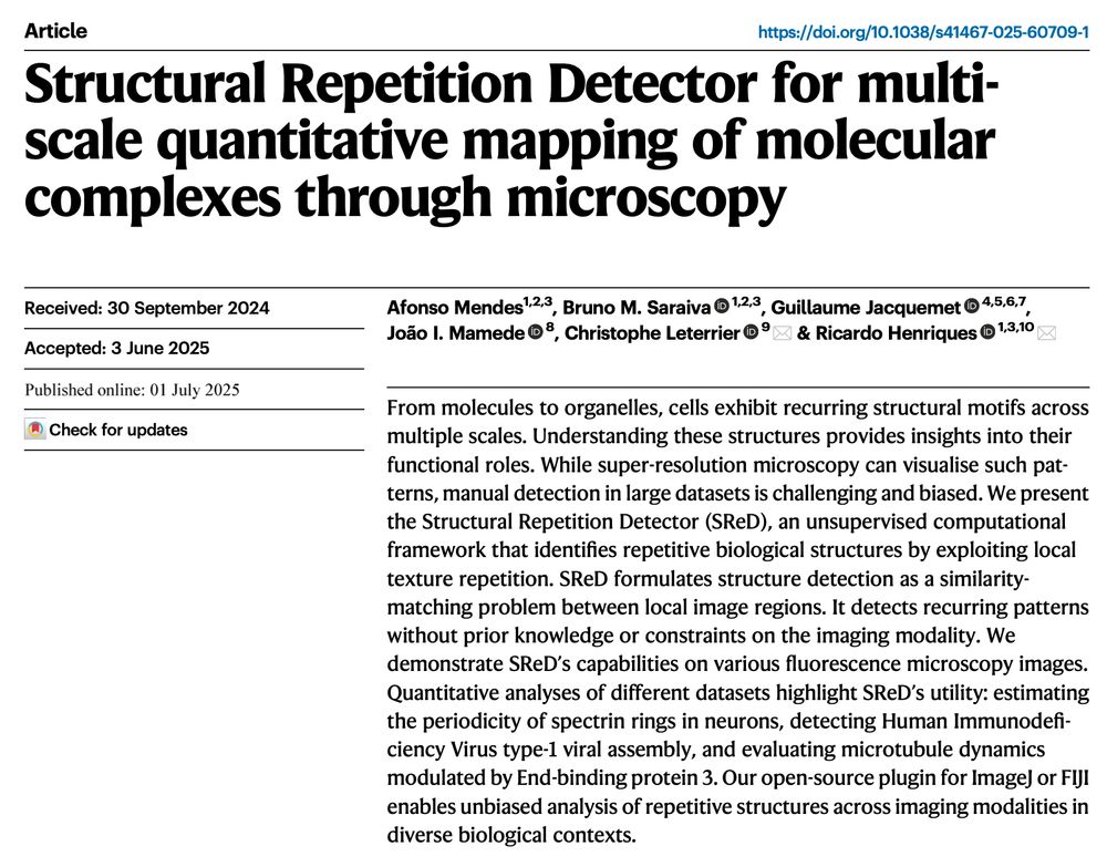
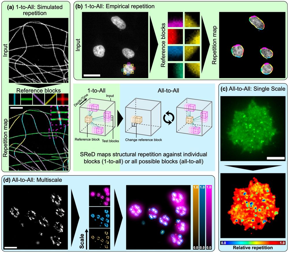
🔬👨💻📰 #SReD is out!
Automated structural detection for #ImageJ & #FIJI, from nano to macro ✨🐘. No training data, no bias - texture analysis with GPU acceleration!⚡️
Brainchild of @afonsomendes92.bsky.social and adventure w @christlet.bsky.social lab + friends.
Check: www.nature.com/articles/s41...
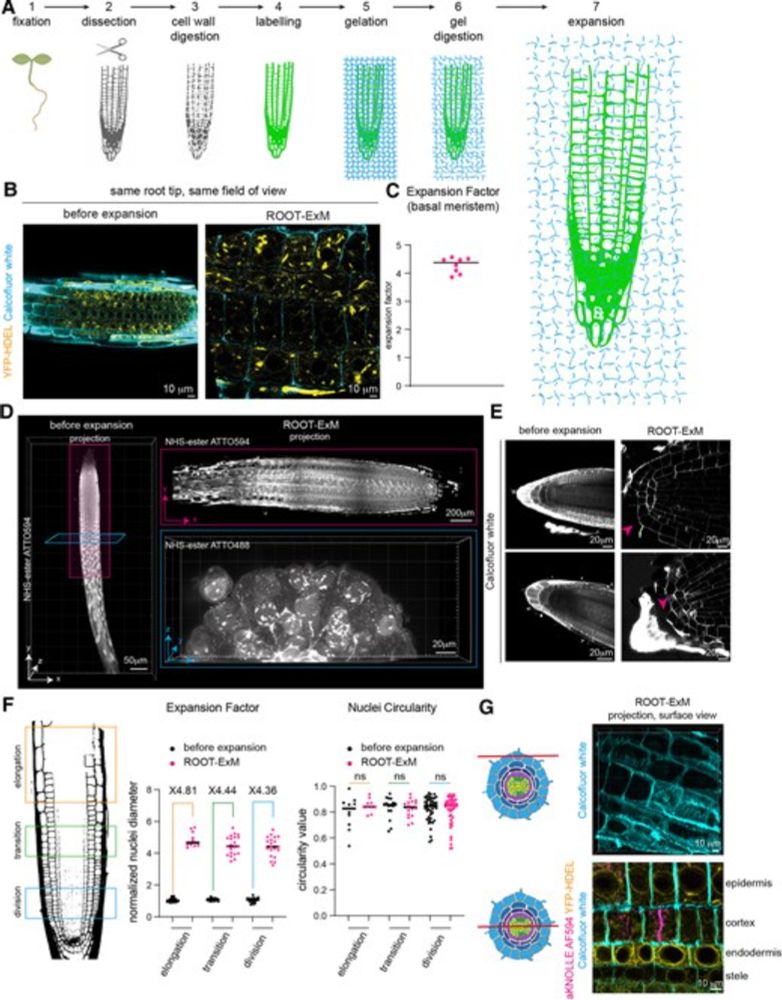
Finally out!, after a few months of delay, our protocol on ExM in plant roots is now available in The Plant Cell
academic.oup.com/plcell/artic...
Beautiful images in a great collaboration of the @bic-bordeaux.bsky.social with @emmanuellebayer.bsky.social and Magali Grison @lbm-bordeaux.bsky.social
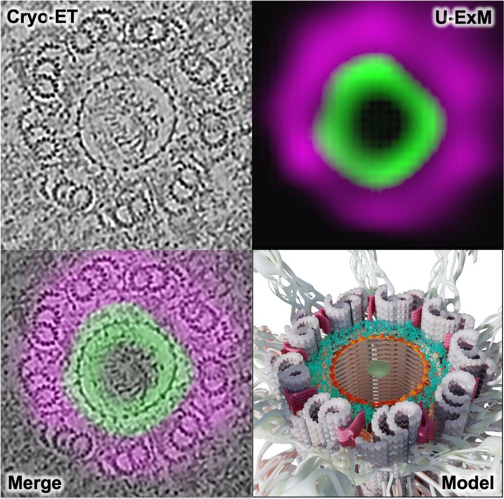
🚨 New preprint!
Using U-ExM + in situ cryo-ET, we show how C2CD3 builds an in-to-out radial architecture connecting the distal centriole lumen to its appendages. Great collab with @cellarchlab.com @chgenoud.bsky.social @stearnslab.bsky.social 🙌. #TeamTomo #UExM
www.biorxiv.org/content/10.1...

Proud to share our latest paper. doi.org/10.1016/j.cr...
Through the dedication of @glynnca.bsky.social and @cryingem.bsky.social we report a thorough method to image molecular organisation within hippocampus tissue.
Structural biology in tissue is well and truly here!
@rosfrankinst.bsky.social
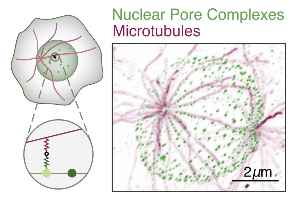
Expansion microscopy image, and accompanying sketch, of S. arctica nucleus with labelled microtubules and nuclear pore complexes.
9/ The observed patterns matched our model, and their parameters place these systems near the predicted optimal filtering regime -- These NPCs may act as efficient spatial thresholding filters! #Microscopy #QuantBio #Microtubules #UExM
18.06.2025 17:07 — 👍 12 🔁 4 💬 1 📌 1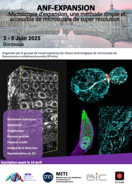
Three full days of #ExM in Bordeaux. Onsite practical course on expansion microscopy. We will be sharing very soon the theoretical and application courses.
01.06.2025 21:18 — 👍 5 🔁 2 💬 1 📌 0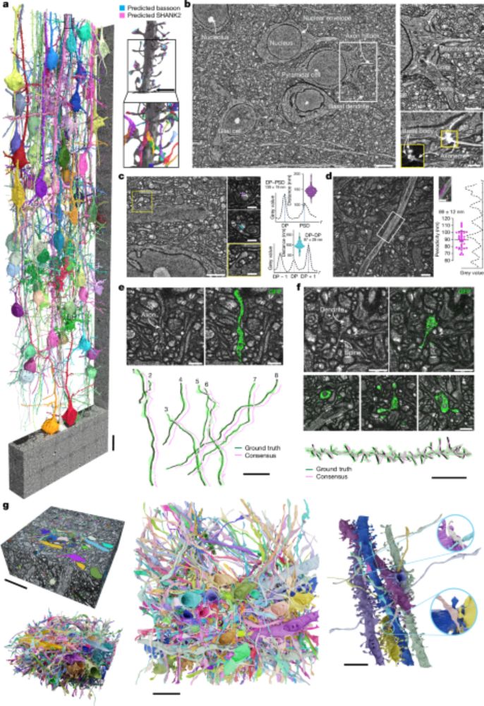
Thanks to expansion microscopy, clever labeling, and modern segmentation approaches, doing connectomics with #light #microscopy has become feasible - huge congratulations Mojtaba & the Danzl lab at @istaresearch.bsky.social !
www.nature.com/articles/s41...
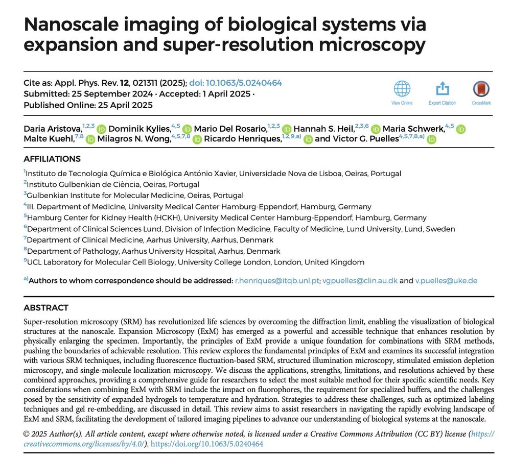
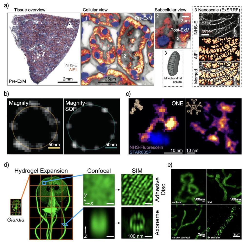
🚨🔬💗Whether investigating cell organelles or mapping proteins, together with Victor Puelles's lab we lay a roadmap for selecting optimal #ExM and #SuperResolution #microscopy combinations. Daria Aristova and Dominik Kylies review with amazing co-authors
pubs.aip.org/aip/apr/arti...
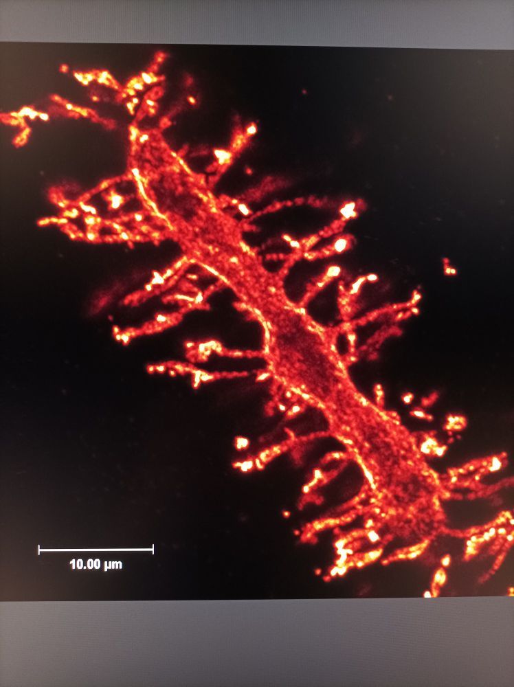
Happy #fluorescentFriday: dendritic spines of Purkinje cell after 10x expansion microscopie #expansionMicroscopie #ExM
25.04.2025 14:25 — 👍 3 🔁 1 💬 0 📌 0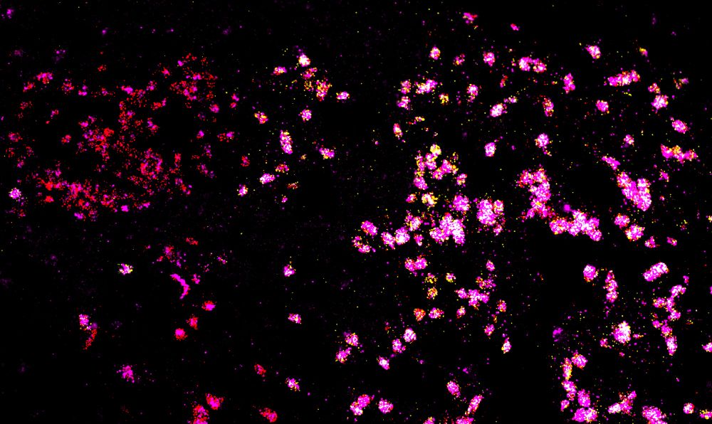
Labeling of RXFP1/DRD1/OPRM1 in tissue section of human brain through the nucleus accumbens using single molecule fluorescence in situ hybridization
I will never tire of looking at these clusters of DRD1🔴/RXFP1🟡/OPRM1🟣 cells - here showing RNAscope in nucleus accumbens of human 🧠. Patterning of them is so neat!
23.04.2025 19:00 — 👍 31 🔁 6 💬 0 📌 0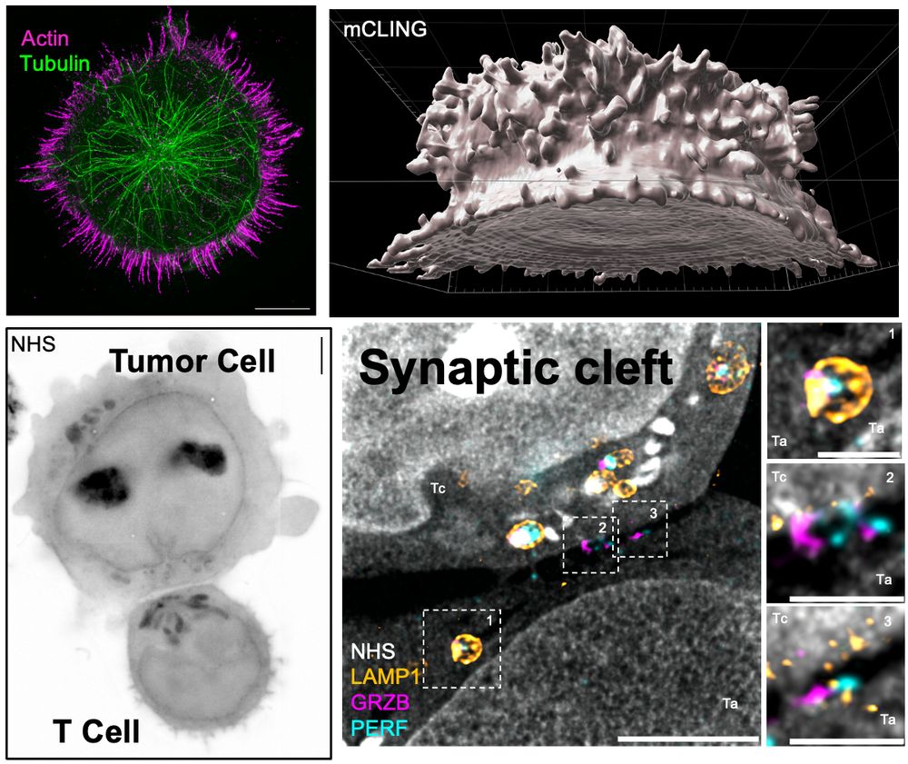
New lab preprint! Unveiling the Molecular Architecture of T Cells and Immune Synapses with Cryo-Expansion Microscopy (Cryo-ExM). A fantastic collaboration with Dr. Benita Wolf. Congratulations to all authors, especially Florent Lemaitre, for leading this amazing work! www.biorxiv.org/content/10.1...
23.04.2025 14:04 — 👍 129 🔁 42 💬 3 📌 4Incredible 4Pi and pan-expansion imaging of the Golgi 👇
29.03.2025 16:03 — 👍 31 🔁 13 💬 0 📌 1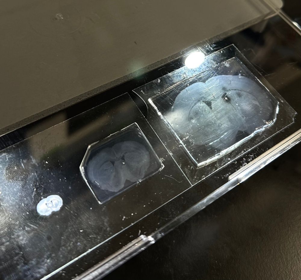
Mouse brain slices, unexpanded and at two different expansion levels, prepared using expansion microscopy.
I don't think I'm ever going to get tired of expansion microscopy, so this makes for a great first post here to look back on in the years to come 😄
Mouse brain slices, unexpanded and at two different expansion levels, skillfully prepared by @damstra.bsky.social and Aashir Meeran at E11 Bio! 🧠
Hi, you can have a look to the ITI Neurostra website neurostra.unistra.fr/en/neurostra...
to reach neuroscientists in Strasbourg!
