A fantastic effort by first authors Huib Rabouw, @janinschoko.bsky.social, @michamuller.bsky.social with lots of help in image analysis by @baarsmatthijs.bsky.social and @jakobpueschel.bsky.social. Also a great collaboration with the groups of @hansclevers.bsky.social and Ron Fouchier!
12.02.2026 07:45 —
👍 4
🔁 3
💬 1
📌 0

Pre-assembly of biomolecular condensate seeds drives RSV replication
Nature - Viral ribonucleoprotein–viral protein networks form pre-replication centres that nucleate viral factories and drive respiratory syncytial virus replication.
Now out in Nature! We visualize infection of the RNA virus RSV in real-time with single-vRNP resolution to understand how RSV establishes viral factories, biomolecular condensates that act as sites of viral replication. A huge collaborative effort led by Dhanushika Ratnayake!
rdcu.be/e1bBW
28.01.2026 20:38 —
👍 91
🔁 35
💬 1
📌 2

Excited to share our new paper! We developed a method to visualize proteasomal degradation at the single–molecule level in live cells, enabling us to dissect distinct modes of substrate engagement, probe co-factor dependence, and study proteasome–ribosome collisions.
www.biorxiv.org/content/10.6...
20.01.2026 12:27 —
👍 48
🔁 15
💬 1
📌 1
The project was led the very talented graduate student @maxmadern.bsky.social and we received a lot of important advice on the proteasome from our collaborator David Haselbach
20.01.2026 08:32 —
👍 3
🔁 0
💬 0
📌 0
In vivo kinetics of protein degradation by individual proteasomes
Protein degradation by the proteasome is central to cellular homeostasis and has been studied extensively using biochemical and structural studies. Despite an in-depth understanding of core proteolytic activity, it has remained largely unresolved how individual proteasomes process substrates inside living cells where many substrate types and co-factors exist. Here, we establish a live-cell single-molecule imaging approach that enables direct visualization and quantification of protein degradation by individual proteasomes. Using this approach, we find that substrate identity, folding and protein-protein interaction have a surprisingly modest impact on processing efficiency, whereas the mode of substrate engagement greatly impacts substrate processing; degradation initiated from protein termini typically proceeds rapidly and with high processivity, whereas internal engagement constitutes a distinct processing mode that exhibits poor processivity and a specific requirement for the AAA+ family ATPase p97/VCP. Furthermore, degradation initiated from opposite termini proceeds with asymmetric rates in a sequence-dependent manner, demonstrating that directionality is an important feature of proteasomal processing in vivo. Notably, poly-glutamine substrates associated with neurodegenerative disease are efficiently degraded from one terminus but resist degradation when engaged from the opposite terminus, highlighting the importance of substrate engagement mode. Together, our results show that different modes of substrate engagement lead to different proteasomal processing outcomes in vivo and revise the prevailing view of the proteasome as a uniform degradation machine. ### Competing Interest Statement The authors have declared no competing interest.
New lab paper!! We develop a technology for real-time, single-molecule visualization of proteasomal substrate degradation in cells. We find that the site of substrate engagement by the proteasome determines decay kinetics, efficiency and co-factor requirement.
www.biorxiv.org/content/10.6...
20.01.2026 08:32 —
👍 47
🔁 15
💬 1
📌 2

Curious about where, how and when #extracellularvesicles fuse with target cells?
A new methodology developed in our group, spearheaded by Jasper van den Ende, provides insight in this process.
EV-FUSIM: an EV-Fusion Spatiotemporal Imaging Method.
www.biorxiv.org/content/10.1...
02.08.2025 20:21 —
👍 7
🔁 2
💬 0
📌 0
The Hubrecht Institute has an open call for it's PhD program. It's a great place to work and do research, so apply!
15.07.2025 08:41 —
👍 1
🔁 0
💬 0
📌 0
Wow, very interesting!
08.07.2025 12:46 —
👍 1
🔁 0
💬 0
📌 0
viruses?
07.07.2025 08:52 —
👍 0
🔁 0
💬 0
📌 0
Can anyone recommend good mRNA therapeutics conferences that are coming up? Ideally conferences that are attended by both industrial and academic researchers?
26.06.2025 08:32 —
👍 1
🔁 0
💬 2
📌 1


It's possible to apply for the Hubrecht Talent Program again!
The HTP aims to promote scientific excellence in the Netherlands by supporting talented minority students in pursuing a career in scientific research.
Read more in the flyer and on www.hubrecht.eu/about-us/hub...
28.04.2025 09:28 —
👍 5
🔁 5
💬 0
📌 1

Marvin Tanenbaum
Marvin Tanenbaum is developing revolutionary imaging techniques that allow him to film molecular processes in living cells at the level of individual biomolecules.This provides fundamentally new insig...
Very honored to receive the Ammodo Science Award for fundamental research! Great recognition for the amazing work done by all our group members, both past and present, over the past 10 years! Also a huge congrats to my co-awardee Leila Akkari!
www.ammodo-science.org/researches/m...
15.04.2025 08:59 —
👍 26
🔁 1
💬 7
📌 0
Sounds really exciting!
28.03.2025 17:42 —
👍 2
🔁 0
💬 0
📌 0
Amazing work led by Dhanushika Ratnayake, and a huge thanks to the large group of fantastic collaborators!
28.03.2025 16:15 —
👍 0
🔁 0
💬 0
📌 0

Pre-assembly of biomolecular condensate seeds drives RSV replication
During infection many RNA viruses, including respiratory syncytial virus (RSV), form specialized biomolecular condensates, inclusion bodies (IBs), where viral transcription and replication occur[1][1]...
Very excited to share a new paper from the lab! We visualize RSV infection with single-vRNP resolution and show how the virus coordinates transcription and replication with the formation of viral factories, biomolecular condensates that act as sites of viral genome control.
doi.org/10.1101/2025...
28.03.2025 16:15 —
👍 28
🔁 10
💬 4
📌 0
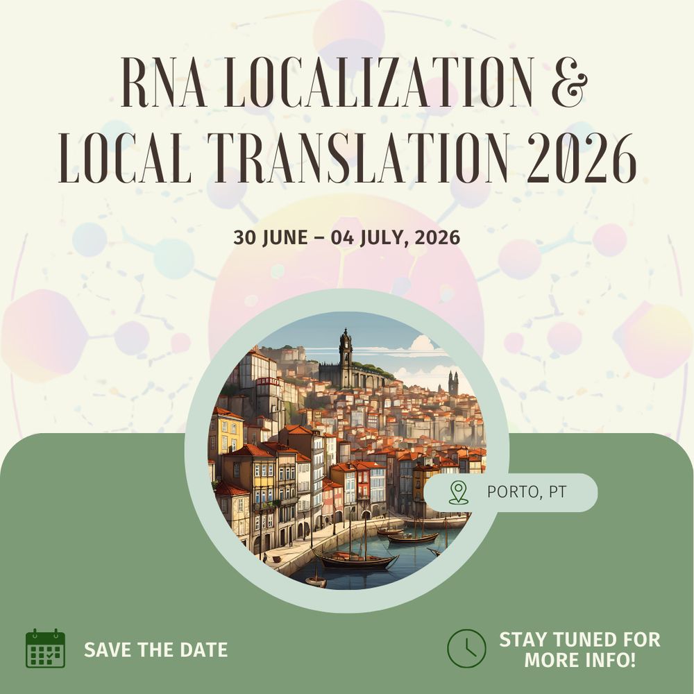
Save the date for the next edition of this wonderful meeting. We look forward to seeing you in Portugal!!
28.02.2025 15:53 —
👍 57
🔁 24
💬 2
📌 1
Super happy to receive a VICI grant from NWO! 5 more years of studying mRNA translation using single-molecule imaging
28.02.2025 14:36 —
👍 40
🔁 0
💬 6
📌 0
This is also our experience. Photobleaching of FPs is often reported at low excitation power (e.g. LED based widefield), and it is often unpredictable how such FPs will behave at high power, for example when using a different microscope modality
18.02.2025 09:18 —
👍 3
🔁 0
💬 0
📌 0
Wow, very nice!
12.02.2025 07:52 —
👍 8
🔁 0
💬 1
📌 0
Amazing work lead by @maxmadern.bsky.social and Sora Yang, and a very nice collaboration with @mariannebauer.bsky.social and Olivier Witteveen. @hubrechtinstitute.bsky.social
03.02.2025 07:38 —
👍 5
🔁 0
💬 0
📌 0

Long-term imaging of individual ribosomes reveals ribosome cooperativity in mRNA translation
Ribosomes cooperate through transient collisions to ensure efficient translation.
Our paper on Stopless-ORF Circular RNAs (socRNAs) is now out in Cell. By high-res tracking and comparing translation by either single or multiple ribosomes, we find that ribosomes cooperate to overcome pausing to ensure fast and efficient translation
www.cell.com/cell/fulltex...
03.02.2025 07:38 —
👍 114
🔁 47
💬 4
📌 6
Wow, mindblowing!
30.01.2025 08:04 —
👍 7
🔁 0
💬 0
📌 0
Very happy to share our preprint on visualizing the life cycle of Influenza viruses using single-molecule imaging! 🥳 We developed two techniques to visualize infections of unmodified influenza viruses in live cells from endosomal release to budding of new viruses. For more details&videos see below ⬇️
21.01.2025 10:16 —
👍 65
🔁 14
💬 2
📌 1
Fantastic, multidisciplinary team effort by Huib Rabouw, Janin Schokolowski and @michamuller.bsky.social with enormous help from @baarsmatthijs.bsky.social and Jakob Puschel and great collaborations with @hansclevers.bsky.social's and Ron Fouchier's groups!
21.01.2025 09:56 —
👍 1
🔁 0
💬 1
📌 0
I love this paper!
17.01.2025 11:14 —
👍 1
🔁 0
💬 1
📌 0


