
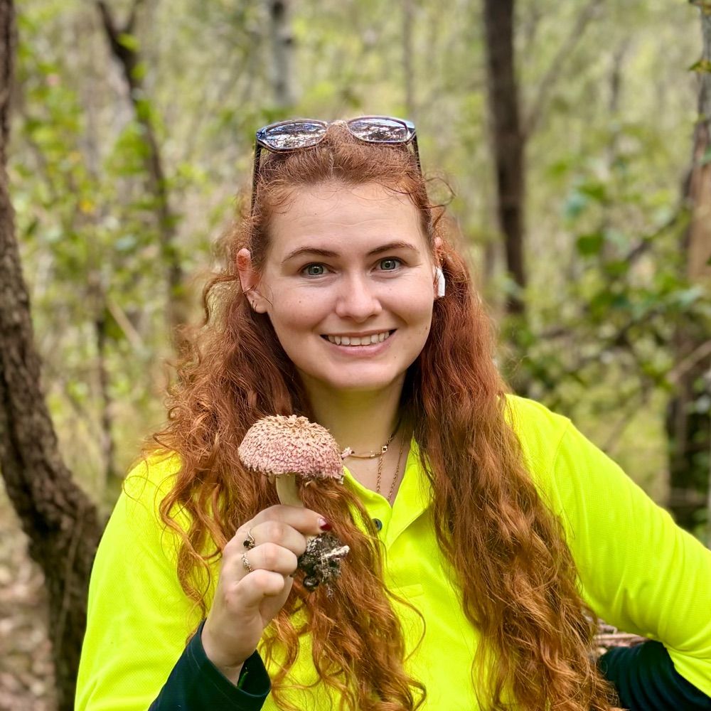
A fabulous find at this year's Fungal Foray 🍄.
www.rnz.co.nz/news/nationa...
@tessmcbride.bsky.social
Biochemist and mycologist studying small secreted proteins and their role in mycorrhizal root formation 🍄 Postdoctoral research fellow at Hawkesbury Institute for the Environment 🌿 Kiwi working in Australia. She/her.
A fabulous find at this year's Fungal Foray 🍄.
www.rnz.co.nz/news/nationa...

This week (11-17 May) sees the 36th annual New Zealand Fungal Foray taking place in the forests near Urenui, Taranaki. Staff at Manaaki Whenua began the Fungal Forays back in 1986 and have forayed across different regions in the country almost every year since then.
buff.ly/HDQDk4r

I just voted! Vote for NZ's Fungus of the Year 2025: interactives.landcareresearch.co.nz/foty/
17.05.2025 22:14 — 👍 0 🔁 0 💬 0 📌 0
Last week, we asked you to guess, based on a series of microscopy images, what samples were successfully sliced using our 7000smz-2 vibratome.
The answer? Plant roots (more specifically eucalypt root) — to investigate fungal colonisation. Did you guess correctly? 🌱
This work was headed by @tessmcbride.bsky.social in Jonathan Plett's Group at @westernsydneyu.bsky.social.
They highlight the highly complex relationship between plants and fungi. Studying the relationship is possible due to the development of effective methods for visualisation and measurement.
A huge congratulations and thanks to the team for preparing this note. #plantsky, be sure not to miss the team's future publications and more stunning plant images by following @tessmcbride.bsky.social!
.
.
.
If you haven’t read it yet, the full application note is available here: lnkd.in/dExESF7G

From asphalt to microscope: the spores from Pisolithus have cute little spiny ornamentations that assist with survival and dispersal 🦔
#fungi #spore #microscopy #mushroom #pisolithus

We're breaking new ground with a brand new vibratome application note — a sample type we don't usually discuss.
Can you guess what sample has been sectioned from these fluorescent microscopy images?
Read the full note here to see if you're right: lnkd.in/dExESF7G

Under the microscope: a cross-section of a eucalypt root colonised by Pisolithus microcarpus 🍄. The fungal nutrient exchange network is clearly visible as it wraps around the outside of the root and in between the root cells 🫚
#mycology #symbiosis #euclaypt #pisolithus #mushroom #fungi #microscopy