Congratulations to Frederic Bonnet and the @mdibl-lmf.bsky.social on these amazing achievements!! 🎉
17.10.2025 19:17 — 👍 5 🔁 2 💬 0 📌 0

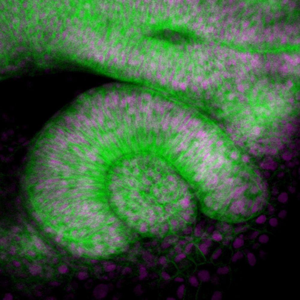
I love Frederic Bonnet's compound eye image that got first place 😍
14.10.2025 16:03 — 👍 11 🔁 2 💬 0 📌 0

Grateful to see one my #superresolution #pollen images won 2nd place at the 2025 @bioimagingna.bsky.social image contest!
www.bioimagingnorthamerica.org/image-contes...
#microscopy #confocal #botany #plantscience #allergies #bioimaging
14.10.2025 15:59 — 👍 65 🔁 12 💬 8 📌 1

This #microscopymonday image of a planarian regenerating its head 7 days after decapitation is from MDI Bio Lab's Disney lab. The translucent area at the top is the blastema—a mass of stem cells that gives rise to new tissues during regeneration. MDIBL Light Microscopy Facility
🧪 🤝 🌎 #microscopy
13.10.2025 14:00 — 👍 3 🔁 1 💬 0 📌 0
🎉 🔬 We’re thrilled to announce the winners of this year’s #NikonSmallWorld in Motion video #Microscopy competition: bit.ly/4mtjUk7
🥇 1st place goes to Jay McClellan of Saranac, Michigan (USA) for his video capturing self-pollination in a thymeleaf speedwell flower.
#Microscope #SciArt #SciComm
24.09.2025 18:34 — 👍 5 🔁 1 💬 0 📌 1
Thanks go out to MDI Bio Lab Digital Image Analyst @hsomers.bsky.social for this #microscopymonday view of an adult #zebrafish (brain vasculature) from the MesoSPIM.
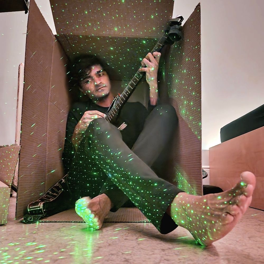
#standwithscience #researchmatters 🧪 🤝 🐟🎸 #microscopy #fluorescentproteins #lightsheet
22.09.2025 12:20 — 👍 14 🔁 5 💬 0 📌 1
YouTube video by BioImaging North America
Exchange of Experience Virtual Group | May 28 2025 | Frederic Bonnet
Speaking with BioImaging North America about the Maine Microscopy Course! @bioimagingna.bsky.social @mdibiolab.bsky.social
www.youtube.com/watch?v=t90L...
#science #education #microscopy #EoE
04.09.2025 18:53 — 👍 3 🔁 2 💬 0 📌 0
One of the people I'm indebted the most for creating stunningly beautiful #mesoSPIM samples was Martina Schaettin - who tragically passed away in March this year. To honor her memory, Elkhan Yusifov created an award - submit your best #microscopy images & videos! www.linkedin.com/company/mart...
22.08.2025 17:32 — 👍 63 🔁 16 💬 2 📌 2

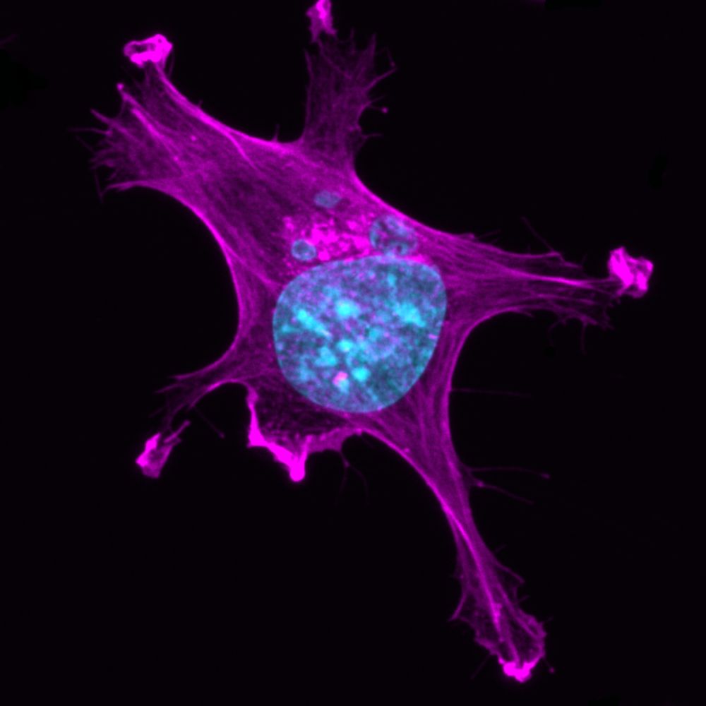
microscope image of fribroblast cell
This mouse fibroblast cell captured by @mdibl-lmf.bsky.social in 2022 is labeled with a #fluorescence to visualize the actin cytoskeleton—a network of actin filaments & proteins that maintains cell shape, enables movement & supports key processes.
#researchmatters #microscopymonday #lightsheet 🧪 🤝
18.08.2025 12:20 — 👍 5 🔁 2 💬 0 📌 0
Hi Dr. Lachie,
I use the AI voice over due to my strong accent. I agree, it doesn't always sound the best. But your data analysis is great.
12.08.2025 14:27 — 👍 0 🔁 0 💬 1 📌 0
YouTube video by LMF@MDIBL
What is optical aberration ?
Do you see chromatic aberration in your images? www.youtube.com/shorts/9lyix...
#science #microscopy #bioimaging #microscopymonday #chromaticaberration #bluesci #mdibl
04.08.2025 12:42 — 👍 3 🔁 1 💬 0 📌 0
YouTube video by LMF@MDIBL
What is Köhler illumination ? #science #microscope #biology #optics
What is Köhler Illumination?
youtube.com/shorts/-1ct0...
#microscopy #microscopymonday #Köhler #Köhlerillumination #brightfield #science
28.07.2025 13:02 — 👍 5 🔁 2 💬 0 📌 0
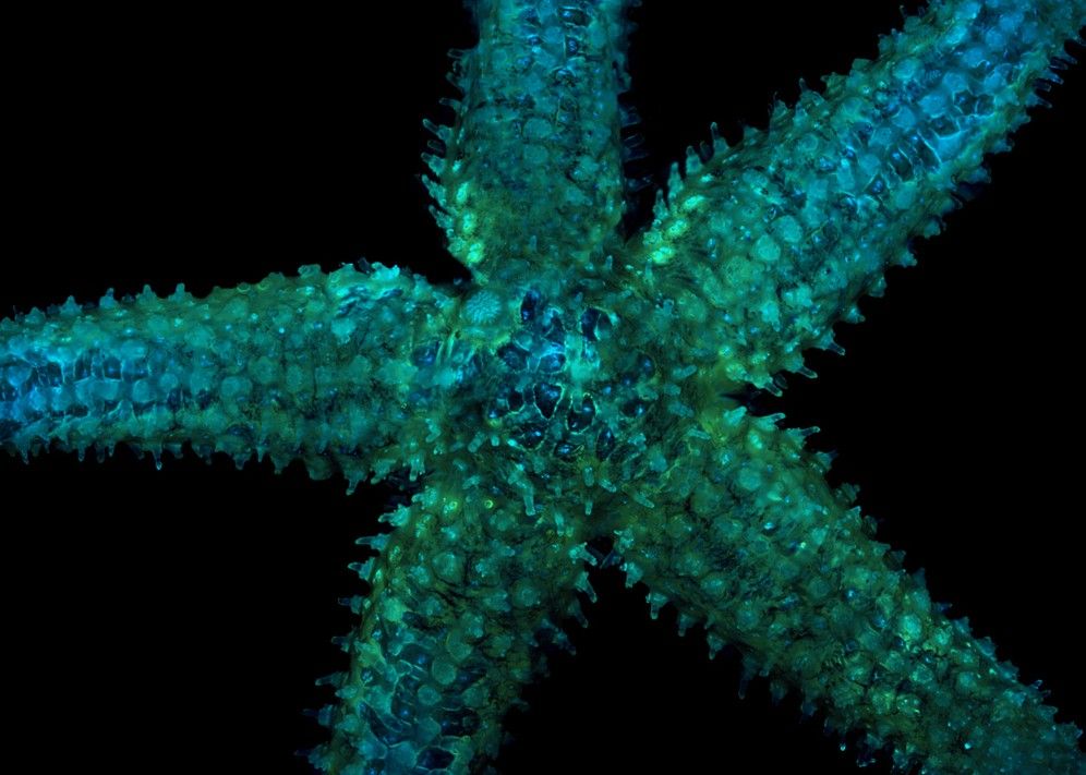
star fish under a microscope
A beautiful #microscopymonday sea star captured by @hsomers.bsky.social in 2022.
Sample: Asterias amurensis (Northern Sea Star)
Label: Nuclear staining with DAPI in Cyan and auto-fluorescence in green
#standwithscience #researchmatters #microscopy #fluorescentproteins #lightsheet 🧪 🤝 #wildlife
28.07.2025 12:13 — 👍 9 🔁 3 💬 0 📌 0
Applications of Human iPSCs and Organoids 2025
Reminder! Application Deadline: August 8, 2025
From mastering iPSC culture to directed differentiation and organoid creation, participants will gain skills they can immediately apply to their own research workflows.
#standwithscience #researchmatters 🧪 🤝 🐟🎸 🍎 👩🔬 🖥️🧬
24.07.2025 11:15 — 👍 5 🔁 2 💬 1 📌 0
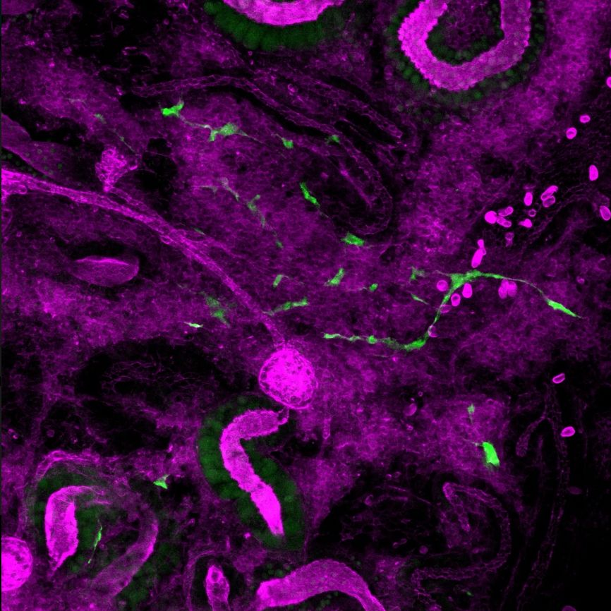
GFP+ stem cells moving through the complex environment of a zebrafish kidney.
This #microscopymonday image shows GFP+ stem cells moving through the complex environment of a #zebrafish kidney.
It was captured in 2023 by Senior Research Scientist Caramai Kamei.
#standwithscience #researchmatters #microscopy #fluorescentproteins #lightsheet 🧪 🤝 🐟🎸
21.07.2025 12:24 — 👍 5 🔁 3 💬 0 📌 0
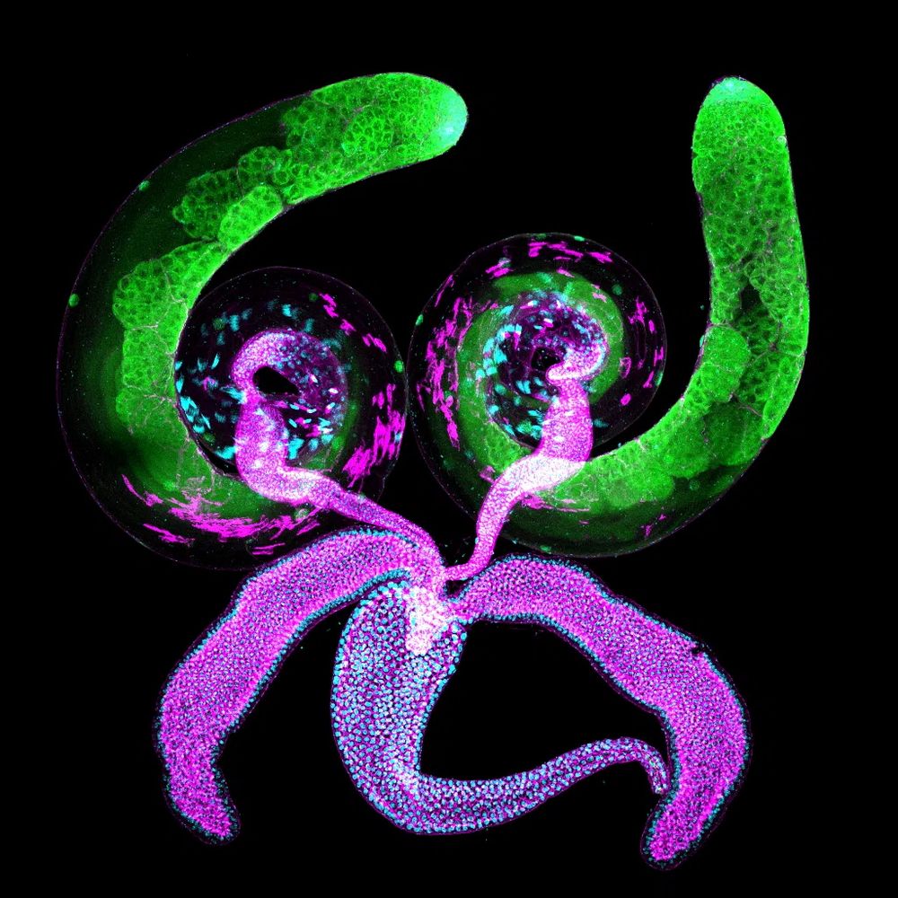
This #microscopymonday image is of a #Drosophila male reproductive system: paired coiled testes, seminal vesicles & accessory glands, and a single ejaculatory duct. Each coiled testis is about 1.5mm. Image captured by MDI Bio Lab Sr. Scientist Travis Carney.
#standwithscience #researchmatters 🧪 🤝
09.06.2025 13:36 — 👍 28 🔁 15 💬 3 📌 1
YouTube video by LMF@MDIBL
What is Numerical Aperture (NA) ?
What is Numerical Aperture?
#microscopy #na #numericalaperture #microscopymonday #science #bioimaging
www.youtube.com/shorts/x1XUs...
21.07.2025 19:09 — 👍 5 🔁 1 💬 0 📌 0

This image from 2024 is by MDI Bio Lab Senior Scientist Travis Carney. It shows the neurons in the brain of a #Drosophila fruit fly larva. The images are color-coded projections, so different focal planes are in different colors.
#standwithscience #researchmatters #microscopymonday #lightsheet 🧪 🤝
30.06.2025 12:14 — 👍 4 🔁 3 💬 0 📌 0
YouTube video by LMF@MDIBL
What is the Point Spread Function (PSF) of a microscope
Do you know the PSF of your microscope?
#PSF #Pointspreadfunction #microscopy #microscopymonday #introduction #science #biology
youtube.com/shorts/K7Xr1...
14.07.2025 12:50 — 👍 1 🔁 1 💬 0 📌 0
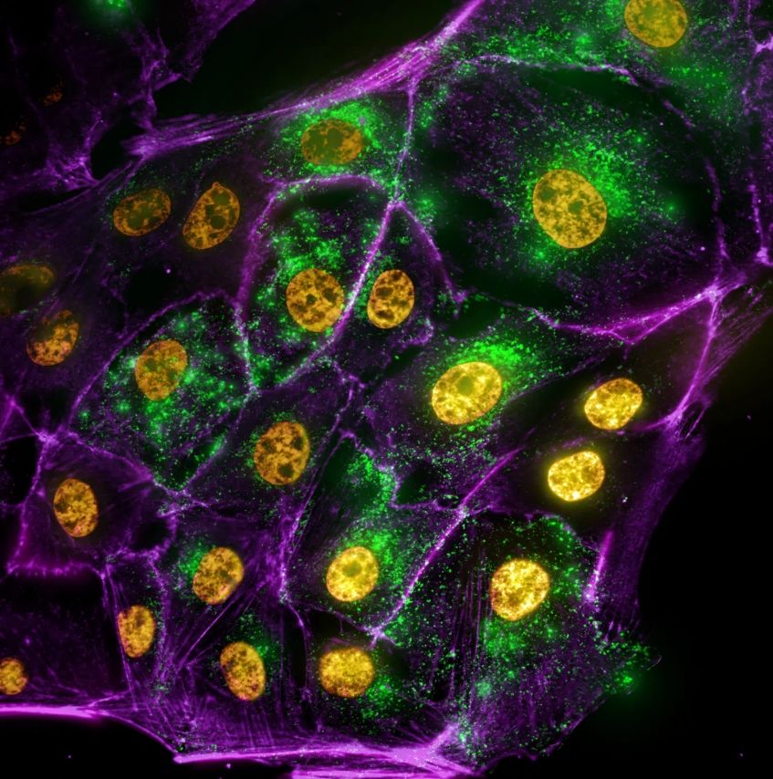
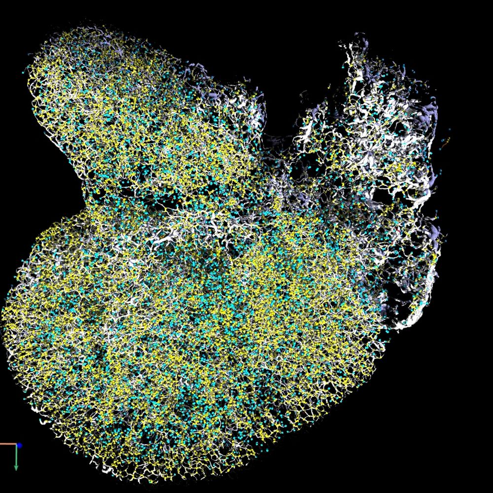
This colorized #microscopymonday image from 2023 by Frederic Bonnet shows kidney cells of a pig: the nuclei in yellow, the cytoplasmic vesicle in green and the membrane in magenta.
#standwithscience #researchmatters @mdibl-lmf.bsky.social
#microscopy #fluorescentproteins #lightsheet 🧪 🤝 🐟🎸 🍎 👩🔬
14.07.2025 12:26 — 👍 3 🔁 1 💬 0 📌 0
YouTube video by LMF@MDIBL
What are best practices for visualizing microscopy images ?
Do you follow best practices for visualizing microscopy images?
#microscopy #microscopymonday #imaging #visualization #science
youtube.com/shorts/_HlXa...
07.07.2025 17:26 — 👍 8 🔁 3 💬 0 📌 0
SGLT2 Inhibition Ameliorates Age-Dependent Renovascular Rarefaction https://www.biorxiv.org/content/10.1101/2025.06.27.654312v1
29.06.2025 04:06 — 👍 3 🔁 2 💬 0 📌 0

Maine Microscopy: Foundations and Fundamentals 2025
Deadline Approaching! July 7, 2025
Maine #Microscopy: Foundations and Fundamentals 2025, August 5-13 at MDI Bio Lab!
Optimize your experiment planning, imaging and analysis skills to strengthen your research and accelerate discoveries in your field.
#standwithscience #researchmatters 🧪 🤝 🍎 👩🔬
25.06.2025 13:08 — 👍 5 🔁 4 💬 0 📌 0
Maine Microscopy: Foundations and Fundamentals 2025
Maine #Microscopy: Foundations & Fundamentals 2025, August 5-13
For Maine grad students, postdoc trainees, research assistants & junior faculty. Understand current technology and how technical decisions impact final image products.
Application Deadline: July 7. Register today!
🧪 🤝 🍎 👩🔬 🖥️ 🧬
13.06.2025 12:11 — 👍 3 🔁 2 💬 0 📌 0

Happy #MicroscopyMonday!
#microscopycommunity - join @bioimagingna.bsky.social May 28, to hear Fredric Bonnet of @MDIBL_LMF speak on “Maine Microscopy: Democratizing Microscopy Education for Local Scientists” as part of the BINA EoE Virtual Group
Learn more and register here: buff.ly/F3rZzx7
26.05.2025 16:06 — 👍 7 🔁 5 💬 0 📌 0
Looking forward to the @mesospim.bsky.social #lightsheet #Microscopy anniversary symposium in October with @preibischs.bsky.social @adamkglaser.bsky.social @christellelangevin.bsky.social & many more! And to the next mesoSPIM decade! 🔬 Thanks @nvladimus.bsky.social & Co for organizing it!
16.05.2025 12:15 — 👍 19 🔁 11 💬 0 📌 0

A leading Light Microscopy Facility in Maine, the goal of MDI Bio Lab's @mdibl-lmf.bsky.social is excellence in research support & training with a strong emphasis on #education.
For instance, this #microscopymonday #zebrafish image is an example of a practice sample.
#standwithscience 🧪 🤝 🐟🎸 🍎 👩🔬
12.05.2025 11:55 — 👍 10 🔁 3 💬 0 📌 0

New chapter from Iain Drummond (MDI Bio Lab) and Heiko Schenk (Hannover Medical School) in Current Topics in Developmental Biology (@elsevierconnect.bsky.social): "Kidney development, injury and regeneration—Zebrafish"
#standwithscience 🧪 🤝 🐟🎸 #SciencePublishing
doi.org/10.1016/bs.c...
01.05.2025 12:09 — 👍 2 🔁 1 💬 0 📌 0

Michael W. Davidson Memorial Award
The BioImaging North America (BINA) Michael W. Davidson Memorial Award is designed to support members who embody the mission and core values of BINA by demonstrating commitment, shared values and the…
Happy #MicroscopyMonday!
Nominate an academic @bioimagingna.bsky.social member for the 2025 Michael W. Davidson Memorial Award!
2025 submissions are now being accepted through May 23, 2025 & details are available here buff.ly/Bh9qo5r .
07.04.2025 13:18 — 👍 9 🔁 4 💬 0 📌 0

Light Sheet Fluorescence Microscopy 2025
Have you been thinking of enrolling the MDI Bio Lab's #LightSheet Fluorescence Microscopy course?
Registration has been lowered to $1,000 — down from $3,500!
This is a great opportunity for researchers/students to enhance their knowledge & skills in cutting-edge #microscopy techniques. 🧪 🤝 🍎 👩🔬
03.04.2025 12:25 — 👍 11 🔁 4 💬 0 📌 3
Neuroscience researcher exploring neurodegenerative diseases through the lens of light microscopy — and always curious about the unseen 🧠✨
Professor, U of Utah. Biology, family, sports, food. Knitter, musician, aspiring weaver. SLC, via LA, the Bay Area, and Boston. Views are my own.
PhD student @MPI Science of Light
BioImage Analyst at @WEHI_research. Foodie, geek, Dad. Functions on coffee and red wine. Bringer of #MicroscopistsNightmares. He/Him
Professor of Nanooptical Imaging at FAU Erlangen-Nuremberg/CITABLE, Associated Group Leader at the MPI for the Science of Light. Glycobiology, optics, philosophy, and music.
Vanderbilt Cell Imaging Shared Resource (CISR). Microscopy for all of VU and VUMC. Light microscopy and electron microscopy. Image analysis. https://medschool.vanderbilt.edu/cisr/
Kirschstein-NRSA Postdoctoral Research Fellow at UVA studying B1 cells in various diseases w focus on fibrosis. Likes making tools. Flow cytometry + microscopy + data wrangler.
Wannabe polymath.
Leftist/anti-authoritarian hot takes are my own.
R&D Scientist@ICOB Imaging Core, Academia Sinica, Taiwan
https://weichenchu.com/
https://eabias.github.io/
https://www.linkedin.com/in/weichen-chu/
https://orcid.org/0000-0002-3447-9043
Neurobiologist @UofSC 🔬 Super-resolution #microscopy, synaptic plasticity, cytoskeleton, #neurodegeneration 🦠
Bluesky feed for Maine Discovery Museum. We help people of all ages to discover the world around them through creative exploration and science.
I study how lymphocytes self-organize into multicellular structures. Also interested in how cells sense and respond to chemical gradients. Currently: Wyss Institute at Harvard. Previously: Comp Bio at Duke. debrajghose.com
Junior Group Leader in Münster, Germany.
Morphogenesis, collective cell migration, cytoskeleton, cell-adhesion and self-organization/emergent behavior in #Drosophila.
Animal photos: 📷 instagram.com/maikscritters 🐸🐍
Volunteer led organization focused on providing microscopy-related activities and events and disseminating microscopy knowledge and skills to anyone interested in the New England region.
Postdoctoral Research Fellow
Baden Lab, University of Sussex — badenlab.org 🇬🇧
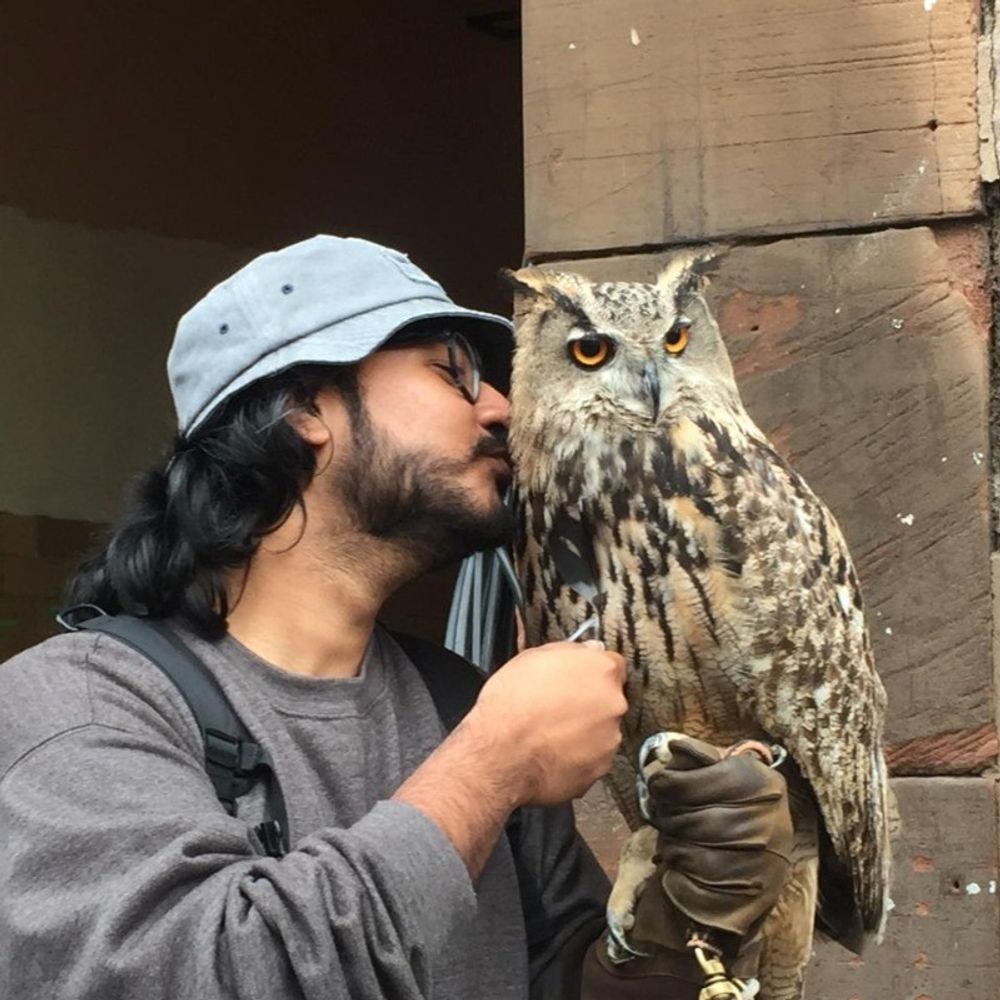
Exploring vision, birds, and viral tools 🐦
Ph.D. in Neuroscience, University of Oldenburg 🇩🇪 — #Magnetoreception
#Birds #Retina #VisionScience #Neuroscience #2PhotonImaging #AAV
PhD in the lab of @vlecaudey.bsky.social at JGU. Developmental Biology in #Zebrafish.
Epithelial cell biologist and physiologist studying the GI tract.
The official account for the Life Science Solutions division of the Nikon Healthcare Business, with a focus on biological microscopy products and services for the life science, biotech/pharma, and clinical laboratory markets.
Director of Data Science & Analytics @syGlass.io, making data beautiful. 🦠🔬📊 Enthusiast of all things microscopy, hiking, and cooking. Board member @NESM.
Harvard | Rochester Institute of Technology | Science Club for Girls
Head of the Centre for Cellular Imaging, Gothenburg, Sweden. President of the Core Technologies for Life Sciences, CTLS, Association
Scientist, Microscopist, community builder, collaborate don’t compete there’s too much work to do.