

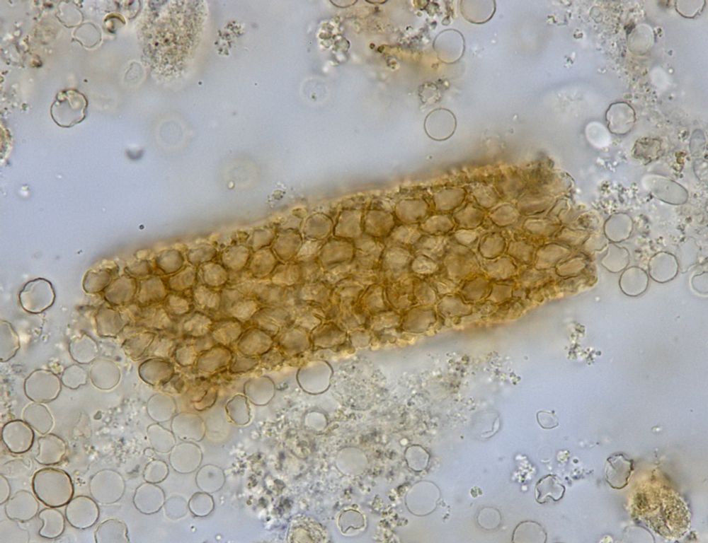
WBC cast with numerous "glitter cells" - phase contrast - from patient with suspected acute interstitial nephritis - #UrineMicroscopy #UrinarySediment
03.12.2025 00:11 — 👍 2 🔁 0 💬 0 📌 0WBC cast with numerous "glitter cells" - phase contrast - from patient with suspected acute interstitial nephritis - #UrineMicroscopy #UrinarySediment
03.12.2025 00:11 — 👍 2 🔁 0 💬 0 📌 0

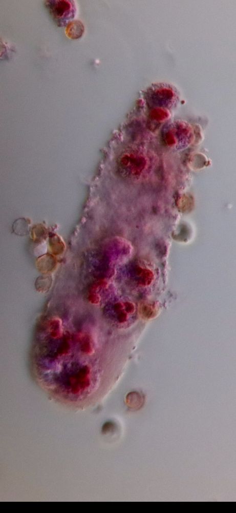
Mixed cellular casts - brightfield with SM stain - #UrinarySediment #UrineMicroscopy
22.11.2025 04:04 — 👍 6 🔁 0 💬 0 📌 0
Bilirubin crystals under polarized light #UrineMicroscopy #UrinarySediment
20.11.2025 14:05 — 👍 0 🔁 1 💬 0 📌 0


30 y/o man, with hx GPA 15 years ago, presents with vasculitic rash, pulm infiltrates, and AKI with rapid worsening. DAH on bronch. #UrineMicrosopy showed RBC cast, WBC cast, and tubular cell cast - brightfield with SM stain. #UrinarySediment
06.11.2025 12:09 — 👍 6 🔁 0 💬 0 📌 0
Mixed cellular cast (RBC’s and WBC’s, and a few tubular epithelial cells) - note the WBC’s are clearly indicated by Prescott-Brody stain whereas the tubular cells are not - in preserved urine sample from a case of GPA, stored at room temperature for 2.5 years! #UrineMicroscopy #UrinarySediment
11.10.2025 17:15 — 👍 0 🔁 0 💬 0 📌 0
RBC cast - brightfield, unstained - in preserved urine sample from a case of GPA, stored at room temperature for 2.5 years! #UrineMicroscopy #UrinarySediment
11.10.2025 17:14 — 👍 0 🔁 0 💬 0 📌 0

Oval fat body and acanthocyte - brightfield with SM stain - in preserved urine sample from a case of GPA, stored at room temperature for 2.5 years! #UrineMicroscopy #UrinarySediment
10.10.2025 00:55 — 👍 1 🔁 0 💬 0 📌 0

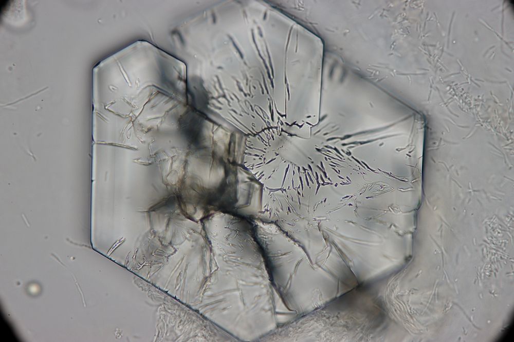
Calcium oxalate dihydrate crystals - brightfield and darkfield, unstained - from pt of @nephrothaniel.bsky.social - #UrineMicroscopy #UrinarySediment
02.10.2025 01:54 — 👍 3 🔁 0 💬 1 📌 0
RBC cast - brightfield, unstained - from patient with hydralazine-induced vasculitis #UrineMicroscopy #UrinarySediment
16.08.2025 01:55 — 👍 4 🔁 0 💬 3 📌 0


RBC's within waxy cast - brightfield and darkfield, unstained - from patient with hydralazine-induced vasculitis #UrineMicroscopy #UrinarySediment
16.08.2025 01:55 — 👍 3 🔁 0 💬 0 📌 0
RBC cast - brightfield, with SM stain - from patient with hydralazine-induced vasculitis #UrineMicroscopy #UrinarySediment
16.08.2025 01:54 — 👍 1 🔁 0 💬 0 📌 0

WBC cast - brightfield and darkfield, with SM stain - from patient with hydralazine-induced vasculitis #UrineMicroscopy #UrinarySediment
16.08.2025 01:53 — 👍 2 🔁 0 💬 0 📌 0
Acanthocyte - phase contrast (original mag x400) - from patient with hydralazine-induced vasculitis #UrineMicroscopy #UrinarySediment
16.08.2025 01:53 — 👍 4 🔁 2 💬 0 📌 0
RBC cast - brightfield with SM stain - from patient with MSSA bacteremia and osteomyelitis - #UrineMicroscopy #UrinarySediment
03.06.2025 03:21 — 👍 5 🔁 0 💬 0 📌 0
RBC cast - brightfield with SM stain - from new case of MPA - #UrineMicroscopy #UrinarySediment
13.05.2025 03:04 — 👍 5 🔁 1 💬 0 📌 0Very long RBC cast - darkfield with SM stain - from new case of MPA - #UrineMicroscopy #UrinarySediment
11.05.2025 02:32 — 👍 5 🔁 1 💬 0 📌 0


RBC casts - brightfield with SM stain - from new case of MPA - #UrineMicroscopy #UrinarySediment
10.05.2025 02:37 — 👍 1 🔁 0 💬 0 📌 0
Acanthocyte - darkfield, unstained - cropped from original x1000 - from case of suspected hydralazine-induced vasculitis - #UrineMicroscopy #UrinarySediment
30.04.2025 02:17 — 👍 6 🔁 0 💬 0 📌 0

RBC casts - brightfield with SM stain - from case of suspected hydralazine-induced vasculitis - #UrineMicroscopy #UrinarySediment
30.04.2025 02:17 — 👍 2 🔁 0 💬 0 📌 0
Vacuolar cast (which probably also contains some RBC's) - brightfield with SM stain - from patient with AKI and suspected hydralazine-induced vasculitis #UrineMicroscopy #UrinarySediment
30.04.2025 02:17 — 👍 1 🔁 0 💬 0 📌 0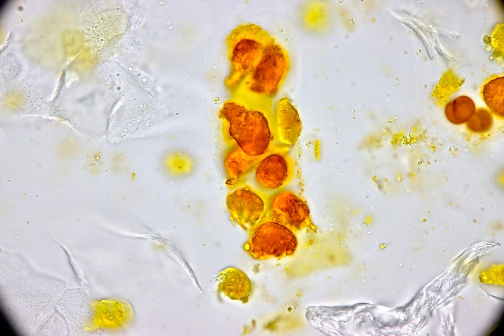
Interested in learning more about the art of microscopic examination of the urinary sediment and its clinical applications? @jrseltzer.bsky.social @swethakanduri.bsky.social and I will be running a Hands-On Workshop at KidneyCon this year. Come join us! @kidneycon.bsky.social May 1-3. Register ASAP
01.04.2025 18:26 — 👍 6 🔁 5 💬 0 📌 1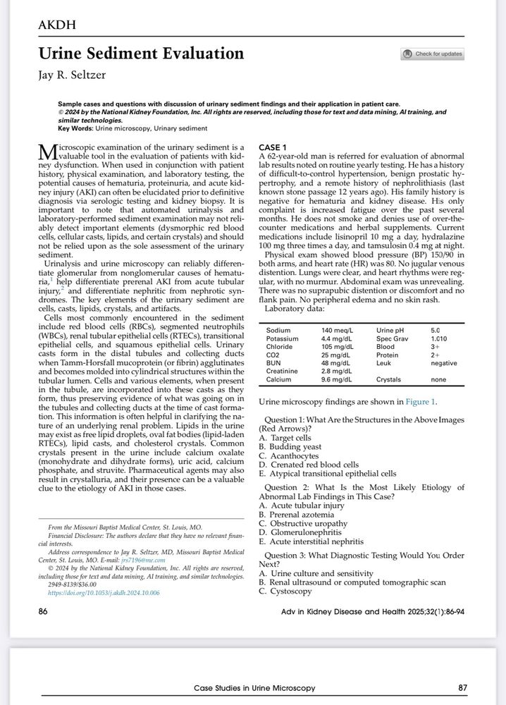
New #UrinarySediment and #UrineMicroscopy case studies in @akdhjournal
Use the link below but be sure to view as a pdf - the online version is not formatted correctly
kwnsfk27.r.eu-west-1.awstrack.me/L0/https:%2F...
Sorry for incomplete link:
Here it is:
kwnsfk27.r.eu-west-1.awstrack.me/L0/https:%2F...


Young male presented with DVT and PE. Found to have UPC 8 gm/gm, serum albumin 2.2, and creatinine 2.3 - #UrineMicroscopy showed this mixed cellular cast containing RBC's, tubular epithelial cells, and WBC's - brightfield with SM stain - #UrinarySediment
27.02.2025 02:11 — 👍 6 🔁 1 💬 0 📌 0


Vacation's over and I'm back in the lab....
Young male presented with DVT and PE. Found to have UPC 8 gm/gm and serum albumin 2.2 and creatinine 2.3 - #UrineMicroscopy showed RBC casts with fibrinous matrix as well as a few lipid casts - brightfield with SM stain - #UrinarySediment

WBC cast - brightfield with SM stain - from patient with recurrent UTI's s/p abx rx over 3 weeks - #UrineMicroscopy #UrinarySediment
30.01.2025 21:00 — 👍 3 🔁 0 💬 0 📌 0
RBC cast - brightfield, unstained - from patient with IRGN - #UrineMicroscopy #UrinarySediment
30.01.2025 20:57 — 👍 4 🔁 0 💬 0 📌 0
RBC's in a waxy cast - brightfield, unstained - from patient with IRGN - #UrineMicroscopy #UrinarySediment
30.01.2025 20:57 — 👍 3 🔁 1 💬 1 📌 0
Registration is open for KIDNEYcon 2025
May 1-3
Little Rock, AR
kidneycon.org/registration
