Historical illustration showing two magnified embryo figures: the upper is a human embryo and the lower is a dog embryo. Both are detailed with visible early anatomical features such as curved shapes and developing structures. The image is from Darwin’s work "The Descent of Man," with the human embryo attributed to Ecker and the dog embryo to Bischoff, highlighting early developmental similarities between species in evolutionary theory. The page header reads "SECOND DAY’S SITTING" with the page number 39.
🧠 Thx 2 @NHM_London 4 contributing this dog & human embryo fig. frm the bk: Homo vs. Darwin. An interesting read.
[Source]
17.02.2026 13:23 —
👍 4
🔁 1
💬 0
📌 0

Illustration of a flying dragon lizard perched on a tree branch, displaying large, colorful skin folds extending from its neck to its limbs. The lizard has a scaly greenish-blue body with brown and black spots on the extended skin, resembling wings used for gliding. It has an open mouth, clawed feet gripping the branch, and a long striped tail hanging down. The tree branch features rough bark and some yellow-green foliage. The artwork mimics a mythical dragon while depicting the real animal’s distinctive gliding adaptations.
🐉 The naturalists' miscellany: .
London: Printed for Nodder co, 1789.. Smaller cousin of fire-breathing dragons? Flying Dragon has folds of skin that it uses 2 glide through the air #bhlib
[Source]
16.02.2026 23:23 —
👍 28
🔁 9
💬 0
📌 0


To a first approximation, all Australian vertebrates are lizards. 🦎
But not just any lizards, Sphenomorphine skinks!
With more than 280 species they are hyper variable. Now, Janne Torkkola has pulled together the biggest phylogeny of the group to date. Read for free:
doi.org/10.1016/j.ym...
1/4
17.02.2026 00:56 —
👍 45
🔁 21
💬 3
📌 2
Time well spent
11.02.2026 22:53 —
👍 1
🔁 0
💬 0
📌 0
These ultra-fine structures shown at 30X magnification are the reason why geckos can adhere to most surfaces, using Van der Waals force.
The power of hairy lizard toes!
(🔬: Power & Syred, SciencePhotoLibrary)
06.02.2026 17:14 —
👍 123
🔁 37
💬 4
📌 1

Also this month, Šulcová et al., studied how teeth attach to jaws across vertebrates. In veiled chameleons, firmly ankylosed teeth formed via a transient cell type at the tooth–bone interface showing both osteoblast- and odontoblast-like features. The authors call these theorised cells ankyloblasts
03.02.2026 12:06 —
👍 1
🔁 2
💬 1
📌 0

Line drawing illustration from 1758 depicting the developmental stages of frogs, from egg clusters at the top through successive tadpole phases in middle rows, to fully formed frogs at the bottom right. At the bottom center, a detailed botanical illustration of a rose plant with leaves and blooming flowers is shown. Each stage and subject is labeled with figure numbers, emphasizing the biological progression alongside the rose plant as a botanical reference. The image is arranged clearly on a plain background with delicate, precise linework.
🌹 Historia naturalis ranarum nostratium: Nürnberg: gedrucht bey Johann Joseph Fleischmann, 1758.
[Source]
25.01.2026 21:23 —
👍 9
🔁 2
💬 0
📌 1

Cricket embryo for #FluorescenceFriday !
(macroH2A in magenta, neurons in yellow and F-Actin in cyan)
16.01.2026 16:52 —
👍 29
🔁 5
💬 0
📌 0

Modesty and candour: the Darwin-Wallace friendship
To mark the 200th anniversary of Wallace’s birth, an article exploring the friendship between Charles Darwin and Alfred Russel Wallace.
08-Jan: Born on this day in 1823, the man who independently of Darwin came up with the idea of natural selection, Alfred Russel Wallace. Here’s a post I wrote about Darwin’s and Wallace’s friendship…
friendsofdarwin.com/articles/dar...
#HistSci
08.01.2026 18:34 —
👍 32
🔁 17
💬 3
📌 1

Photo of a Mourning Gecko (Lepidodactylus lugubris) facing left on a branch with the sky in the background.

Photo of a Bynoe's gecko (Heteronotia binoei), facing left on a lichen-covered rock with the dark sky in the background.

Photo of an Indopacific slender gecko (Hemiphyllodactylus typus), facing right, on a large green leaf.
‘tis the season to celebrate the many animal species that reproduce via parthenogenesis - or reproduction from an unfertilized egg. Pictured here are 3 species of all-female, parthenogenetic gecko: Mourning Gecko; Bynoe's gecko; & Indopacific slender gecko
#MerryChristmas #Herpetology 🎄🧪🦎
25.12.2025 01:46 —
👍 64
🔁 12
💬 2
📌 0

Molecular basis for de novo thymus regeneration in a vertebrate, the axolotl
The molecular, cellular, and functional restoration of the axolotl thymus after de novo regeneration is described.
Can't believe my postdoc paper is finally out. Christmas came early this year, holy moly 🎄
Molecular basis for de novo thymus regeneration in a vertebrate, the axolotl | Science Immunology www.science.org/doi/10.1126/...
05.12.2025 21:17 —
👍 55
🔁 23
💬 4
📌 0
Very excited to share that our latest paper is out in Science! We show that the type specimen of Nanotyrannus—an isolated skull—is fully grown, showing that it is not a juvenile Tyrannosaurus rex but a distinct species (1/12)
www.science.org/doi/10.1126/...
04.12.2025 19:01 —
👍 98
🔁 42
💬 1
📌 6

Tetrapod vocal evolution reveals faster rates and higher-pitched sounds for mammals
Abstract. Using the voice to produce sound is a widespread form of communication and plays an important role across diverse species and contexts. Variation
Now out in Evolution @journal-evo.bsky.social
Tetrapod vocal evolution reveals faster rates and higher-pitched sounds for mammals 🐘🦉🐸.
Mammalian hearing likely allowed the rapid diversification of their vocalizations.
Open access here:
doi.org/10.1093/evol...
#bioacoustics #animalcommunication
01.12.2025 15:24 —
👍 46
🔁 20
💬 1
📌 1

Chromosome-scale Genomes Show Rapid Diversification and Ancient Gene Flow Among Bear Species
Abstract. Reconstructions of evolutionary history can be restricted by a lack of high-quality reference genomes. To date, only four of the eight species of
@tywooldr.bsky.social et al. present assemblies for three bear species — the sun, sloth, and Andean bears — and use a whole-genome alignment of all bear species and other carnivores to reconstruct the evolution of Ursidae.
🔗 doi.org/10.1093/gbe/evaf188
#genome #evolution
20.11.2025 16:50 —
👍 25
🔁 12
💬 0
📌 0
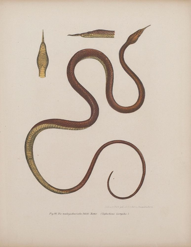
Illustration of a long, slender amphibian identified as the Malagasy dagger-tooth snake (Xiphorhina Longaula). The body is coiled in an S-shape, showing smooth, scaly skin in brown and yellow tones. Two detailed close-ups of the head display its pointed snout and scale pattern. The drawing emphasizes the snake’s elongated, narrow form and distinctive head shape, highlighting its natural texture and coloration for scientific study.
🐸 Bilder-Atlas zur wissenschaftlich-populären Naturgeschichte der Amphibien in ihren sämmtlichen Hauptformen
Wien: Kaiserl. Koenigl. Hof- und Staatsdr., 1864
[Source]
14.11.2025 13:23 —
👍 17
🔁 2
💬 0
📌 0

A developmental series of anole lungs, progressing from early embryo (left) to late embryo (right)
Taken together, we show that there are distinct processes by which different squamate species build their lungs. Notably, the luminal pressure-driven stress ball morphogenesis in anoles appears to be derived, potentially due to the fact anoles develop in ovo so much faster than other squamates!
14.11.2025 17:57 —
👍 2
🔁 0
💬 0
📌 0

Immunofluorescence labeling E-cadherin (green) and alpha-smooth muscle actin (magenta), and phosphorylated myosin light chain (white) in the developing lungs of veiled chameleon
Notably, these structures aren’t being sculpted by smooth muscle like in mammal lungs. Rather, they are enriched for pMLC in the apical region of the epithelium, suggesting an apical-constriction mediated outgrowth. This developmental process is what bird lungs use to branch!
14.11.2025 17:56 —
👍 1
🔁 0
💬 1
📌 0

EdU assays of chameleon lungs through development. EdU signal (white) is localized to the growing tips of the transitional chambers and diverticulae.
We also found some very interesting aspects of chameleon lung development. Chameleon lungs exhibit multiple chambers and these bizarre projections called diverticulae. These chambers and diverticulae seem to grow out via concentrated cell proliferation.
14.11.2025 17:54 —
👍 1
🔁 0
💬 1
📌 0

Simulation of a epithelium and smooth muscle mesh that either increases via luminal pressure or epithelial proliferation. The latter predicts a thinner epithelium and immunfluorescence of E-cadherin (green) and alpha-smooth muscle actin (magenta) recapitulates what the simulation predicted!
We decided to computationally simulate our hypothesized process: anoles expand their epithelium via luminal pressure while chams and geckos expand their epithelium via proliferation. This simulation suggested the epithelium would be thinner in anole lungs, and sure enough, that’s what we found!
14.11.2025 17:53 —
👍 1
🔁 0
💬 1
📌 0

Immunofluorescence labeling E-cadherin (green) and EdU (white) in the developing lungs of chameleons, leopard geckos, and brown anoles.
This suggests geckos and chams don’t drastically increase luminal pressure to push the epithelium through the smooth muscle. EdU shows more cell proliferation in the epithelium of gecko and cham lungs than anoles, suggesting proliferation pushes the epithelium through the mesh in geckos and chams.
14.11.2025 17:50 —
👍 1
🔁 0
💬 1
📌 0
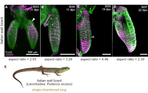
Immunofluorescence image labeling E-cadherin (green) and alpha-smooth muscle actin (magenta) of their developing lungs of Italian wall lizard
What we found in leopard gecko and chameleon lungs was also consistent in our opportunistic sampling of Italian wall lizard embryos.
14.11.2025 17:45 —
👍 2
🔁 0
💬 1
📌 0

Immunofluorescence labeling E-cadherin (green) and alpha-smooth muscle actin (magenta) of their developing lungs of brown anole (top) and leopard gecko (bottom)

Immunofluorescence labeling E-cadherin (green) and alpha-smooth muscle actin (magenta) of their developing lungs of veiled chameleon
We found that smooth muscle meshes show up in all three species, but what is really striking are the differences in inflation between anoles and the other two species. Aspect ratios are significantly different through development of the anole lung, but not in leopard geckos or chameleons.
14.11.2025 17:44 —
👍 1
🔁 0
💬 1
📌 0

A phylogeny of squamates demonstrating the diverse morphologies that are present in each clade with schematics of some of these distinct morphologies. The species we studied have either single-chambered lungs (brown anole & leopard gecko) or transitionally multi-chambered lungs with distal diverticulae (veiled chameleon)
We aimed to investigate whether stress ball morphogenesis is conserved in other squamates. Additionally, we wanted to see if more complex lungs exhibit distinct patterns of lung morphogenesis. To do this, we looked at embryonic lungs of leopard geckos, brown anoles, and veiled chameleons!
14.11.2025 17:42 —
👍 1
🔁 0
💬 1
📌 0