Huge shout out to @jenevievenorton.bsky.social, Erika, Sarah, @noahgurley.bsky.social, Rebecca, Emmanuel, Taino, the Jones Lab ...and of course my great mentor and PI Mark Peifer @peiferlabunc.bsky.social !!!
30.01.2025 16:24 — 👍 3 🔁 2 💬 0 📌 0
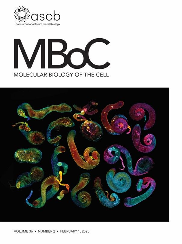
Wow! We got the cover! #MBOC #ASCB #CellBio
So proud of #TeamTestis. Screening <270 lines for shape defects was a huge team effort!
I created this collage from the team’s images from the past 2 years! Depth-color-coded with Fiji. Inspired by late 19th century lithographic animal illustrations.
30.01.2025 16:17 — 👍 112 🔁 17 💬 8 📌 3

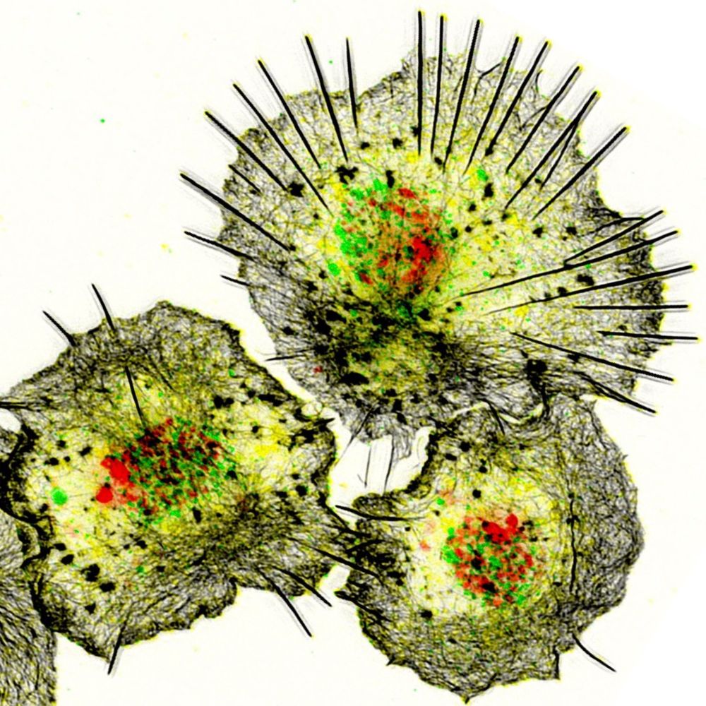
Immunofluorescence images of embryos showing that CnoRA1 domain is important for dorsal closure, head involution and epidermal integrity.
Wild-type (WT) embryo. Some cells are rounded up to divide (white arrows), but they rapidly return to columnar architecture. (B) cnoΔRA1. Groups of cells near the ventral midline fail to resume columnar architecture (red arrows). (C–E) Stage 11. (C) Wild-type embryo. (D,E) cnoΔRA1. Failure to resume columnar architecture becomes more apparent (red arrows). (F–H) Stage 14. (F) Ventral view of a wild-type embryo. Dorsal closure is completed (white arrow), head involution is underway (yellow arrow) and segmental grooves are shallow (green arrowheads). (G) cnoΔRA1. Dorsal closure failed, exposing underlying tissues (red arrow). There are gaps in the head epidermis (yellow arrows) and deep segmental grooves remain (green arrowheads). (H) Ventral view, cnoΔRA1. Holes in the epidermis (red arrows) and deep segmental grooves (green arrowheads) are observed. (I–J) Closeups, wild-type stage 10 (I–I″) and 11 (J). Dividing cells (green arrows) and forming tracheal pits (cyan arrows) are observed. Arm, Baz and Cno remain enriched at AJs. (K–K‴) Stage 10/11 cnoΔRA1 mutant. Cells that retained columnar architecture retain junctional Arm, Baz and CnoΔRA1 (yellow arrows). However, in some cells AJs are fragmented (red arrows) and in less epithelial regions Arm, Baz and Cno are strongly reduced (cyan arrows).
@emily-mcparland.bsky.social, @noahgurley.bsky.social, Kevin Slep, @peiferlabunc.bsky.social and colleagues dissect the differential roles of the dual Ras-association domains of Drosophila Canoe in linking cell junctions to the cytoskeleton.
journals.biologists.com/jcs/article/...
17.12.2024 13:06 — 👍 14 🔁 6 💬 1 📌 0
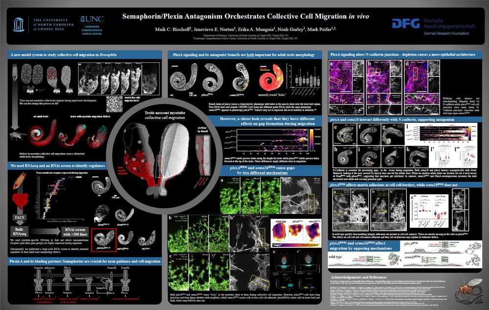
Next to my talk (12/16 4 pm room 33B), I will present a poster: "Semaphorin/Plexin Antagonism Orchestrates Collective Cell Migration in vivo". Tuesday, Dec 17 11:15 AM.
P2780/Board No. B397
I am so excited. I just love ASCB meetings!
#cellbio2024 #cellbio #devcell @ascbiology.bsky.social #ASCB
12.12.2024 15:05 — 👍 17 🔁 6 💬 2 📌 0

#FluorescenceFriday for my first post, of course. Developing chicken embryo transfected with pCIG on the left (green) and pCI H2B-RFP on the right (red) and co-stained for SOX10 (magenta) and PAX7 (cyan).
23.11.2024 01:46 — 👍 16 🔁 1 💬 1 📌 1
#CellPolarity lab using #3Dculture to study how cells collectively move in cancer. Professor, University of Glasgow. Senior Group Leader, @cruk-si.bsky.social. UKRI Future Leader Fellow.
A biophysicist with a background in mathematical modelling, scientific programming, and data analysis. Staff scientist at the Institute of Cell Biology in Bern🇨🇭
https://macdobry.net
https://github.com/dmattek
Diligently working towards the infinite replacement organ paradigm.
Building Robots That Help People Learn Things Quicker
NRSA-funded postdoc in the Stappenbeck Lab at Cleveland Clinic studying how epithelial interactions can shape immunity. Views my own.
Investigating Human Endogenous Retroelements at the Interface of Epigenetics & Diseases-INHERITED
Alum: Feschotte lab, Cornell University New York, Clinical Neuroscience, MPI Gottingen, Izsvak lab, MDC Berlin, Uni of Bath UK, CCMB Hyderabad, JNU New Delhi
Harvard Neuro PhD Candidate in Goodrich Lab | Gilliam Fellow | Glia in PNS Development Researcher | Scientist with Tourette's & Disability Advocate
I am a senior scientist in the Cell & Tissue Dynamics research group at Oslo University Hospital, Norway.
My main interests are mechanical and biochemical processes that regulate cell migration during tissue development, wound healing & cancer metastasis.
Delphine Gourdon - Prof. in Biomedical Engineering - Univ. of Glasgow 🧪
#Mechanobiology #BreastCancer #Biomaterials #ECMatrix #TumorMicroenvironment #Tribology #WomenInSTEM
www.gourdonlab.org
We are a Glasgow-based multidisciplinary team of bio- and cell engineers.
We focus on understanding the interactions between materials, proteins and cells to gain insight into engineering cell behaviour and translating technologies into better healthcare.
Postdoc | Princeton University
Active soft matter, Fluid mechanics, Biophysics.
https://scholar.google.de/citations?hl=en&user=QLNh8EwAAAAJ&view_op=list_works&sortby=pubdate
Postdoc at @MITCheme• PhD @MatarLab @ImperialChemEng @nnf_lab @MITMechE• Computational Non-Newtonian Fluid Dynamics • Biomechanical Modeling • Cell Mechanics • NPs-cellular membranes modeling
Enjoying a mash-up of biophysics and development biology with plenty of cytoskeleton fun in the Grill Lab at MPI-CBG Dresden. Interested in climbing, outdoors and raising two decent human beings.
Professor of Cancer Cell and Metastasis Biology. CRUK Senior Fellow. Institute of Cancer Research. London.
https://www.icr.ac.uk/our-research/researchers-and-groups/professor-victoria-sanz-moreno
Prof. Shashank Shekhar @EmoryUniversity, Atlanta, USA
Biophysicist interested in Actin dynamics and ciliary flows.
Departments of Physics, Biochemistry and Cell Biology.
Lab website : www.shekharlab.org
Chemotaxis. Math. Computers. Cells. Machine learning.
Edgar C. Hendrickson Chair in Biomedical Engineering - Professor of Biomedical Engineering and General Surgery at The Ohio State University.
Nanotechnology, non-viral gene and cell therapies in regenerative medicine and cancer.
🎓 PhD Student @rouxlab.bsky.social
🔬 Exploring forces in stem cells and development | Biophysics
👨🎓 Alumni: @xaviertrepat.bsky.social & @mhverlhac.bsky.social
⛷️ Hockey, mountains, and skiing enthusiast
📍 Based in Geneva 🇨🇭
Cell and developmental biology at the University of Sheffield unravelling the mysteries of planar polarity using Drosophila