Top: Experimental set up. Single pulses of TMS were applied to the hand area of the right primary motor cortex with an inter-pulse interval randomized between 6 and 10 s. Simultaneously, the neuronavigated coil position (yellow), electrocardiogram (red), respiratory signal (blue), electrogastrogram (green), and electromyography from the left hand (gray) were recorded. The figure shows traces of a 20-s time segment from one participant of the cardiac (raw), respiratory (filtered), gastric (filtered), and EMG (raw) signals. The experimental measure was the Motor Evoked Potential amplitude measured on a hand muscle (first dorsal interosseous), analyzed against the phase of the cardiac, respiratory, and gastric rhythms. Note that the three rhythms have very different periods (~1 s for the heart, ~ 5 s for respiration, and ~20 s for the gastric rhythm). Bottom: Artwork illustrating how rhythms of the internal organs interact with the moment-to-moment fluctuations observed in corticospinal motor excitability. Image depicts an outline of a brain with representations of the three rhythmic organs influencing the motor system: the heart, lungs, and stomach. Image credit: Tahnée Engelen.
How do internal bodily rhythms influence #brain activity & motor function? @tahnee-engelen.bsky.social &co show that #cardiac, #respiratory & #gastric rhythms independently modulate motor excitability, revealing distinct #interoceptive profiles across individuals @plosbiology.org 🧪 plos.io/4nMtpLT
13.11.2025 10:17 — 👍 35 🔁 20 💬 3 📌 2

Incredible study by Raut et al.: by tracking a single measure (pupil size), you can model slow, large-scale dynamics in neuronal calcium, metabolism, and brain blood oxygen through a shared latent space! www.nature.com/articles/s41...
25.09.2025 08:53 — 👍 67 🔁 17 💬 1 📌 1

Villringer et al. Figure 1. Conceptual framework for brain–body states

Villringer et al. Figure 2 Brain–body micro-, meso-, and macro-states can be distinguished on the basis of their duration and reversibility
'Brain–body states as a link between cardiovascular and mental health'
by Arno Villringer, Vadim Nikulin & Michael Gaebler @mbe-lab.bsky.social @michaelgaebler.com @mpicbs.bsky.social sky.social
www.cell.com/trends/neuro...
23.09.2025 20:13 — 👍 36 🔁 13 💬 1 📌 2
Long-term ECG data have great potential to expand research into diurnal variation of heart rate variability, beyond short-term measures of resting state. Data processing, however, is labor intensive. Led by Maximilian Schmaußer, we compared the performance of open-source tool boxes 1/2
15.09.2025 19:10 — 👍 5 🔁 3 💬 1 📌 0
Thanks, Emilia! Systole performed best overall, though all tools produced outliers we needed to double-check. Happy to share more if you're interested!
05.08.2025 07:51 — 👍 0 🔁 0 💬 0 📌 0
Three days left to participate in round one of our Delphi study. 🔥
For more infos, follow the link! #hormones #psychoneuroendocrinology 🧪
12.06.2025 11:24 — 👍 5 🔁 6 💬 0 📌 0

Our new paper on fully automated ECG processing pipelines is out!
We benchmarked NeuroKit2, RHRV, and Systole against the manual gold standard (Kubios) using 48h ECG recordings.
⬇️ Abstract
www.researchgate.net/publication/...
26.05.2025 09:33 — 👍 2 🔁 0 💬 1 📌 0

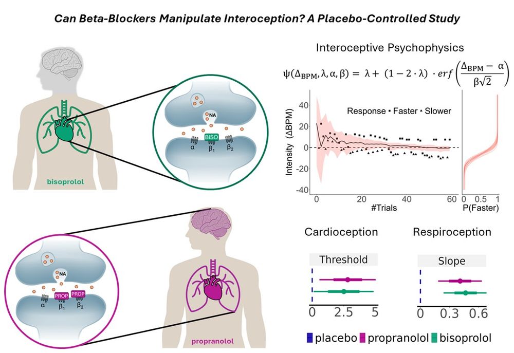
A graphical abstract titled "Can Beta-Blockers Manipulate Interoception? A Placebo-Controlled Study." The image illustrates how bisoprolol and propranolol affect interoception. On the left, a green figure represents bisoprolol, showing a zoomed-in synapse where it selectively blocks β1 receptors. On the right, a purple figure represents propranolol, blocking both β1 and β2 receptors, affecting both brain and body. A psychophysics graph models interoceptive responses, showing response intensity over trials. Below, line plots compare placebo, propranolol, and bisoprolol effects on interoception—cardioception (threshold) and respiroception (slope)—indicating drug effects on bodily awareness.
Can we enhance interoception by controlling the heart? Thrilled to share our new study, led by @ashleytyrer.bsky.social , where we use computational modeling to show that blockading peripheral noradrenaline uniquely alters awareness of heart rate & breathing! www.biorxiv.org/content/10.1... 🧵👇
10.03.2025 15:29 — 👍 146 🔁 48 💬 2 📌 7


Fascinating results on distinctive peripheral beta-blockade effects on cardiac and respiratory interoceptive sensitivity and metacognition presented by @micahgallen.com @the-ecg.bsky.social at the #MindBrainBody Symposium 💊🫀🫁
#MBBS25
12.03.2025 12:27 — 👍 18 🔁 4 💬 1 📌 1
Clinical Psychology and Psychotherapy Section @uniwuerzburg.bsky.social || Headed by Katja Bertsch ||Translational Research with a Focus on Social Interaction Processes and Mental Health
PhD Student @ UCL Institute of Cognitive Neuroscience |
UCL-Wellcome PhD in Mental Health Science |
Brain-body interactions in hallucinations and schizophrenia 🫀🧠
The Institute for Mind and Brain at the University of South Carolina works to understand the biological bases of the mind, brain, and cognition and intersections with health, aging, and neurodevelopment.
The European Society for Cognitive and Affective Neuroscience aims to promote scientific enquiry within the field of human cognitive, affective and social neuroscience, particularly with respect to collaboration and exchange of information between research
Clinical Psychologist at King’s College London and South London and Maudsley NHS Foundation Trust. Interested in experimental methods and research into and treatment of anxiety.
Assistant Prof @Northeastern. Psychology + Biology. Neuroimaging, brain metabolism + mental health. Director of IASLab with Lisa Feldman Barrett & Karen Quigley
https://www.affective-science.org/
http://www.jordan-theriault.com/
Shared account: Biological Psychology and Neuropsychology section of the German Psychological Society (DGPs) and German Society for Psychophysiology (DGPA)
https://www.dgps.de/fachgruppen/fgbi/
https://www.dgpa.de/index.php
Die Interessengruppe Offene & Reproduzierbare Forschung (IGOR) in der @dgps.bsky.social Fachgruppe Biologische Psychologie & Neuropsychologie.
Website: https://www.dgps.de/fachgruppen/fgbi/aktivitaeten-der-fachgruppe/igor/
Clinical Social Neuroscientist & Psychotherapist @tudresden.bsky.social & head of @kanskelab.bsky.social
https://tud.link/0p4j
Biopsychology Lab at University of Cologne - Decision Neuroscience - Gambling - Reinforcement Learning
Spitzenmedizin. Tag für Tag. Hand in Hand. - 🛑 Derzeit inaktiver Account der Uniklinik Köln. Impressum: http://uk-koeln.de/impressum
#UniKöln #UniCologne // Impressum: http://ukoeln.de/3JGDD
Mastodon ➡ @UniKoeln@Wisskomm.social
Professor of Medicine, McMaster University
Executive Director of the Firestone Institute for Respiratory Health
Board of Directors Lung Health Foundation
#macrophages #aging #respiratoryinfections #vaccinations #immunity #inflammation #scicomm
Neuroscientist | Professor of Medical Psychology at U Bonn | PI Neuroscience of Motivation, Action, & Desire Lab at U Bonn & Tübingen
aka @cornu_copiae
Postdoc in cognitive neuroscience 🧠 at the University of Jyväskylä 🇫🇮 . Previously Ecole Normale Superiéure 🇫🇷 & Maastricht University 🇳🇱
Interested in brain-body interactions, interoception, emotions
Website: www.tahneeengelen.com
PhD candidate studying perception of naturalistic facial expressions across lifespan | Former opera singer | Interested in multimodal communication (vocal/facial) & MSI, affective breathing, interoception 🫁🫀
PI @bodybrainbehaviour.bsky.social at IBB Muenster (GER) | Here for brain rhythms, body rhythms, and predictive processing in health and disease | ERC StG: 'DYNABODY' (2025)
We are a computational neuroscience lab co-located at Aarhus University and Cambridge Psychiatry. Our research investigates how our decisions, emotions, and conscious perception are shaped by visceral and embodied processes.
https://www.the-ecg.org/
Postdoc @HHU Düsseldorf, formerly MPI_CBS Leipzig & Sussex Centre for Consciousness Sci, Sussex Uni | Consciousness, adaptive behaviour, learning, brain-body interaction, taVNS


