Integrating SAMER Retrospective Motion Correction into 3D Deep Learning Image Reconstruction for High-Quality, Fast, and Robust Brain MRI by Daniel Polak (Siemens Healthineers, Erlangen, Germany), et al.
Patient motion is one of the most common sources of image degradation in clinical neuroimaging, manifesting as blurring, ringing, or in severe cases ghosting/folding artifacts. Radiologists are often forced to either interpret images despite these artifacts – risking missed or misinterpreted pathologies – or to request costly and time-consuming repeat scans.
In pediatric patients, motion is typically more severe, often necessitating anesthesia, which increases both procedural risks and costs.
Motion artifacts also reduce the reliability of automated quantitative clinical tools, such as those used for brain morphometry and the identification or segmentation of hemorrhages, edema, and tumors. Such tools are increasingly important in the screening, monitoring, and treatment of neurodegenerative diseases such as Alzheimer’s.
Scout Accelerated Motion Estimation and Reduction (SAMER) is a retrospective motion correction technique for brain imaging that enables fast motion estimation and artifact correction without the need for external tracking hardware. Clinically evaluated in both adult and pediatric patient populations, SAMER has significantly reduced the number of non-diagnostic motion cases.
More recently, SAMER has been integrated into a deep learning-based image reconstruction framework to support highly accelerated, motion-robust 3D brain imaging.
In this article, the authors review the combined DL-SAMER technique and demonstrate its effectiveness in vivo using MPRAGE and SPACE acquisitions at R = 6 acceleration.
Shout out to the co-authors:
Dominik Nickel, Daniel Nicolas Splitthoff, Bryan Clifford, Yan Tu Huang, Wei-Ching Lo, Shohei Fujita, Susie Y. Huang, John Conklin, Lawrence L. Wald, Stephen Cauley
DL-SAMER: A new deep learning + retrospective motion correction method for fast, motion-robust 3D brain #MRI — effective even in challenging pediatric cases.
Check it out 👉 marketing.webassets.siemens-healthineers.com/7a1c195a6a39...
#NeuroSky #RadSky #MagnetomWorld
@harvardmed.bsky.social
22.05.2025 08:30 — 👍 7 🔁 2 💬 0 📌 2

📣 Speaker spotlight for #ESMRMB2025: Sebastian Weingärtner! 📣
Join Dr. Weingärtner to learn about quality assessment of quantitative MRI in "Cycle of Quality"!
Register here to experience an ESMRMB meeting to never forget: buff.ly/GHbFWHc
22.05.2025 08:02 — 👍 3 🔁 2 💬 0 📌 0

As the ISMRM & ISMRT Annual Meeting approaches, many of us are preparing for long flights — or planning to join virtually from around the world. Wherever you are attending from, the ISMRM edition of MAGNETOM Flash offers a timely opportunity to engage with key developments in MRI.
Featureing an editorial by Jeong Hee Yoon, MD (Seoul National University Hospital, Republic of Korea), this edition explores the ongoing evolution of MRI in both clinical relevance and technical innovation.
From accelerated body imaging and developments in ultra-high-field MRI to advanced reconstruction methods and functional whole-body applications, the field is moving rapidly toward greater precision and efficiency.
This issue also highlights important directions in sustainable imaging, reproducible research, and real-world AI integration, as well as innovations that address long-standing challenges such as motion and the need for sedation in pediatric exams.
A heartfelt THANK YOU to all the authors and to everyone who contributed to this edition!
Headed to #ISMRM25 or joining virtually?
The new edition of MAGNETOM Flash covers key advances in #MRI — from reproducible research to AI: www.magnetomworld.siemens-healthineers.com/publications...
@ismrm.bsky.social @ismrt.bsky.social @tomhilbertmri.bsky.social #RadSky #NeuroSky #MedPhys #OncoSky
04.05.2025 11:09 — 👍 5 🔁 2 💬 0 📌 0
YouTube video by EPFL School of Engineering
Génie Électrique et Électronique SEL à l'EPFL
youtu.be/lKR9FDVNZsI?...
So nice to see our collaboration with EPFL engrained in this video for the school of electrical engineering. We have had so many excellent students, like Mathieu, who have helped to push MR technology to the next level.
08.04.2025 18:14 — 👍 3 🔁 1 💬 1 📌 0

Extending conventional DWI to include advanced diffusion encoding and image readouts brings the field closer to realizing the promise of a “virtual microscope.”
These advancements rely on high-performance MRI systems, with particular emphasis on the gradient system.
Importantly, a vast majority of advanced DWI methods gain marked improvement in data quality from ultra- strong gradients and more efficient readout strategies, and the exploration of biomarkers can be extended to include experimental designs that were previously either too slow or yielded insufficient SNR.
Such developments are particularly promising for prostate imaging, offering high-fidelity high-resolution images that reflect ever more subtle features of tissue microstructure—enhancing detection, diagnostics, and treatment monitoring.
The authors present early insights into the novel information made accessible by new hardware, and how this drives further development in the context of prostate cancer imaging. As powerful MRI systems become commercially available worldwide, strong-gradient technology will facilitate new imaging biomarkers, likely playing a fundamental role in everyday clinical diagnosis and advancement of individualized precision medicine.
#MRI of the #Prostate: The Promise of Ultra-Strong Gradients and Advanced Microstructural Imaging by Dr Molendowska, @deekayjay.bsky.social, et al (Lund University & CUBRIC)
Learn more marketing.webassets.siemens-healthineers.com/f0c8b5c08fab...
#dMRI #DWI #Microstructure
@tomhilbertmri.bsky.social
08.04.2025 05:17 — 👍 2 🔁 2 💬 0 📌 1
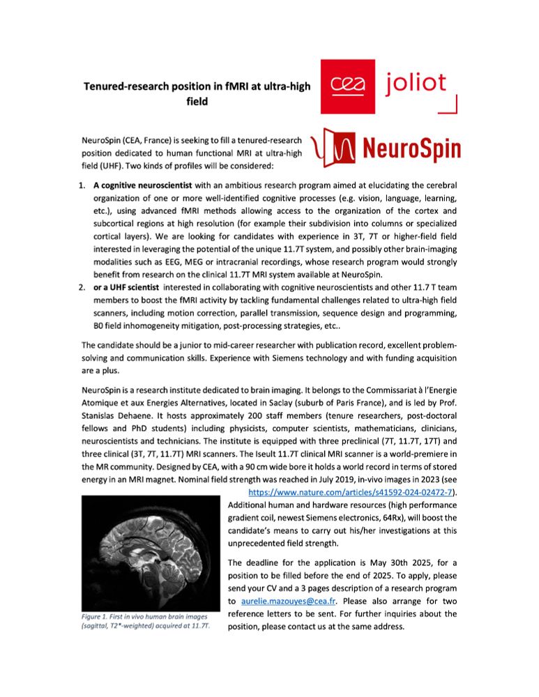
Tenured-research position opened here in Paris : A great opportunity to leverage the full potential of the new 11.7T MRI scanner at Neurospin @inserm.fr @cea.fr @unicog.bsky.social
🧠🟦 / 🧠🤖 / 🧠🩺
#AcademicSky
15.03.2025 09:23 — 👍 45 🔁 29 💬 0 📌 1

With optimized imaging protocols, the enhanced signal-to-noise ratio and spatial resolution of second-generation 7T MRI offers new diagnostic advantages that enable high-precision radiological imaging as part of routine clinical protocols.
This has tangible therapeutic implications, such as the identification of additional demyelinating lesions on 7T MRI in multiple sclerosis patients, which could fulfill the McDonald criteria for spatial dissemination and guide therapeutic decisions, or the detection of epileptic foci leading to surgical intervention.
The authors show the technical setup of a MAGNETOM Terra.X in Geneva, and share a few cases from routine clinical use.
Unlocking New Frontiers in Neurological Imaging: The Power of Second-Generation Clinical #7T #MRI by Professor Felix Kurz, et al. (HUG, Switzerland).
marketing.webassets.siemens-healthineers.com/b005273b6da8...
#NeuroSky #MagnetomWorld #Epilepsy #MultipleSclerosis #UHF @tomhilbertmri.bsky.social
18.02.2025 07:34 — 👍 10 🔁 7 💬 0 📌 1
Incredible how the 7T technology has managed to translate a lot of research into clinical routine.
18.02.2025 07:58 — 👍 3 🔁 0 💬 0 📌 0

On today’s International Childhood Cancer Day let me highlight Michael Kean’s comprehensive look at Scanning Faster: Application in Pediatric Neuroimaging.
The need for faster MR scanning is driven by muliple factors in both clinical and research settings. These factors include patient compliance, increasing pressure on the MR facility to scan more patients in a shorter time amid rising healthcare costs and long waiting lists, the integration of newer sequences into the protocol, and minimizing sedation or anesthesia times.
Faster scanning techniques can be successfully integrated into routine pediatric protocols significantly reducing scan times. While no single solution fits all clinical protocols, the tools are available to customize your sequences to match the requirements of your department.
The figure shows how to put it all together into a clinical neuro-oncology protocol. Recurrent supra tentorial Ewings Sarcoma.
29A: T2 TSE DL p3, 0.2 × 0.2 × 2.5 mm3, 2:20 min
29B: T2 FS FLAIR DL, 1:43 min
29C: SMS RESOLVE p2s2 DWI, b-value 1000 s/mm2
29D: CS FS T1 SPACE BB, 0.8 mm isotropic, 4:12 min
Scanning Faster: Application in #Pediatric Neuroimaging by Michael Kean, Chief #MRI Technologist at The Royal Children’s Hospital/@mcri.bsky.social, Melbourne, Australia.
Read the full article: marketing.webassets.siemens-healthineers.com/5cfbee50efc2...
#MagnetomWorld #NeuroSky #ChildhoodCancerDay
15.02.2025 10:02 — 👍 7 🔁 4 💬 0 📌 0

Coronary magnetic resonance angiography (CMRA) could potentially offer a safe, non-invasive alternative for the anatomical assessment of coronary artery disease (CAD), free from ionizing radiation and iodinated contrast agents.
However, image acquisition with conventional free-breathing CMRA frameworks is limited by long and unpredictable scan times, whilst image degradation due to respiratory motion remains a challenge.
The authors present a CMRA framework, designed to overcome some of these challenges by incorporating a highly undersampled Cartesian acquisition with a 2D image navigator to enable 100% respiratory scan efficiency, 2D translational motion correction, and 3D non-rigid motion estimation, which is then fully reconstructed using a 3D patch-based low-rank regularization framework.
This framework has been validated against coronary computed tomography angiography (CCTA) in a single-center trial of 50 patients with suspected CAD. Diagnostic image quality was obtained in 95% of all coronary segments.
The sensitivity, specificity, and negative predictive value were as follows:
- per-patient, 100%, 74%, and 100%;
- per-vessel, 81%, 88%, and 97%; and
- per-segment, 76%, 95%, and 99%, respectively.
February 14 is the Day of the Coronary Patient—an opportunity to repost High Spatial Resolution Coronary Magnetic Resonance Angiography by Reza Hajhosseiny, @mri-cprieto.bsky.social, et al (@kingscollegelondon.bsky.social).
marketing.webassets.siemens-healthineers.com/4dd530cb46a9...
#CardioSky #MRI
14.02.2025 07:13 — 👍 10 🔁 2 💬 2 📌 0
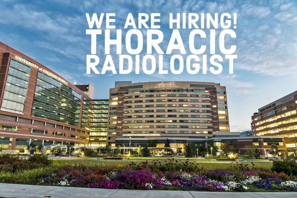
As we continue to expand, we are looking for a Thoracic Radiologist to join our wonderful team!
Work in a progressive department and live in a fantastic city!
DM me if interested!
03.01.2025 06:47 — 👍 3 🔁 2 💬 1 📌 0
If it is an organic chemist that can do image processing, why not :)
12.12.2024 17:30 — 👍 0 🔁 0 💬 1 📌 0

Looking for a job at a beautiful lake close to the mountains? Join us at the Swiss Innovation Hub of Siemens Healthineers! We are looking for a software developer to join our efforts to innovative and create new research applications in the field of MRI. jobs.siemens-healthineers.com/careers/job?...
12.12.2024 07:49 — 👍 11 🔁 5 💬 1 📌 0
Hi -- I have a postdoc position open in Computational MRI -- looking for experience and interest in MRI acquisition (in addition to reconstruction). Offer competitive pay. Opportunity to collaborate with Center for GenAI and Dell Med School. Bonus if you get your app in before Dec 1!
25.11.2024 21:26 — 👍 16 🔁 11 💬 0 📌 1

Ultra-high-field magnetic resonance imaging (UHF MRI) at 7T holds promise for tackling these obstacles, given its capacity to generate higher-resolution images and offer distinct contrasts due to a higher susceptibility effect and different relaxation time constants.
GRE T2*-weighted imaging (T2*WI) is a fast, convenient, noninvasive, and feasible technique that exhibits advantages in clinical settings. T2*W sequences make it easier to identify lesions in the local magnetic field, such as bleeding and hemosiderin accumulation. Structures with slightly different magnetic properties introduce detectable field variations at high B0, which leads to a noticeable increase in lesion identification at 7T.
Learn more about
- SWI in Wilson disease
- SWI in primary angiitis of the central nervous system
- T2*-weighted MRI in multiple sclerosis
- Other biomarkers such as motor band sign in amyotrophic lateral sclerosis (ALS) patients
Insights from 7T: GRE T2*-weighted Sequences Uncover Biomarkers in Neurological Diseases by Jing Jing, MD; et al (Tiantan Neuroimaging Center of Excellence, Beijing, China). Learn more at
marketing.webassets.siemens-healthineers.com/536502fb9ca6...
#MagnetomWorld #UltraHighField #7T #MRI #NeuroSky
26.11.2024 08:57 — 👍 7 🔁 2 💬 0 📌 0
When you give a lab meeting presentation sitting down
26.11.2024 15:32 — 👍 89 🔁 4 💬 1 📌 1
[clears throat, first BlueSky post]
The 2025 Gordon Research Conference on *Tissue Microstructure Imaging* program is out!
This unique meeting format offers a unifying look at tissue microstructure based on #MRI, #US, #optics, #EM , #x-ray, #photoacoustic, etc.
Join us July 12-18 & repost! More 👇
21.11.2024 09:00 — 👍 39 🔁 22 💬 1 📌 1
Hi @mrifranz.bsky.social, could you add me please? 🙏
20.11.2024 07:24 — 👍 2 🔁 0 💬 0 📌 0
Thanks to a nice crowd effort, the starter pack is now much larger! So great to see a fantastic network of #MRI researchers! Share and suggest people to add here below!
go.bsky.app/EVqd6pe
20.11.2024 05:42 — 👍 37 🔁 21 💬 13 📌 1

The innovative design and operational strategies at this high-efficiency MRI facility have significantly enhanced patient throughput and patient satisfaction.
By addressing key bottlenecks in the MRI #workflow, focusing on shortening the time spent during key periods in the MRI examination, including the patient preparation, table turnaround, and gradient times, substantial improvements in performance metrics have been achieved.
The optimized facility’s ability to reduce appointment slots from 45 to 30 minutes, while improving on-time performance and patient experience, showcases the promise of this multi-faceted approach. As healthcare systems worldwide grapple with increasing demand for MRI services, the Assembly Row model offers a blueprint for optimizing imaging workflows without compromising image quality or patient care.
Building the Future of #MRI: #Workflow Optimization and Patient-Centered Design at Mass General Brigham’s High-Efficiency Outpatient Facility by Dr Susie Huang, et al. (MGH, Boston, USA).
Learn more & download #MSK protocols www.magnetomworld.siemens-healthineers.com/clinical-cor...
#MagnetomWorld
19.11.2024 08:40 — 👍 4 🔁 3 💬 0 📌 0

EPI-based sequences remain the clinical workhorse and default method for DWI in many applications. However, their image quality could be compromised and diagnosis could be challenged in regions or situations where strong B0 inhomogeneities are present.
The turbo gradient spin echo (TGSE) BLADE DWI sequence samples a train of spin echoes and gradient echoes, and can generate distortion-free DWI images. Several studies have compared the image quality and diagnostic performance of TGSE BLADE DWI and EPI-based DWI, e.g. for cholesteatoma, orbital tumors, sinonasal lesions, and cerebellopontine angle tumors. The results show that TGSE BLADE DWI yields superior diagnostic quality to EPI-based methods in these challenging applications.
Distortion-free Diffusion-weighted Imaging Based on a TGSE BLADE Sequence: Technique and Clinical Application by Kun Zhou, PhD et al. (Siemens Healthineers)
Learn more at marketing.webassets.siemens-healthineers.com/81a78a5b3994...
#magnetomworld #MRI #DWI
12.11.2024 07:55 — 👍 6 🔁 2 💬 0 📌 0

SWI is a useful MRI technique for the detection and characterization of microhemorrhages and small venous structures. In general, SWI profits from high magnetic field strength with respect to susceptibility contrast and spatial resolution.
However, methods based on 3D EPI have been implemented at 0.55T and result in similar detection rates of microbleeds compared to 1.5T. Recently, a deep learning (DL)-based reconstruction has been added to this 3D-segmented EPI sequence, enabling susceptibility-weighted images with higher spatial resolution and increased sharpness.
Learn more about this research sequence that offers great advantages for diagnostic neuroimaging at lower field strengths, and enabled the authors to visualize a larger volume of the brain parenchyma more accurately, and to avoid common pitfalls and mimics.
Susceptibility-Weighted Imaging at Lower Field Strength with Deep Learning Image Reconstruction by Johanna M Lieb, M.D.; et al. (University Hospital Basel, Switzerland).
Learn more at marketing.webassets.siemens-healthineers.com/4aee99d7d80c...
#MagnetomWorld #MRI #Below1T #DeepResolve
14.11.2024 06:21 — 👍 5 🔁 2 💬 0 📌 0
Hiii, could you add me please too?
17.11.2024 19:54 — 👍 1 🔁 0 💬 0 📌 0
Structural biologist by day 🧠 manga artist by night 🎨 PhD loading ~90% 👨👩👧 mom 📍Wien, AT
linkedin.com/in/anna-santa-molbio
researchgate.net/profile/Anna-Santa-2?ev=hdr_xprf
MRI hardware nerd. Low-field, high-field, mid-field—it’s all cool!
Electrical Engineer. BS+PhD at Texas A&M, postdoc at KCL, now at NIST/CU Boulder.
My posts are my own, and don’t reflect the views of my employers.
Global Healthcare Services Manager @ Hyland | Passionate about healthcare, digital health, AI & cloud | BSc, PRINCE2, MSP, L6σ Green Belt | Opinions are my own | https://linktr.ee/paulcochrane
Neuroscientist and connectome researcher. Human & chimpanzee brain connectivity / microstructure using diffusion MRI. Learning & brain plasticity @ MPI CBS, Leipzig
MRI physicist and amateur photographer
MRI Clinical Scientist @ Philips Healthcare | MR Physicist, Neuroimager, Pulse Programming Enthusiast
Assistant Professor at Instituto Superior Técnico, Universidade de Lisboa, Portugal
The ELH is a center for research, development and application of ultra-high field magnetic resonance (UHF-MRI).
https://hahn-institute.de
NMRCoRe is the KU Leuven based NMR/X-Ray platform for Convergence Research and the NMR Infrastructure hub of the international Centre for Molecular Water Science
https://set.kuleuven.be/nmr
Fitness Expert | Writer | Brown University Public Health Alum
domenicangelino.com
instagram.com/domenichealth
Deputy NMR Facility Manager (Molecular Sciences Research Hub, Imperial College London) 🏳️🌈
MRI Physicist working in industry. I’m interested in collaborating with academic researchers on clinical research projects.
Le GERM, asso. 1901, rassemble la communauté française des chercheur·es, enseignant·es-chercheur·es, étudiant·es et ingénieur·es en résonance magnétique. Prochain congrès: https://germ2025.sciencesconf.org/
Sinon c'est là: http://germ-asso.fr/
I usually do diffusion MRI stuff.
European. Likes MRI, endurance sports and thinking about sustainability (also of democracy...)