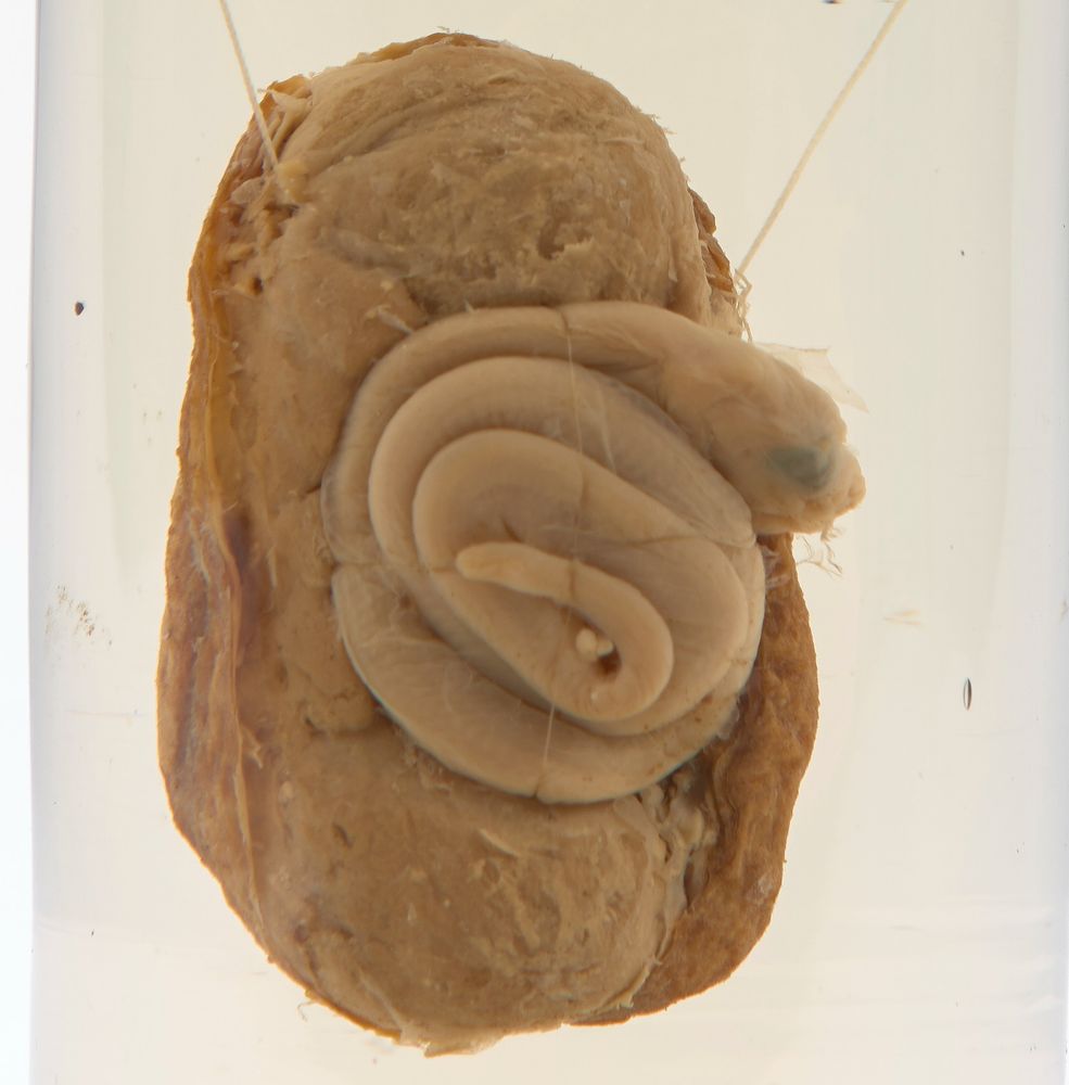
A close-up view of a preserved rattlesnake embryo
This specimen from the 1700s reveals what's inside one of these internal eggs, showing a developing rattlesnake embryo.
Credit
Rattlesnake egg, 1760 - 1793, Hunterian Collection. RCSHC/3316
#rattlesnake #rattlesnakeegg #historyofscience #museum #freemuseum
31.07.2025 15:57 — 👍 1 🔁 0 💬 0 📌 0
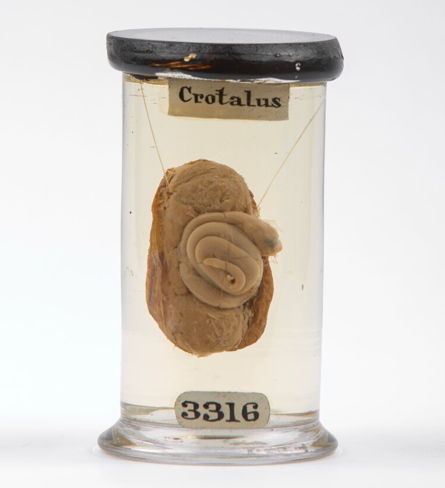
A glass container filled with clear liquid containing a specimen of a rattlesnake embryo. The label at the top of the container reads "Crotalus" and the label at the bottom reads "3316". The embryo is coiled and visible within an internal egg structure.
Did you know that rattlesnakes give birth to live young? 🐍
Unlike many reptiles, rattlesnakes don't lay eggs. Instead, their embryos develop in eggs inside the mother's body, a process called ovoviviparity. The young are then born fully formed.
1/2
31.07.2025 15:57 — 👍 6 🔁 1 💬 1 📌 0
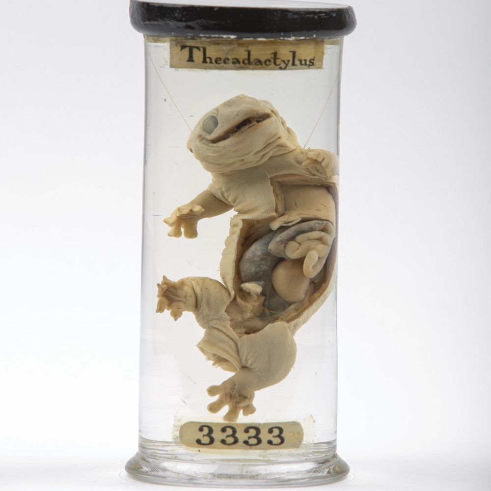
Glass specimen pot labelled '3333' and 'Thecadactylus' containing a gecko specimen with a dissected abdomen suspended in fluid
This is a turnip-tailed gecko, named after the shape and size of its tail (which this one is actually missing!)
It was originally prepared by John Hunter (1728-93), to reveal the development of the ovum in the ovaries.
RCSHC/3333
#historyofmedicine #historyofscience #museum
24.07.2025 11:00 — 👍 4 🔁 1 💬 0 📌 1
What can a jar tell us about the history of medicine?
These beautiful drug jars, some over 400 years old, originate from across Europe. They once held remedies ranging from oil of roses to senna laxatives.
Comment below if you want more information on any particular jar!
23.07.2025 16:16 — 👍 2 🔁 0 💬 0 📌 0
Job closing on Friday! 👇👇👇
21.07.2025 12:21 — 👍 0 🔁 0 💬 0 📌 0
An excellent question that the team here at the museum were also pondering - perhaps that's why it didn't catch on!
17.07.2025 16:24 — 👍 1 🔁 0 💬 0 📌 0
This model bed is a prototype from 1873 that allowed a patient's bedding to be changed without disturbing them.
It was designed by surgeon William Winchester, who also designed a clockwork amputation saw.
Learn more with Hunterian Museum Curator Alice
17.07.2025 14:26 — 👍 11 🔁 3 💬 1 📌 1
Hi Jacq - yes all eyes were made to order, but she also had thousands of pre-made examples to use as guides for size and colour.
15.07.2025 14:38 — 👍 1 🔁 0 💬 0 📌 0
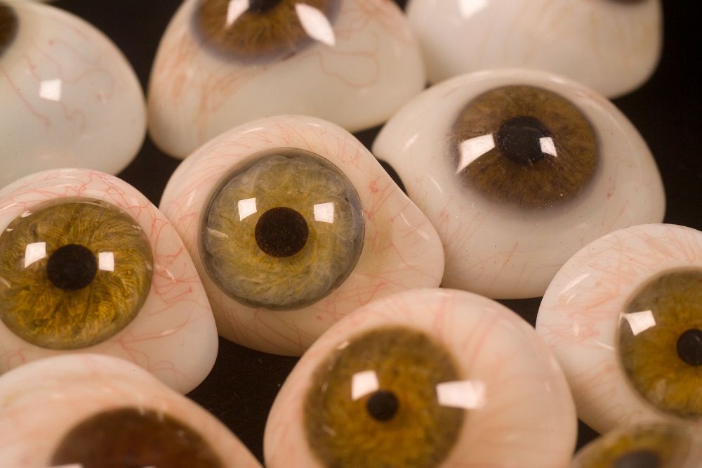
Glass eyes lying on a black background. The eyes are half globes that closely resemble a human eye, including the black pupil, coloured iris (in shades of blue, green and brown) and the white sclera with the details of red blood vessels.
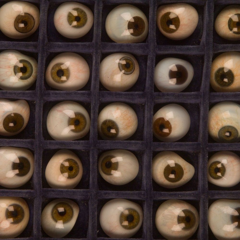
Glass eyes lying in a grid in a blue velvet case. The eyes are half globes that closely resemble a human eye, including the black pupil, coloured iris (in shades of blue, green and brown) and the white sclera with the details of red blood vessels.
These glass eyes were made by Mollie Surman of Kingston, Surrey in the mid 1900s.
Surman was from a family of glassblowers and made her first 'eye' when she was 12.
The main eye would be blown from white glass before other colours of glass were added in to make the iris, pupil, and veins.
15.07.2025 12:58 — 👍 19 🔁 6 💬 1 📌 1
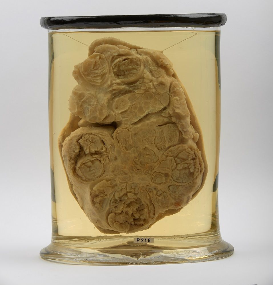
Glass specimen pot, labelled 'P 216', containing a large solid specimen suspended in fluid. The specimen's internal structure is formed of multiple circular compartments of different sizes.
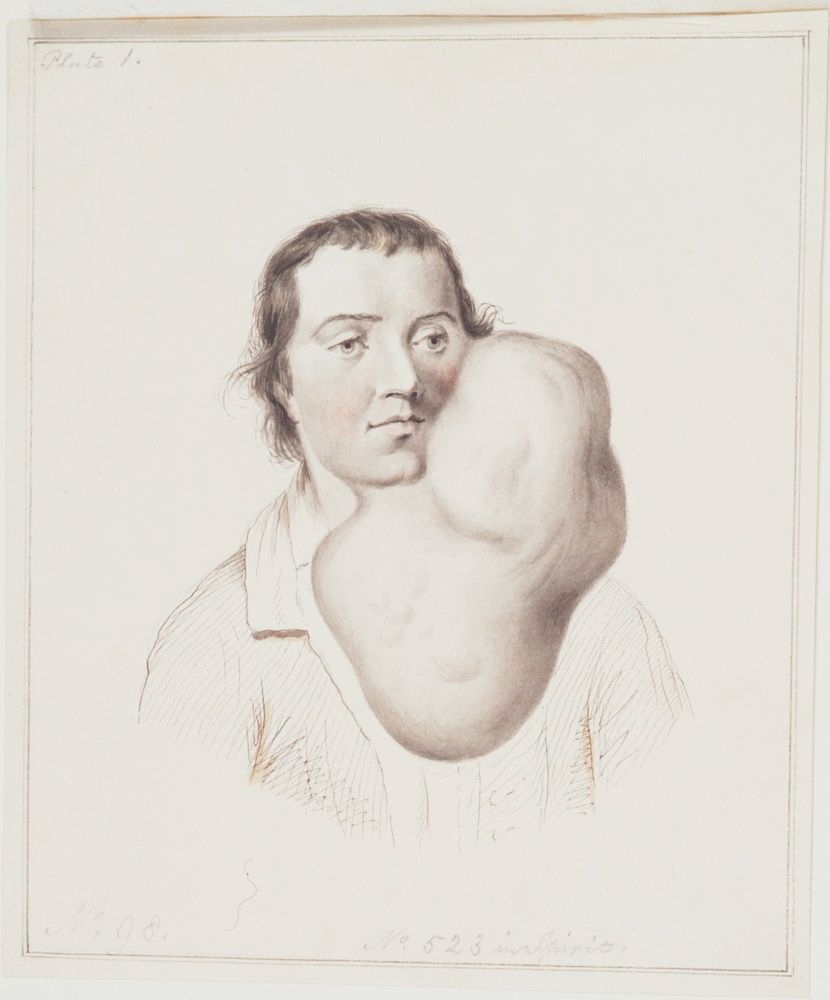
A drawing of a man with large tumour on the left side of his face and jaw, extending well below his chin
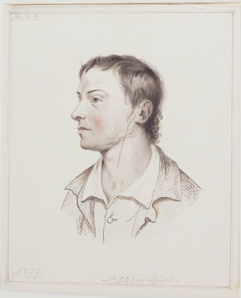
A drawing of a man looking to his right, with a scar extending from his ear to the middle of his neck
This specimen is only part of a 4kg benign tumour (a salivary adenoma) that was removed from 37-year-old John Burley on 24 October 1785.
The operation by John Hunter took just 25 minutes and Burley made a complete recovery.
The drawings show him both before and after the operation.
14.07.2025 09:47 — 👍 106 🔁 16 💬 1 📌 1
Sorry!
14.07.2025 08:58 — 👍 1 🔁 0 💬 1 📌 0
Thank you Lindsey!
14.07.2025 08:54 — 👍 0 🔁 0 💬 0 📌 0
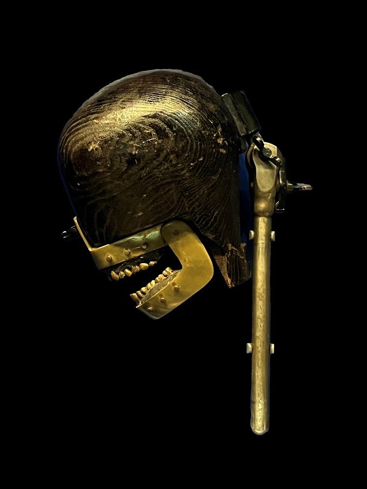
Wooden head shaped object with brass jaws and teeth mounted on a metal stand
This dental phantom was made around 1900 for dental students to practice their skills before treating real patients. A hat-maker’s dummy has been added to represent the head of the patient. The jaws contain removable brass teeth allowing various dental procedures to be practiced. (RCSIC/Z 149)
10.07.2025 15:27 — 👍 85 🔁 34 💬 8 📌 11
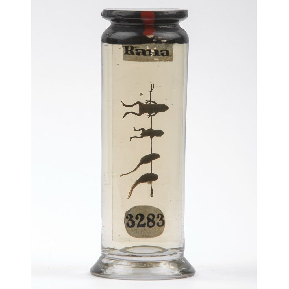
Glass Specimen pot, labelled '3283' and 'Rana', containing four individual specimens threaded onto a piece of wire in a vertical column suspended in fluid. The specimens show the development of a frog, at the bottom is a tadpole, at the top is a young frog.
These are specimens of the common frog, showing the different stages of growth from a tadpole to a young frog. At the bottom is the tadpole stage with a tail and no limbs. As it grows the tail reduces and legs begin to appear, as shown in the top specimen. (RCSHC/3283)
10.07.2025 15:23 — 👍 22 🔁 4 💬 3 📌 1
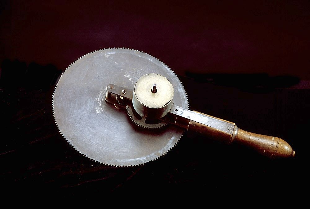
Surgical instrument consisting of a round, toothed blade attached to a wooden handle. The blade is made of a rusted metal.
This is a clockwork surgical saw from c.1850. Once wound up it would spin at a high speed, but there was no way to control the speed or stop it once it was going. This made it incredibly dangerous for both the patient and the surgeon. Luckily for everyone, it did not catch on.
10.07.2025 09:49 — 👍 46 🔁 11 💬 5 📌 2
After Hewson's death, John Hunter purchased many of his slides. Today, 97 of them survive and are part of the Hunterian Collection. This particular slide shows the blood vessels in a section of chorion - part of the amniotic sac - from a human pregnancy. (RCSHC/Hewson/G2)
2/2
10.07.2025 09:34 — 👍 6 🔁 0 💬 0 📌 0
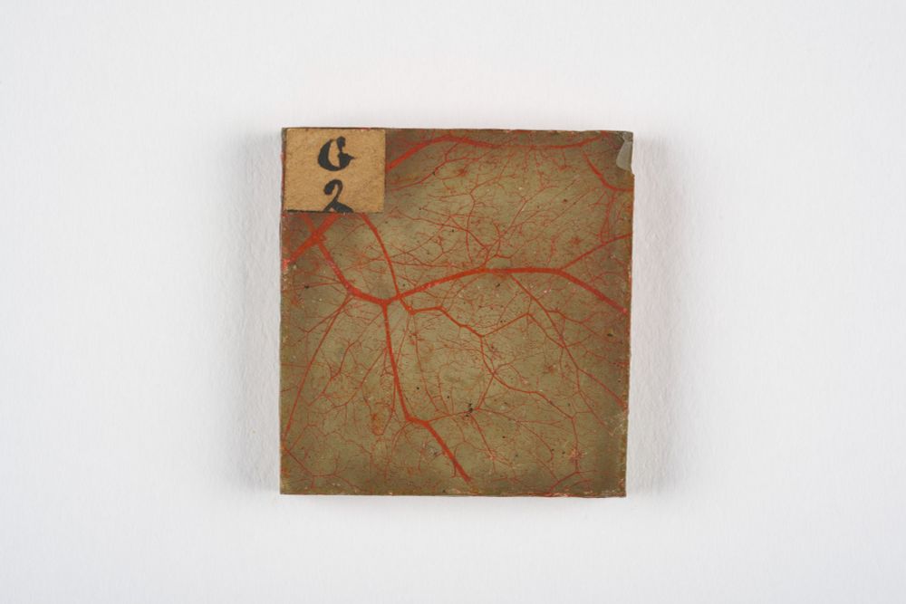
A square microscope slide with a beige background and an overlying network of red blood vessels. In the top left corner, a small label reads "G 2" in handwritten text. The slide shows signs of age, with minor chips and wear along the edges.
This microscope slide is over 250 years old and was made by William Hewson (1739–1774).
Hewson was a surgeon anatomist who made important discoveries about blood and the lymphatic system. He died from sepsis in 1774, aged 35, after injuring himself during a dissection.
1/2
10.07.2025 09:34 — 👍 12 🔁 2 💬 1 📌 0
Vacancy - Museums Engagement Manager
📢Come and work at the Hunterian Museum 📢
RCS England Museums is seeking a dynamic, highly motivated and empathetic individual to develop, deliver and promote inspiring public and professional Museum engagement programmes, on site and online.
👇👇👇
hunterianmuseum.org/news/vacancy...
07.07.2025 09:05 — 👍 7 🔁 8 💬 0 📌 2
Due to ongoing essential maintenance work, the Hunterian Museum will remain closed until Friday 4 July.
The Museum will reopen on Saturday 5 July.
Our apologies for any inconvenience caused.
02.07.2025 11:12 — 👍 0 🔁 0 💬 0 📌 0
Credit
Top - The head of a Roman snail, 1760 - 1793, Hunterian Collection. RCSHC/302
Middle - A Roman snail with the shell removed, mounted to show the mouth, 1760 - 1793, Hunterian Collection. RCSHC/301
Bottom - A Roman snail with the shell removed, 1760 - 1793, Hunterian Collection. RCSHC/1391
15.05.2025 15:30 — 👍 1 🔁 0 💬 0 📌 0
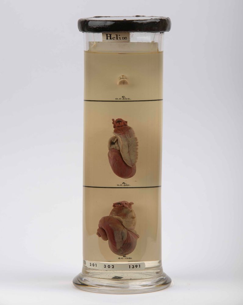
A cylindrical glass container filled with a clear liquid, containing three preserved snail specimens positioned at different levels. Each specimen is labeled with letters 'a', 'b', and 'c'. The container has a black lid and a base marked with the numbers 301, 302, and 1391. The label at the top reads 'helix'
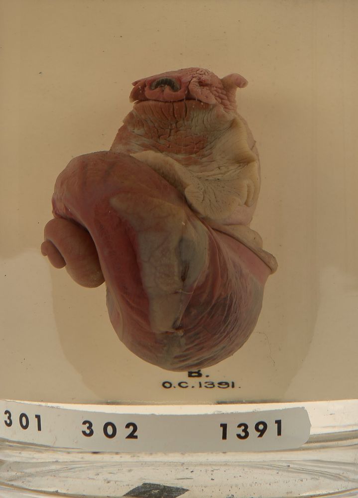
A close up of the bottom specimen - A Roman snail with the shell removed
Ever wondered what's inside a snail's shell?
Inside a snail's shell is most of its body, curled up, with the shell acting as a protective shield for its internal organs.
These are Roman snails, named because they were a delicacy for ancient Romans. They remain popular in French cuisine today.
15.05.2025 15:30 — 👍 4 🔁 1 💬 1 📌 0
Looking forward to it!
You can book your ticket for the Hunterian Event here: hunterianmuseum.org/events/natur...
23.04.2025 15:07 — 👍 4 🔁 2 💬 0 📌 0
2. Illustration of hernia from 'A series of engravings : accompanied with explanations, which are intended to illustrate the morbid anatomy of some of the most important parts of the human body; divided into ten fasciculi' by Matthew Baillie, Public Domain Mark. @wellcomecollection.bsky.social 7/7
15.04.2025 15:03 — 👍 1 🔁 0 💬 0 📌 0
Credit 1. Section of small intestine and mesentery involved in a strangulated hernia, 1760 - 1793, Hunterian Collection. RCSHC/P 1090
6/7
15.04.2025 15:03 — 👍 0 🔁 0 💬 1 📌 0
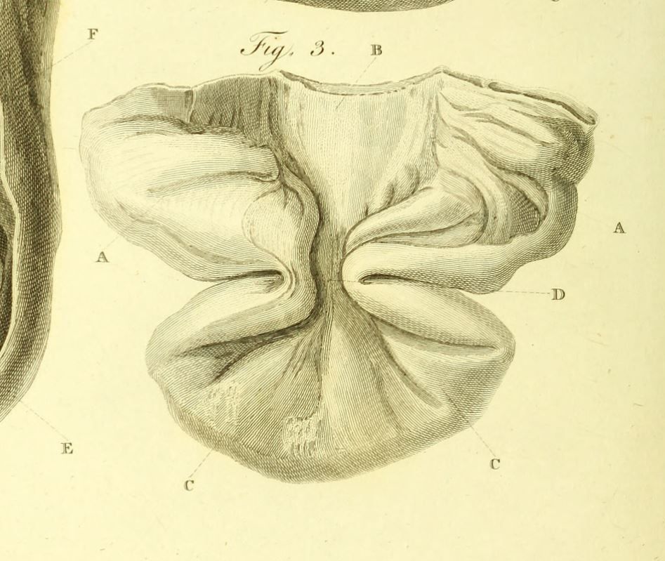
The specimen was illustrated and described by Matthew Baillie, John Hunter's nephew, in his 1793 book The Morbid Anatomy of Some of the Most Important Parts of the Human Body, the first systematic study of human pathology. 5/7
15.04.2025 15:03 — 👍 3 🔁 0 💬 1 📌 0
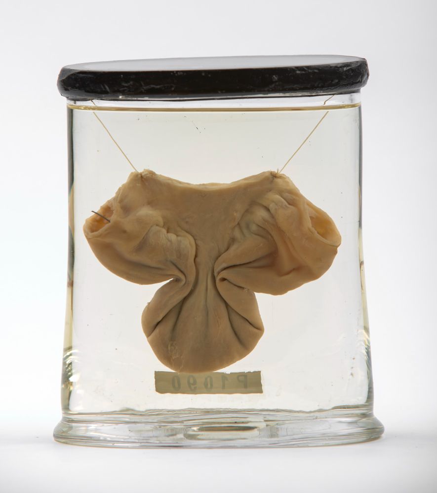
This specimen is from the 1700s and shows a section of intestine that was trapped in a strangulated hernia. It still shows a constricted 'neck'. Nothing is known about the individual who the specimen came from. 4/7
15.04.2025 15:03 — 👍 1 🔁 0 💬 1 📌 0
Intestinal hernias can be a surgical emergency if they become obstructed, preventing the movement of intestinal contents, or strangulated, where the blood supply is cut off causing tissue death. 3/7
15.04.2025 15:03 — 👍 0 🔁 0 💬 1 📌 0
Hernias can be reducible, which means they can be pushed back into their normal position, or irreducible/incarcerated, where the hernia becomes stuck. 2/7
15.04.2025 15:03 — 👍 0 🔁 0 💬 1 📌 0
What is a hernia?
A hernia occurs when a part of the body protrudes into an abnormal position, ending up somewhere it shouldn't be. For example, this can happen when a section of intestine passes through a weakness in a person's abdominal muscles. 1/7
15.04.2025 15:03 — 👍 0 🔁 0 💬 1 📌 0
Nature’s Memory: Book Launch
NEW EVENT
📅 Thurs 1 May, 18:30 - 20:00
Join us for an evening talk about the hidden stories behind the world’s iconic natural history museums, celebrating the launch of Jack Ashby’s new book, Nature’s Memory: Behind the Scenes at the World’s Natural History Museums.
tinyurl.com/5n834ub8
11.04.2025 15:35 — 👍 7 🔁 4 💬 0 📌 0
Historian of Britain and colonialism, material culture, the EIC. Also works on equalities, museums, open access & research policy. Download the EIC @ Home open access volume here: https://www.uclpress.co.uk/products/88277 (or individual chapters via JSTOR)
We're renowned for Romans, cuckoo about clocks and wild for animals! Custodians of Colchester Castle, Hollytrees Museum and Colchester's Natural History Museum.
Official page of the American Museum of Natural History in New York City. Open daily, 10 am–5:30 pm.
https://linktr.ee/amnh
One of the finest collections of natural history literature, art and online resources.
Come and research with us Tuesday - Thursday 10.00 - 16.00 (by appointment).
linktr.ee/nhmlibraryarchives
(Natural History Museum, London and Tring)
We’re the museum looking deeper into the Earth’s past to shape a new future where both people and planet thrive.
Protecting the planet, it’s in our nature. 🌍
Assistant Professor in Swarthmore’s History Department studying how Africans survived enslavement, epidemics, and empires in the early modern Atlantic World.
Insta: @bydreamphd
The RCN (@rcn.org.uk) Library and Museum is Europe’s largest nursing resource for practice and history. Use our services online or visit our libraries, exhibitions and events in person.
www.rcn.org.uk/library
Online|Belfast|Cardiff|Edinburgh|London
Philosophically-bent, historian/childhood studies PhD, Wellcome Trust Fellow, history of medicine, material culture/moral babies & children/amateur thespian/tea snob
Based in the School of Philosophy, Religion and History of Science at University of Leeds
Established in 1957 at the University of Leeds. We are a group of researchers exploring the development and meanings of science, technology and medicine.
British historian, historian of medicine, & lover of fashion. Author of Consumptive Chic: A History of Beauty, Fashion and Disease & Remembering Anne Beach: Love, Scandal & Sickness in 18th century Britain (forthcoming UTP).
Art historian of medicine
Asst Prof, Health Humanities & Bioethics @ Uni of Rochester
plastic surgery illustration, anonymity, humor
Book: https://boydellandbrewer.com/book/putting-plastic-surgery-on-paper/
@drawingbloodpod.bsky.social co-host
she / her
Medical Heritage Museum Curator at TCD, queer #deathpositive artist, childfree cat-lady, in Dublin, Ireland. Aspiring sociologist at UCD writing on death, mortality, & display.
🇵🇸 🏳️⚧️🏳️🌈 🐈⬛🪶📚📷💀🎨
https://www.evi-numen.com/
www.thanatography.com
Views, my own.
@ukri.org AHRC Standard Research Grant project (2024-28) exploring historical & contemporary understandings of the human hand. Hosted by @lcflondon.bsky.social & @lancasteruni.bsky.social & co-led by @jbhist.bsky.social & medhistoryman.bsky.social
Historian at Lancaster University and Co-Investigator on the AHRC-funded @victorianhand.bsky.social project. Blissfully married to my PI @jbhist.bsky.social. Author of two books: https://bit.ly/3968wLB & https://bit.ly/3KwWtVS
The Royal College of Physicians museum, heritage library, archive & garden in Regent's Park | History, medicine, art & modernist architecture | Exhibitions & events
The Society for the Social History of Medicine (SSHM) has pioneered interdisciplinary approaches to the history of health, welfare, medical science and practice https://sshm.org/
#histmed
Welcome to the home of human ingenuity. We curate a world-renowned collection & organise exhibitions and events for over 3 million visitors a year. Read our community guidelines: https://www.sciencemuseumgroup.org.uk/smg-social-media-community-guidelines
Archives & Special Collections and Pathology Museum at City St George's, University of London. Custodians of Jenner's cow Blossom
Explore nearly 900 years of history through the archives and objects cared for by Barts Health NHS Trust Archives. Part of Barts Health NHS Trust. Find out more at www.bartshealth.nhs.uk/barts-health-archives
World's first bricks and mortar museum dedicated to vaginas, vulvas and the gynae anatomy.
Website: https://www.vaginamuseum.co.uk/
Other social links: https://beacons.ai/v_museum


