Just one week to get your abstracts in for A12 - "Digital Resilience: Immortalising biodiversity through 3D anatomical models"! You'll present alongside our keynote speaker @jaimiagray.bsky.social who will talk about how making CT data and derivatives FAIR can level the playing field for biologists.
27.02.2026 11:12 —
👍 3
🔁 2
💬 0
📌 1

Priority deadline (Feb 16) for this years Fish Class at Friday Harbor is coming up quick! Get your applications in for what will be an amazing summer filled with great science, people, and tools!
Financial aid is available!!
@cmdonatelli.bsky.social @fishguy.bsky.social @karlycohen.bsky.social
06.02.2026 20:26 —
👍 8
🔁 12
💬 1
📌 0

A view of Friday Harbor Labs dock in the early morning with blowing sea fog in the background.
Want to do a postdoc at Friday Harbor Labs? We have two positions open. Apply by March 1st. I am happy to discuss potential projects with fish people.
apply.interfolio.com/180092
🧪
28.01.2026 18:16 —
👍 31
🔁 31
💬 1
📌 3

This Thursday's IOB cover spotlight is on
The Diffusion Diaries: Diffusible Iodine-Based Contrast-Enhanced Computed #Tomography for #Vertebrate Natural History #Specimens
@jaimiagray.bsky.social Gray , et al
doi.org/10.1093/iob/...
#morphology #science #biology #vertebrates
11.12.2025 12:01 —
👍 5
🔁 3
💬 0
📌 0

brightfield images of three lizard embryos of approximately the same developmental stage. Below each embryo image is an immunofluorescence image labeling E-cadherin (green) and alpha-smooth muscle actin (magenta) of their developing lungs
New paper out in @devdynamics.bsky.social on lizard lung development!
Project co-led w/ Kaleb Hill and also w/ @tonygamble.bsky.social @shylonatasha.bsky.social @aussiebiologist.bsky.social @bezbez.bsky.social @celestemnelson.bsky.social
anatomypubs.onlinelibrary.wiley.com/doi/10.1002/...
14.11.2025 17:35 —
👍 34
🔁 14
💬 1
📌 0
YouTube video by Gray's Vertebrate Anatomy
Quick guide to exploring CT scans on MorphoSource
youtu.be/Q3OePjtZgsI
10.11.2025 17:41 —
👍 13
🔁 6
💬 0
📌 0
Excited to be a co-author on this one in @currentbiology.bsky.social! @tillramm.bsky.social did a fantastic job bringing it all together. A mountain dragon was the *very first* CT scan I ever worked on as an undergraduate, so it holds a special place in my ❤️
29.10.2025 15:32 —
👍 13
🔁 3
💬 0
📌 0

Colored rendering of a CT scan of Bellator militaris, in anterior and anterolateral view

Colored rendering of a CT scan of Bellator militaris, in dorsal, left lateral, and ventral views
When you visit Friday Harbor Labs, @fishguy.bsky.social always has cool fish to discuss 🐟 Last week we pondered the horned searobin (Bellator militaris), & what its spines could be for. CT scan available on MorphoSource for you to explore
www.morphosource.org/media/000027546
Images made in 3D Slicer
13.10.2025 17:41 —
👍 44
🔁 14
💬 1
📌 0

Enophrys taurina CT scan.

Enophrys taurina CT scan


Just wrapped up a week of SlicerMorph/MorphoDepot/MorphoCloud work with @jaimiagray.bsky.social, @cmdonatelli.bsky.social and a great team of folks.
This is Enophrys taurina with a damaged preopercular spine. Fighting or defense?
🧪🐡
11.10.2025 22:25 —
👍 35
🔁 9
💬 0
📌 0

3D rendering of a whole body CT scan of a chimpanzee, showing the skeleton with a density map where yellow = most dense material, blue = medium density material, purple = low density material. Top shows 3 images of the whole body rendered in a series rotating from front to back. Bottom shows a close up of the head and shoulders. The "oVert" symbol is in the top right corner. In the bottom left corner it reads "Pan troglodytes; YPM:VZ:015959; MorphoSource ID: 0000058004"
"What makes us human, I think, is an ability to ask questions, a consequence of our sophisticated spoken language" - J. Goodall.
This #oVertTCN chimpanzee CT scan is available for exploration on MorphoSource: www.morphosource.org/concern/medi...
03.10.2025 15:14 —
👍 50
🔁 5
💬 0
📌 0

3D rendering of a CT scan of Brachycephalus nanicus, with the skeleton rendered in light brown and skin rendered in transparent purple. Top = dorsal view, bottom = ventral view. In the bottom right corner there is an image of the frog (a small brown frog) sitting on a coin.

3D renderings of the skeleton of Brachycephalus nanicus, rendered in light brown. Top left = whole skeleton in dorsal (left) and ventral (right) views. Top right = skull in dorsal (left), ventral (middle), and right lateral (right) views. Bottom from left to right, 3D renderings of the hand, foot, spinal column, and should girdle (top) / pelvic girdle (right lateral).
Meet the newest species of Brazilian flea-toad 🐸 Brachycephalus nanicus! 🐸
B. nanicus is <1cm long & dwells under the leaf litter in the cloud forests of Serro do Mar, southeastern Brazil
I CT scanned this specimen for the osteological description
Out today in Zootaxa: mapress.com/zt/article/v...
09.09.2025 17:28 —
👍 42
🔁 10
💬 0
📌 0

Two sleepy lizards side by side, photographed on the side of a rural road in South Australia.

A sleepy lizard standing peacefully in the sun, facing the camera

A sleepy lizard facing the camera with it's mouth open and tongue out.
They are also called shingleback/bobtail/two-headed skink/pinecone lizard/stumpy lizard, and more. They're heavily armoured, with a peaceful demeanor (but can be a sassy when bothered). They tend to pair up & stay with the same mate year after year, & you can often find pairs hanging out together ❤️
14.08.2025 17:08 —
👍 3
🔁 0
💬 0
📌 0

A 3D rendering of a CT scan of Tiliqua rugosa, facing the front (top) and facing the back (bottom), rendered in yellow (most dense parts), pink (medium density parts), and blue (least dense parts).
A 3D rendering of a CT scan of the head Tiliqua rugosa, in dorsal view (top) and ventral view (bottom), rendered in yellow (most dense parts), pink (medium density parts), and blue (least dense parts).
Hey it's #WorldLizardDay 🦎
Here's one of my faves & one I saw *a lot* of growing up in rural Australia. Tiliqua rugosa has many common names, but where I'm from we call them "sleepy lizard" or "stumpy tail".
Download the #oVertTCN CT scan from MorphoSource here: www.morphosource.org/concern/medi...
14.08.2025 17:08 —
👍 13
🔁 2
💬 1
📌 0
I got my hands on this @floridamuseum.bsky.social specimen freshly fixed in 2021, & created skeletal & soft tissue CT scans. You can download them here (including colored skull model): www.morphosource.org/concern/biological_specimens/000408235
View the exploding skull model here: skfb.ly/pzUFC
11.08.2025 19:13 —
👍 3
🔁 1
💬 0
📌 0

Image of a 3D model of a veiled chameleon skull, with bones colored to demonstrate skull anatomy, on a black background. Model is shown in lateral (top), antero-lateral (middle), and anterior (bottom) views. A small image of the whole museum specimen is shown with a scale bar in the bottom right corner.
Climbing into my #ColorsOfSkullAnatomy collection: a veiled chameleon (Chamaeleo calyptratus) 🦎 this large male was getting up there in age when he was collected, as shown by his worn down teeth and bone pathologies.
Find the model here:
www.graysvertebrateanatomy.com/veiled-chame...
11.08.2025 19:13 —
👍 33
🔁 9
💬 2
📌 1
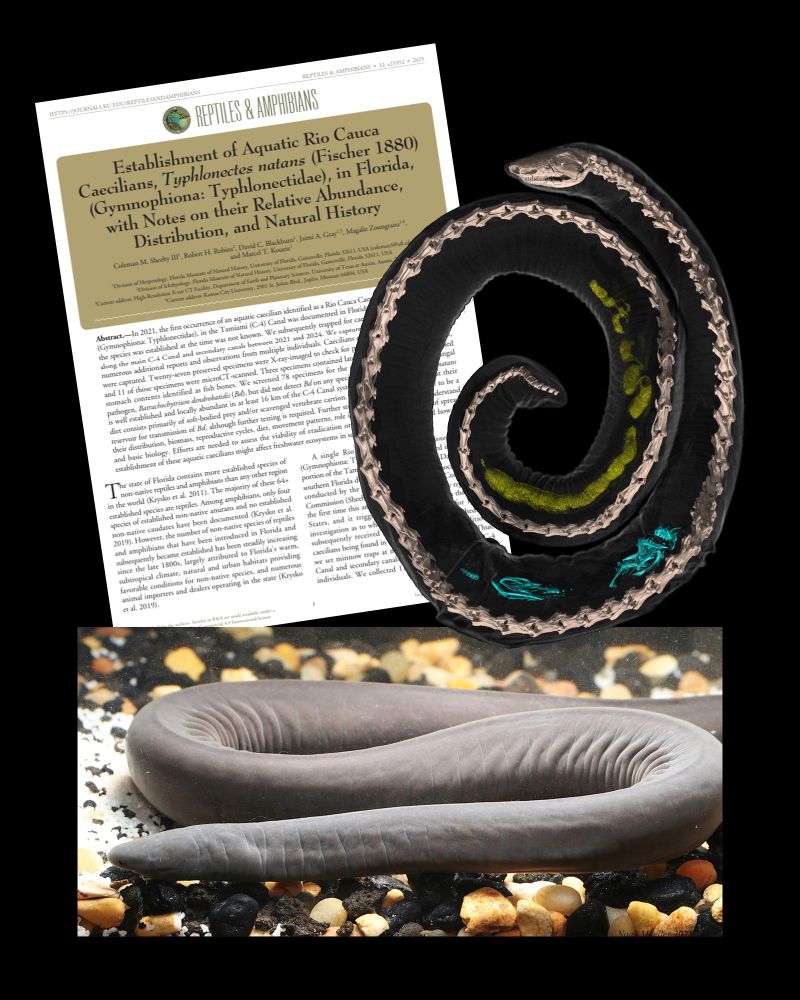
top left - front page of the PDF of the paper entitled " Establishment of Aquatic Rio Cauca Caecilians, Typhlonectes natans (Fischer 1880) (Gymnophiona: Typhlonectidae), in Florida, with Notes of their Relative Abundance, Distribution, and Natural History. Top right - a 3D rendering of a CT scan of a caecilian, with bone colored skeleton and transparent body, with fish bones inside (rendered in blue) and radiodense digested food (rendered in yellow). Bottom - photograph of a live Rio Cauca Caecilian.
It appears that these aquatic caecilians are doing pretty well down there and definitely reproducing in Miami, with a whopping 115 individuals collected using cans of chicken vienna sausages 🌭
26.07.2025 23:11 —
👍 14
🔁 1
💬 1
📌 0
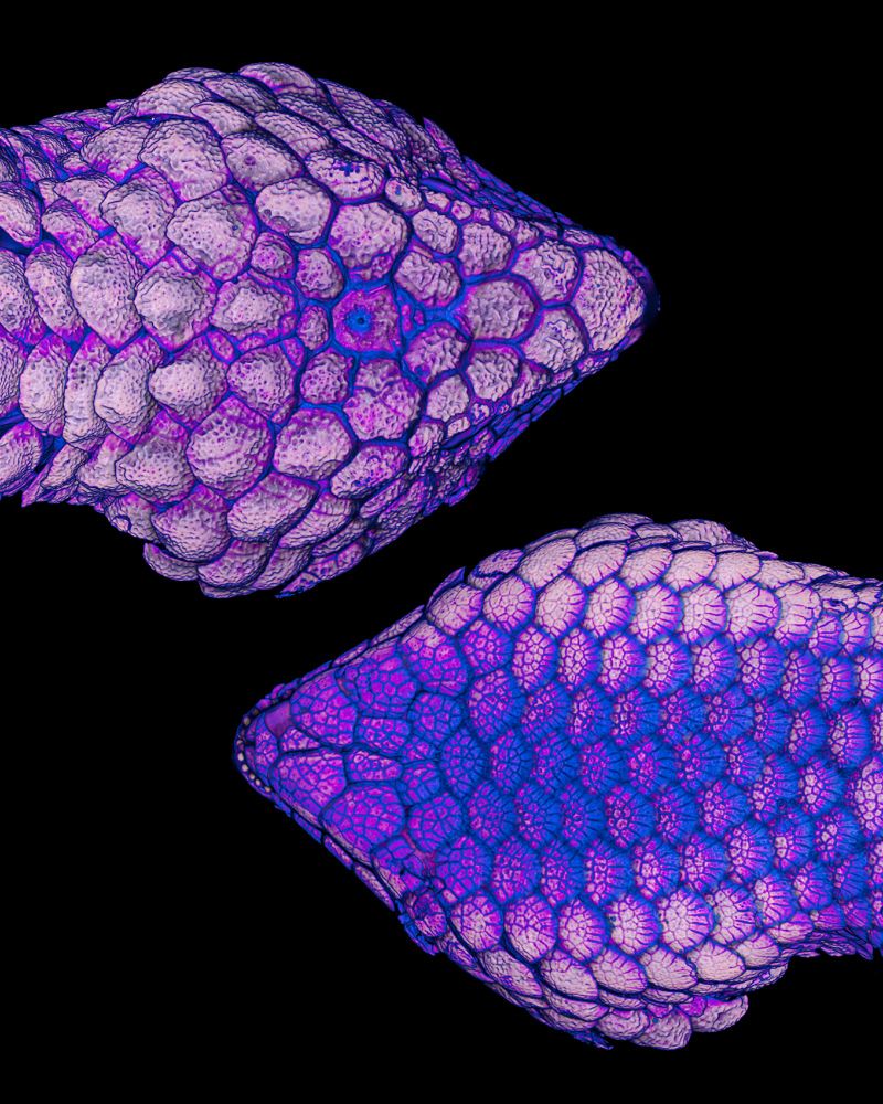
A 3d rendering of a CT scan of two Rio Cauca caecilian with transparent body and bone colored skeleton, showing baby caecilians inside, each rendered in a different color. There are three baby caecilians in the top individual, rendered in green, pink, and purple, and one baby caecilian (rendered in blue) plus one unidentified object (rendered in yellow) in the second individual.
I had so much fun being part of this project! On my scheduled scanning days @ UF Coleman would hand me a bucket of caecilians collected from the Tamiami canal. I put each under the X ray beam to look for babies or interesting food items. If I saw anything of interest, I CT scanned them.
26.07.2025 23:11 —
👍 15
🔁 1
💬 1
📌 0
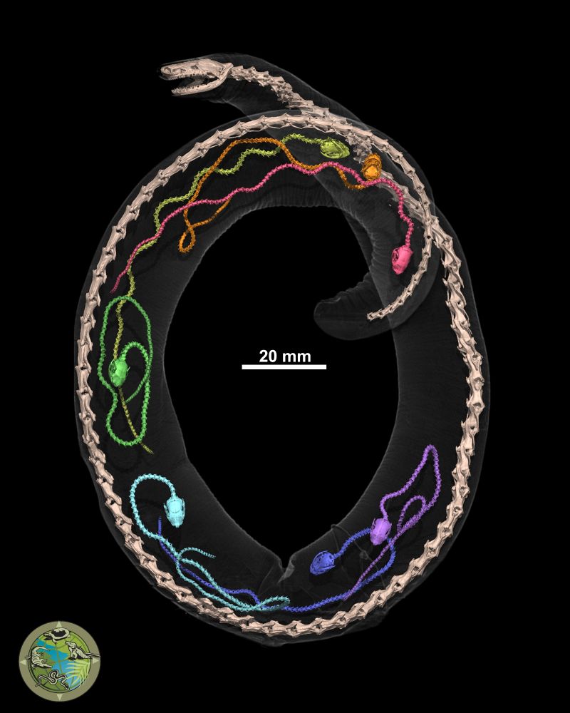
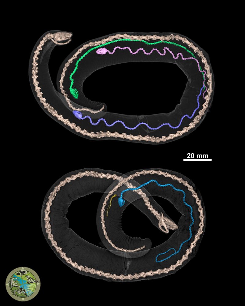
A 3d rendering of a CT scan of a Rio Cauca caecilian with transparent body and bone colored skeleton, showing 7 baby caecilians inside, each rendered in a different color - counter-clockwise from tail to head - red, orange, yellow, green, light blue, dark blue, purple.
Say hello to Florida's newest established species, Typhlonectes natans - the Rio Cauca Caecilian! You can read about their relative abundance, distribution, & natural history, in our brand new paper:
journals.ku.edu/reptilesanda...
Here is one individual I CT scanned that had 7 babies inside!
26.07.2025 23:11 —
👍 188
🔁 55
💬 6
📌 6
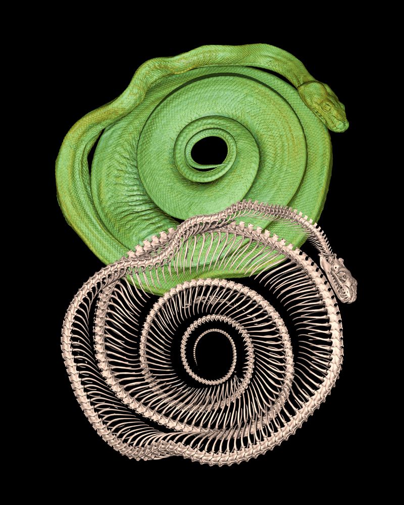
3D rendering of a diceCT scan of a coiled green tree python, rendered in bright green. Slightly overlapping is a 3D rendering of the python's skeleton, rendered in a light brown color.
It's #WorldSnakeDay so here is a green tree python I CT scanned for #oVertTCN 🐍
Get the datasets (both #diceCT and regular CT) on MorphoSource: www.morphosource.org/concern/biol...
💚💚💚
16.07.2025 21:04 —
👍 37
🔁 5
💬 0
📌 0
Now Jaimi Gray is up! "The Diffusion Diaries: Using diffusible iodine-bases contrast-enhanced Computed Tomography for High-throughout imaging of 3D anatomy of natural history specimens" #JMIH25 #ASIHin3D
12.07.2025 15:34 —
👍 10
🔁 3
💬 1
📌 0
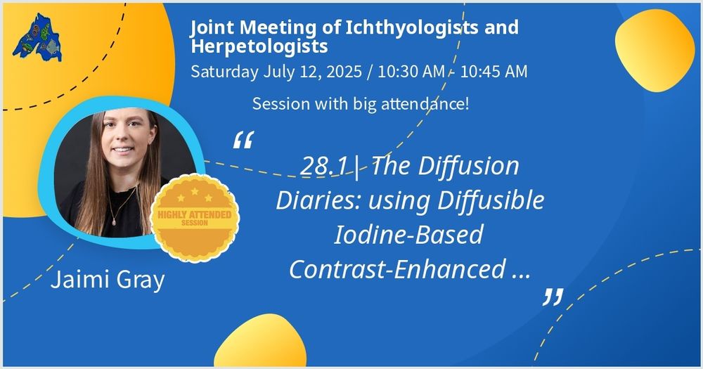
Display slide that reads "Joint meeting of Ichthyologists and Herpetologists
Saturday July 12, 2025 / 10.30am - 10.45
Session with big attendance
'28.1 | the Diffusion Diaries: using Diffusible Iodine-Based Contrast-Enhanced...'
Jaimi Gray"
Catch me at 10.30am in the Science in 3D #ASIHin3D symposium at #JMIH25 today! Amphibians 🐸 Reptiles 🦎 CT scanning ☢️ Iodine 🧪 Anatomy 🧠
12.07.2025 13:42 —
👍 14
🔁 2
💬 0
📌 1
Picture of a powerpoint slide that reads "Science in 3D"
#ASIHin3D begins! An absolute banger line up of speakers today (including me @ 10.30). LET'S GO 🥳 #JMIH25
12.07.2025 13:32 —
👍 3
🔁 1
💬 0
📌 0
"Early bird gets the sperm" direct quote by @drscanley.bsky.social during Katherine Greenwald's amazing keynote on unisexual salamanders... And so #JMIH25 begins
10.07.2025 14:40 —
👍 17
🔁 2
💬 1
📌 0
Alert for those also arriving to St Paul for #JMIH25 on very little sleep and are also into sharks (I think there's a few of you 😉), the Lost Fox has a very strong coffee option called the "Hammerhead".... 🦈
09.07.2025 16:54 —
👍 7
🔁 1
💬 0
📌 0
Got it!
09.07.2025 13:42 —
👍 1
🔁 0
💬 0
📌 0
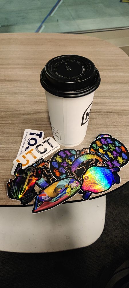
A coffee cup sitting on a table at an airport. Next to the coffee cup lies a pile of colorful stickers showcasing 3D renderings of CT scans
En route to the Twin Cities MN for #JMIH2025 ✈️ I'm bringing plenty of stickers for y'all so you have an excuse to hit me up and talk fish and herps 🐟🐢🐠🐡🐸🦎🐍🐊
Catch my talk in the #ASIHin3D symposium on Saturday...
09.07.2025 10:16 —
👍 16
🔁 2
💬 2
📌 0
I made these, they haven't been used for teaching yet though: bsky.app/profile/jaim...
26.06.2025 18:22 —
👍 0
🔁 0
💬 1
📌 0
You can buy models like this online, but they are pricey (100s of $ each or >$1000 a set). I made these using NSF funded *free* data (from the oVert project), a few bucks worth of PLA filament, and sandpaper & paint (a combo of automotive and acrylics).
26.06.2025 15:53 —
👍 8
🔁 0
💬 1
📌 0
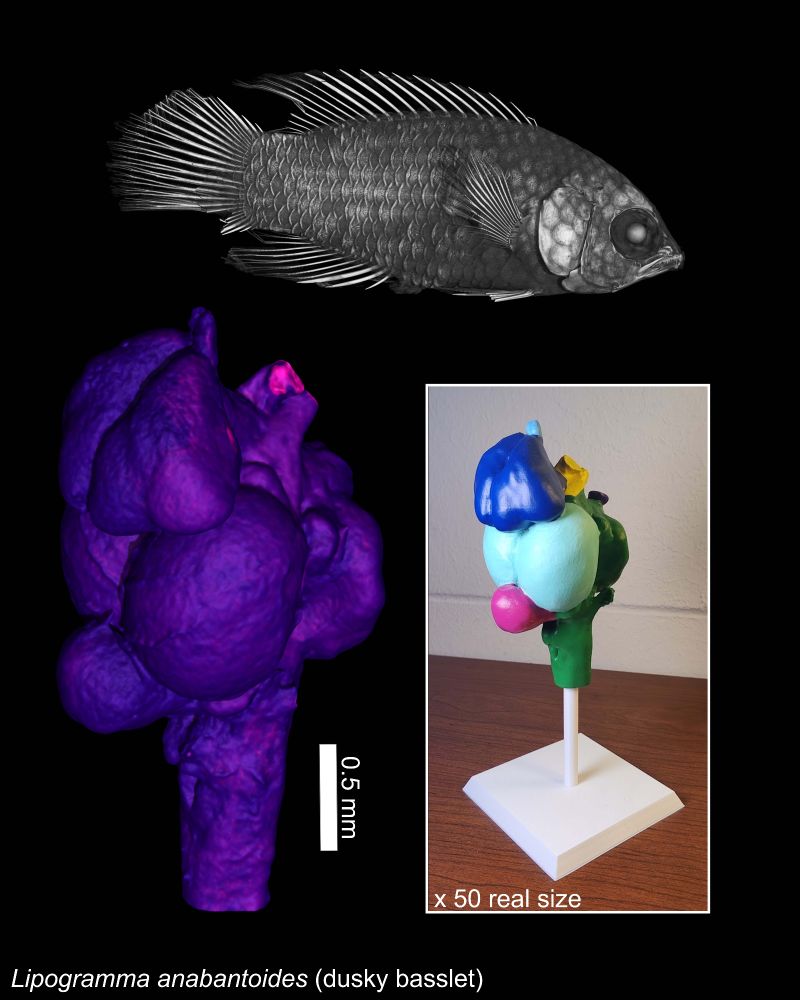
Top: black and white 3D rendering of a dusky basslet fish
Bottom left: purple/pink 3D rendering of the brain of the fish, with a scale bar representing 0.5 mm
Bottom right: A photograph of a 3D printed and painted anatomical model of the fish brain on a white stand, with colors demonstrating anatomical regions.
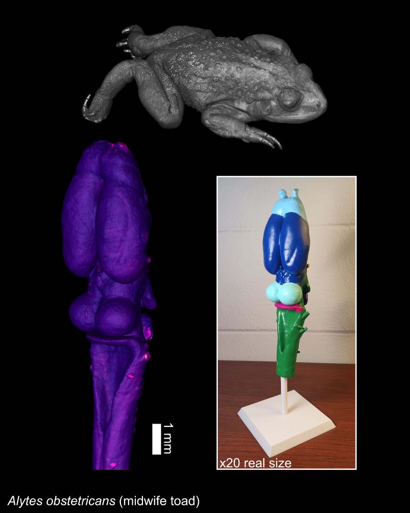
Top: black and white 3D rendering of a midwife toad
Bottom left: purple/pink 3D rendering of the brain of the frog, with a scale bar representing 1 mm
Bottom right: A photograph of a 3D printed and painted anatomical model of the frog brain on a white stand, with colors demonstrating anatomical regions.
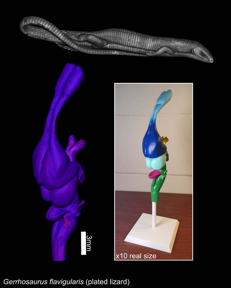
Top: black and white 3D rendering of a plated lizard
Bottom left: purple/pink 3D rendering of the brain of the lizard, with a scale bar representing 3 mm
Bottom right: A photograph of a 3D printed and painted anatomical model of the lizard brain on a white stand, with colors demonstrating anatomical regions.
I used my 3D printer to create educational anatomical models of the brains of fish, frog, & lizard 🐟🐸🦎 The (color-blind friendly) colors demonstrate anatomical regions, and the 🧠 are many times larger than their real size. They can be removed from their stands for closer examination/comparison 🧪
26.06.2025 15:53 —
👍 15
🔁 2
💬 1
📌 1