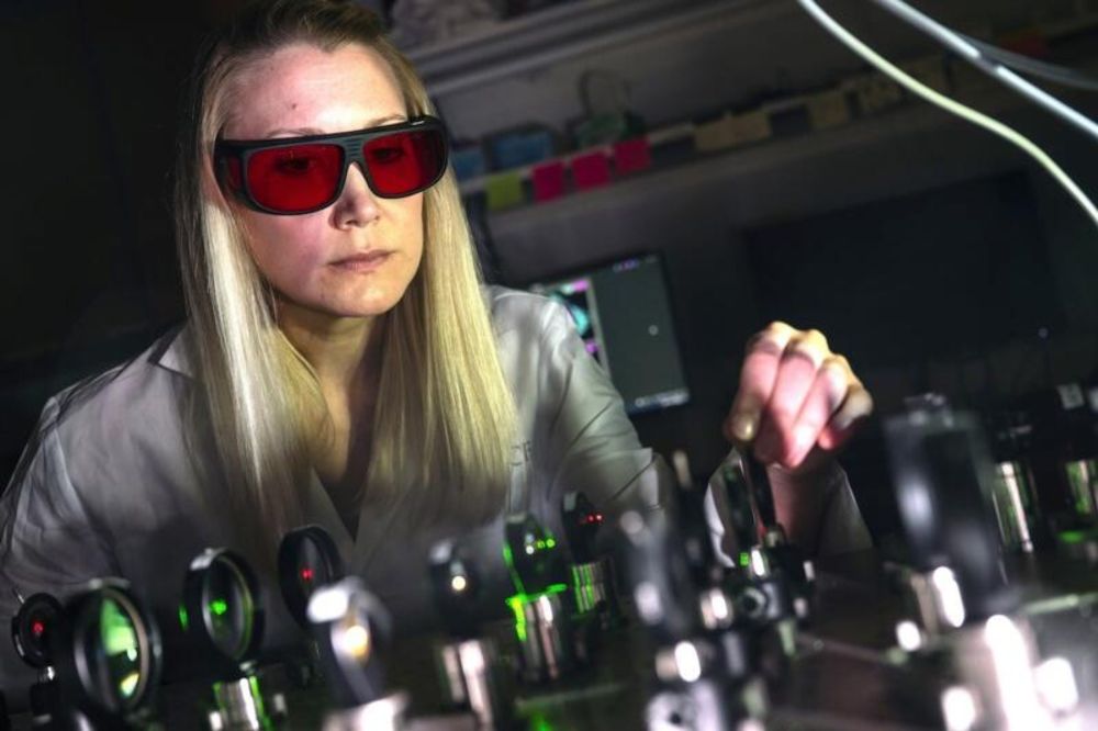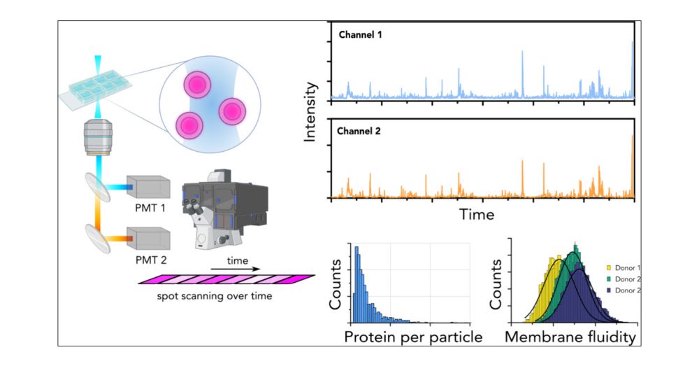
👏 Congratulation to the 🏆 winners of Student Award held by #PicoQuant during 30th anniversary of our annual workshop on Single Molecule Spectroscopy and Ultra Sensitive Analysis in the Life Sciences. #WS30
We were proud to once again support the next generation of scientists. #PicoQuantStudentAward
26.09.2025 11:12 — 👍 8 🔁 2 💬 0 📌 0

🚨
New Preprint!
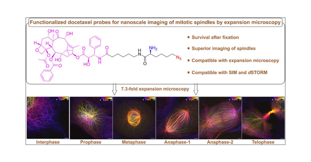
Ex-dSTORM resolves fine details of the molecular architecture of CCPs, the 8-nm periodicity of microtubules, and the docking site of synaptic vesicles at the presynapse of hippocampal neurons.
▶️ www.biorxiv.org/content/10.1...
19.08.2025 15:32 — 👍 49 🔁 20 💬 1 📌 0
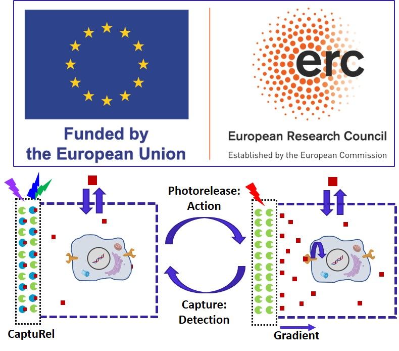
Great news: I received ERC Advanced grant #ERCAdG @erc.europa.eu CaptuRel! It aims at intelligent nanomaterials undergoing bioinspired cycle of capture and photorelease of bioactive molecules for sensing and controlling (bio)chemical gradients. Thanks to my Team, @cnrs.fr and @unistra.fr!
17.06.2025 10:41 — 👍 48 🔁 4 💬 0 📌 1
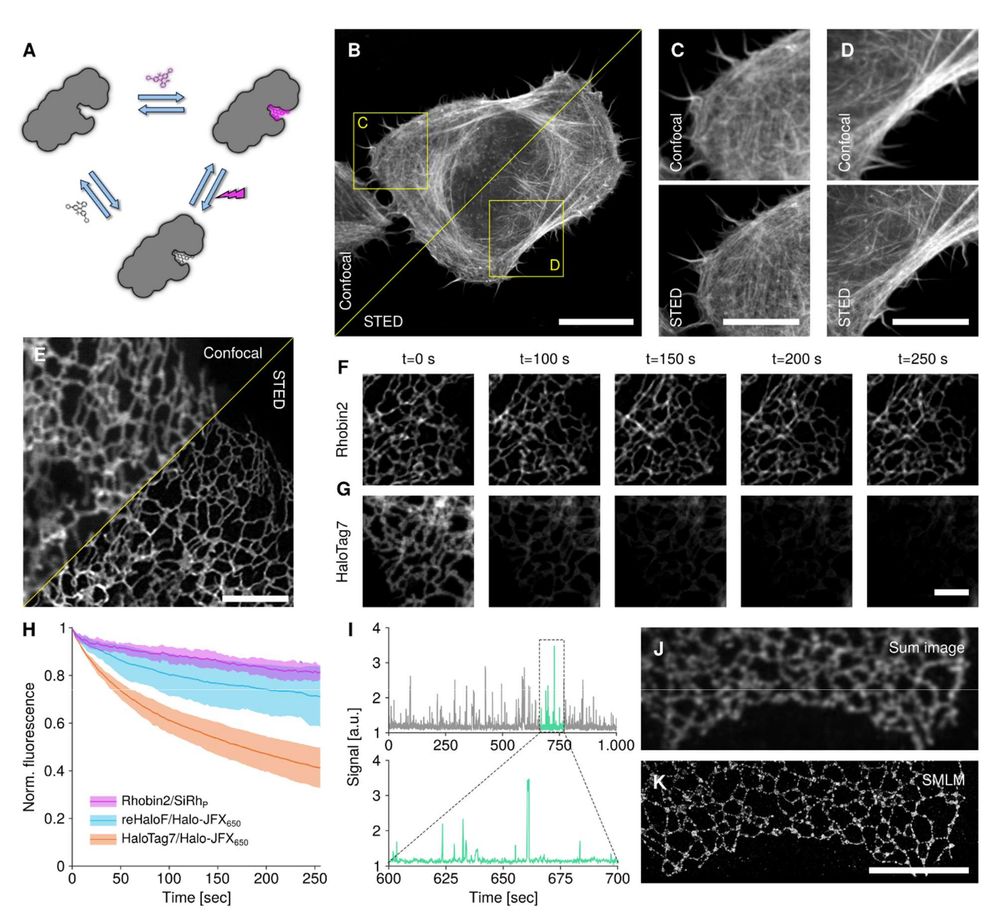
Fig. 4 Super-resolution microscopy in live and fixed cells with Rhobin.
(A) Rhobin transiently binds rhodamine molecules and thereby constantly replenishes signal lost
through photobleaching of dyes. (B-D) No-wash STED microscopy of live U2OS cells transiently
expressing LifeAct-Rhobin2 labeled with 100 nM JFX650. (C, D) Zoom-ins of regions highlighted
in (B) imaged either with confocal or STED microscopy. Scale bar: 20 μm (B) and 10 μm (C, D).
(E-H) Rhobin enables timelapse STED microscopy of fast ER dynamics with minimal signal loss.
U2OS stably expressing N-terminal tag fusions of SEC61B were stained with 1 μM Halo-JFX650
(HaloTag7, reHaloF) or 2 μM SiRhP (Rhobin2) and imaged at a frame rate of 0.387 fps for at least
100 time points. (F, G) Selected frames from timelapse acquisitions of Rhobin2:SEC61B labeled
with SiRhP (F) or HaloTag7:SEC61B labeled with Halo-JFX650 (G). Scale bar: 2 μm. (H) Signal
.CC-BY-NC 4.0 International licenseavailable under a
(which was not certified by peer review) is the author/funder, who has granted bioRxiv a license to display the preprint in perpetuity. It is made
The copyright holder for this preprintthis version posted June 25, 2025.;https://doi.org/10.1101/2025.06.24.661379doi:bioRxiv preprint
22
loss during timelapse imaging. Mean ± SD signal inside cells over time for indicated constructs.
(I-K) PAINT-type super-resolution microscopy with Rhobin. Rhobin2-SEC61B was transiently
expressed in U2OS cells and labeled with 5 nM SiRhP after chemical fixation. Imaging under no-
wash conditions and near-TIRF illumination. (I) Repeated, but transient binding of SiRhP
molecules to Rhobin2 at low nanomolar concentrations can be observed as intensity bursts in
intensity time traces extracted from a diffraction-limited area. See movie S3 for raw data of
binding-induced blinking. (J) Sum image across 10,000 frames of an image stack, simulating a
diffraction-limited image. (K). Reconstructed image from localized molecules in the full stack.
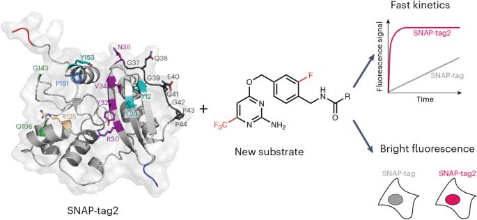
De novo designed bright, hyperstable rhodamine binders for fluorescence microscopy by Bo Huang and team: www.biorxiv.org/content/10.1...
26.06.2025 13:50 — 👍 69 🔁 19 💬 1 📌 2
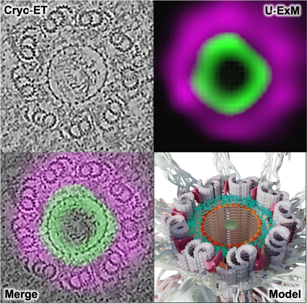
🚨 New preprint!
Using U-ExM + in situ cryo-ET, we show how C2CD3 builds an in-to-out radial architecture connecting the distal centriole lumen to its appendages. Great collab with @cellarchlab.com @chgenoud.bsky.social @stearnslab.bsky.social 🙌. #TeamTomo #UExM
www.biorxiv.org/content/10.1...
19.06.2025 08:58 — 👍 119 🔁 46 💬 0 📌 2
Very excited to present our latest work: SPINNA, an analysis framework and software package for single-protein resolution data! 🖥️🤩
We can directly quantify stoichiometry and oligomerization from super-res (DNA-PAINT, RESI) images!! 🧬🎨
07.05.2025 14:56 — 👍 30 🔁 11 💬 0 📌 0
Thanks so much!
01.04.2025 23:54 — 👍 0 🔁 0 💬 0 📌 0
🔬Happy to share our new preprint, where brightness is used to identify single molecule emission in 2D/3D, with a single camera ! Part of the PhD work of @laurent-le.bsky.social and Surabhi, great collaboration with @emmanuelfort.bsky.social from @instlangevin.bsky.social @ismolab.bsky.social #SMLM
04.03.2025 08:58 — 👍 63 🔁 22 💬 0 📌 0

🧪Paris Jussieu right now #standupforscience
07.03.2025 12:41 — 👍 55 🔁 6 💬 1 📌 2

Science, research & public health face unprecedented attacks in the U.S.
We stand with our American colleagues & support the Stand Up for Science call.
Join the global march tomorrow, incl. in Paris! 🧪✊
📍 March 7, 13:30 – Place Jussieu
🔗 More info: standupforscience.fr
#StandUpForScience
06.03.2025 14:24 — 👍 264 🔁 82 💬 3 📌 5
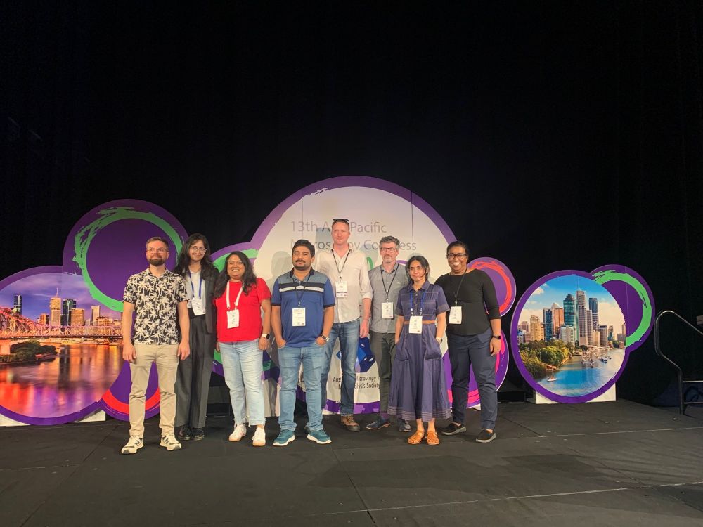
Group photo of eight colleagues representing the dept, standing in front of the conference mural
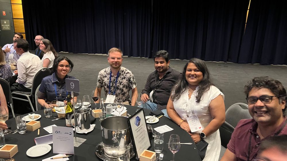
Some colleagues sitting around a dinner table
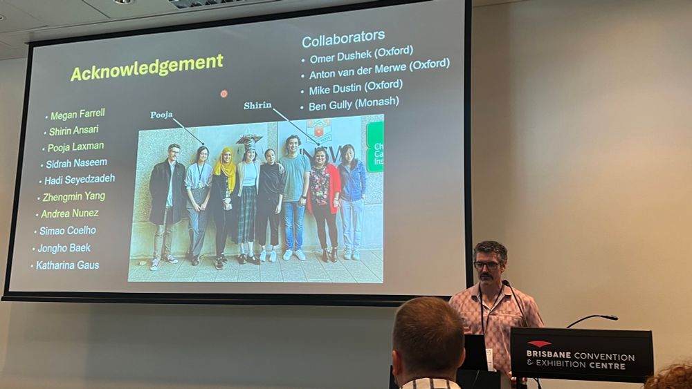
A conference speaker standing at a lectern podium
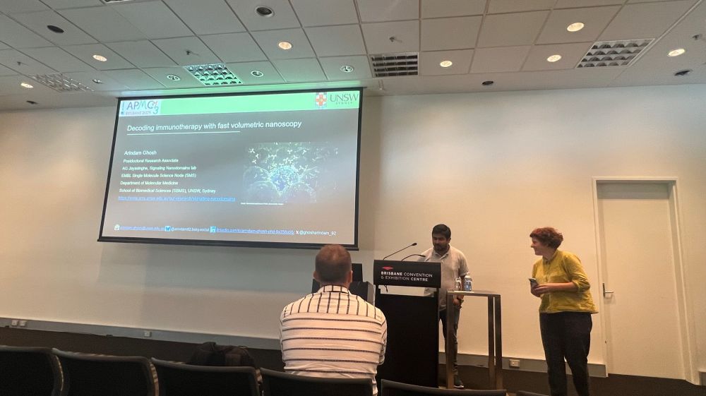
Arindam Ghosh being introduced to the symposium by Liz Hinde.
Aaaand that’s a wrap at Asia Pacific Microscopy Congress 2025 in sunny Brisbane, attended by a big team from Single Molecule Science. Lots of exciting new 🔬 tools and translation in biomedical investigations. @ijayas.bsky.social @arindam92.bsky.social
07.02.2025 10:19 — 👍 15 🔁 5 💬 2 📌 0
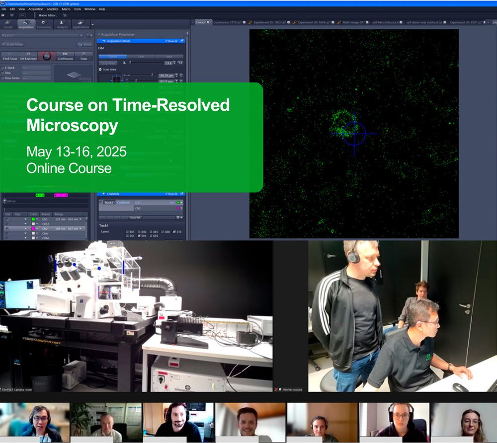
Want to explore the world of time-resolved fluorescence #microscopy? Sign up for our online course and benefit from theoretical as well as practical sessions in an interactive format. Topics: data analysis, FCS, FRET, FLIM, and more. May 13-16, 2025. ➡️ www.microscopy-course.org
04.02.2025 10:52 — 👍 5 🔁 2 💬 1 📌 0
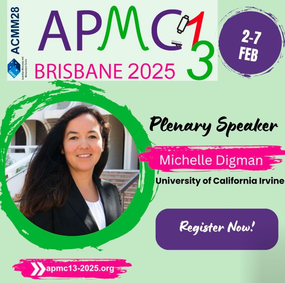
Thrilled and honored to present a plenary talk at the 13th Asia Pacific Microscopy Congress (APMC13) in Brisbane, Australia, on February 7, 2025, hosted by the Australian Microscopy and Microanalysis Society (AMMS).
02.02.2025 19:16 — 👍 5 🔁 2 💬 0 📌 0

🧵Prepint alert! Optimizing Multifunctional Fluorescent Ligands for Intracellular Labeling | tinyurl.com/3n55hvsc. With Jason Vevea, Ed Chapman, and @so-lets-kilab70.bsky.social, we combined dye chemistry, HaloTag, microscopy and cell biology to make protein purification and manipulation tools.
28.01.2025 05:17 — 👍 66 🔁 25 💬 2 📌 2
If you have a passion for single-molecule super-resolution microscopy🔬, there are great news to share: the SMLMS conference series will continue this year and will be held in Bonn/Germany Aug 27th to 29th ✨- please save the date and join us, we are looking forward to meeting you there👇🏻
15.01.2025 08:44 — 👍 18 🔁 5 💬 0 📌 0
How can you enhance the visibility of your next publication or increase your chances of getting your next research proposal accepted? How to get the illustration you need in just a few days? How to find an illustrator who is on the same wavelength with you?😉
12.01.2025 19:32 — 👍 3 🔁 1 💬 0 📌 0
Wow! Alexey @alexeychizhik.bsky.social, you never stop to amaze me. What a wonderful piece of sciart. Many congratulations 👏👏👏👏
24.01.2025 10:21 — 👍 5 🔁 0 💬 1 📌 0
PhD at Johns Hopkins Biophysics | IISERM'18
Postdoc @UoD @CeTPD with Alessio Ciulli
Structural & chemical biology in Targeted Protein Degradation
I am a cell and developmental biologist at the Living Systems Institute (LSI) in Exeter. We work on spreading Wnt signals and characterising cytonemes in zebrafish embryos, human neurons, and cancer using high-res imaging and mathematical modelling.
BioF:GREAT is an NSF BioFoundry located at UGA's Complex Carbohydrate Research Center focused on democratizing glycoscience by increasing accessibility to resources, education, and training.
Postdoc in the Mir Lab at UPenn/CHOP. Interested in all things transcription and microscopy 🔬
Associate Professor at UGAs Complex Carbohydrate Research Center. Lead of the Plant Biopolymers Group. Plant biochemist, gardener, and cat enthusiast with a focus on plant cell wall biology.
Mitochondriac 🔬 Postdoc @EPFL Manley lab of Experimental Biophysics 🧪 Ph.D. @HelsinkiUni 🇦🇷🇫🇮🇪🇺🇨🇭🏳️🌈 (he/él)
linktr.ee/jclandoni
Our groundbreaking SAFe-nSCAN imaging platform takes biological imaging to a whole new level, advancing spatial biology into the nano-era.
Funded by @horizoneu.bsky.social
We are mononuclear phagocytes enthusiasts and we organise biennial international meetings on the topic in France...
https://www.cfcd.fr
PhD student at Karolinska Institutet and Scilifelab | Cell physics
https://www.csi-nano.org/
A career network featuring science jobs in academia and industry.
Visit our platform at www.science.hr
I love science and biology+music
SFwriter, Concept artist.
Southern part of Iran.
Ph.D. Student, Nobel laureate Carolyn Bertozzi and Longzhi Tan Labs, Stanford University🌲, Departments of Chemical and Systems Biology|Neurobiology|Sarafan ChEM-H, Passionate about Chemical Biology🧪, Neuroscience 🧠and Artificial Intelligence🤖
Decoding how the gut thinks 🦠🪱🧠💪
Neuroscientist with interests in
#EnergyMetabolism #EntericNeurons #Fats
@crick.ac.uk @institutducerveau.bsky.social
Researcher in MeLiS - Lyon. Likes centrioles, cilia and anything graviting around those
Cutting-edge research, news, commentary, and visuals from the Science family of journals. https://www.science.org
scientist in #cryoem and professor @uni-wuerzburg.de
Chemical biologist @ Max Planck Institut for Medical Research in Heidelberg


