Studying vision across light levels? Interested in rod photoreceptors and related (patho)physiology? Matteo Rizzi, Kate Powell and I wrote a review on rod photoreceptor activity at daylight doi.org/10.1016/j.vi... . Free access link here kwnsfk27.r.eu-west-1.awstrack.me/L0/https:%2F...
18.12.2025 09:50 — 👍 17 🔁 7 💬 1 📌 1
Hey, @drdorotask.bsky.social is here on Bsky!
Such cool work.
We think that retinas degenerate with time and that if all of us live long enough, we’ll get AMD or other neurodegenerative diseases.
But Greenland sharks manage to live for hundreds of years and repair their eyes.
How?
07.01.2026 04:15 — 👍 50 🔁 11 💬 2 📌 0

Vision scientist Pete Williams talking at the lectern.
It’s @petetheteapot.bsky.social at the Retinal Ganglion Cell meeting talking about his work in gene therapy targeting neuroprotection and neurodegeneration.
11.12.2025 08:14 — 👍 17 🔁 1 💬 2 📌 0
"All existing prosthetic vision systems — penetrating electrodes, cortical surface grids, retinal implants, optogenetics, or sensory substitution — deliver signals that differ drastically from natural retinal coding. The brain must reinterpret foreign codebooks under bandwidth and noise constraints"
11.12.2025 07:42 — 👍 5 🔁 2 💬 1 📌 1

Vision scientist Michel Cayouette speaking at the lectern.
It is @michelcayouette.bsky.social speaking at the Retinal Ganglion Cell Meeting.
11.12.2025 16:51 — 👍 12 🔁 2 💬 1 📌 0

Vision scientist Richard Eva talking at the lectern.
It’s @richardevalab.bsky.social speaking at the Retinal Ganglion Cell Meeting about his work in retinal neuroprotection and optic nerve regeneration.
11.12.2025 17:05 — 👍 4 🔁 2 💬 0 📌 0

Vision scientist John Ash talking at the lectern.
It’s @john-ash8831.bsky.social talking at the Retinal Ganglion Cell Meeting about his work in retinal neuroprotection with combination therapy.
11.12.2025 17:12 — 👍 15 🔁 2 💬 0 📌 0

An image of a whole frog eye that has been labelled with a photoreceptor marker (cyan) and a nuclear marker (orange). The retinal is the striped tissue and the lens is the large round tissue with the orange interior. You can also see the optic nerve at the top middle of the picture (12:00).
Frog eye, frog eye. Can I have a confocal microscope to play on for 12 hrs a day again please? 👉👈🥺 🐸
09.12.2025 19:58 — 👍 49 🔁 13 💬 2 📌 0
Depends upon application for sure. But I made @webvision.bsky.social CC BY-NC for that content. www.webvision.pitt.edu
08.12.2025 19:55 — 👍 3 🔁 1 💬 0 📌 0

Freshly out at @natcomms.nature.com ! Our @univie.ac.at @awi.de @viennabiocenter.bsky.social @ercgrantees.bsky.social research into neurogenic plasticity of adult worm brains, and similarities in stem cells supporting growth of camera-type eyes. www.nature.com/articles/s41... [1/7]
01.12.2025 10:51 — 👍 39 🔁 15 💬 1 📌 3

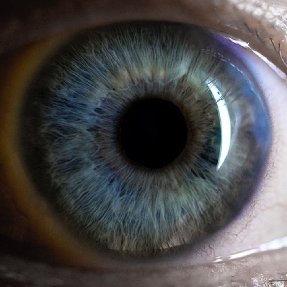
Buenos días, ojazos 👀
09.12.2025 07:20 — 👍 18 🔁 2 💬 2 📌 0

A great talk by @cuilab.bsky.social at the Retinal Ganglion Cell meeting.
09.12.2025 08:14 — 👍 13 🔁 1 💬 0 📌 1

It’s @a-alexandris.bsky.social talking at the Retinal Ganglion Cell Meeting about his work in axonal degeneration.
09.12.2025 09:00 — 👍 7 🔁 2 💬 0 📌 0
Trying to find some protocols for cone isolation from frogs that don't involve transgenics or freezing. There is a surprising dearth of information out there for cone isolation? Seems to be mostly rods. Is anyone familiar with any simple-ish protocols? 🧪 👀 #xenopus
05.12.2025 01:43 — 👍 10 🔁 8 💬 5 📌 0

S-cone-specific circuitry in the outer plexiform layer of a cone-dominant mammal | PNAS
In the vertebrate retina, short wavelength-sensitive S-cones and their downstream
interneurons play unique roles in both image forming and non-imag...
Extremely proud to share our publication on S-cone circuitry in the ground squirrel, newly available this week in PNAS. We've been staring at these reconstructions for a long time, and I'm excited for others to see the results. 1/n
www.pnas.org/doi/10.1073/...
03.12.2025 15:35 — 👍 14 🔁 6 💬 1 📌 2
Just a heads up: I just got a phishing attempt pretending to be from the #VisualSystemDevelopment #GRC. It looked pretty convincing at first glance, so be careful!
27.11.2025 19:36 — 👍 8 🔁 7 💬 2 📌 1
That image is from 1961 and an idealization. Here is an actual trajectory of fixational eye movements. The dots are 2 ms apart. If a midget ganglion cell, with single-cone receptive field, fires at 100 Hz, then every spike reports about a different cone. How can we ever read anything?
07.11.2025 18:23 — 👍 31 🔁 8 💬 3 📌 1
#FluorescenceFriday
A sky of light within ✨
This is the ganglion cell layer of the avian retina. The nerve fiber layer lies beneath like a soft green current, carrying quiet signals forward.
Sometimes to see the universe with its beautiful scattered stars, one only has to look inside.
06.11.2025 18:39 — 👍 22 🔁 4 💬 0 📌 0
Hey folks, this is a very cool opportunity for peeps to do an amazing postdoc in Sydney, Australia in translational vision research.
26.11.2025 00:48 — 👍 23 🔁 11 💬 0 📌 2
Excellent work @victorcalbiagueg.bsky.social !!!
17.11.2025 15:55 — 👍 11 🔁 4 💬 0 📌 0
Axonal pathfinding of zebrafish retinal ganglion cells forms the optic nerve. Credit to Dr. Matthew Bostock @houartlab.bsky.social. #ZebrafishZunday 🧪
16.11.2025 20:30 — 👍 178 🔁 58 💬 4 📌 2

Ending my #FluorescenceFriday with this beautiful avian retina image depicting the beautiful stratification in the avian eyes.
This is where complexity meets art.
#retina #avian #bird #plexiformlayer
14.11.2025 16:19 — 👍 71 🔁 12 💬 3 📌 2
Fantastic new paper from @pierre-mattar.bsky.social lab!
28.10.2025 20:22 — 👍 10 🔁 4 💬 1 📌 0

A picture of a crowd of people at the Foundation Fighting Blindness Vision Walk.
Nice turnout for the @fightblindness.bsky.social #VisionWalk in Pittsburgh.
#ShareYourVision
01.11.2025 14:17 — 👍 18 🔁 2 💬 0 📌 0

#FluorescenceFriday 🌿🔬 post:
Avian retina transduced with my AAVs carrying the #GluSNFR biosensor.
Bright blobs at the top mark photoreceptor terminals; meanwhile below, an RGC connects beautifully with bipolar cells, giving a glimpse of retinal circuitry in action.
#Retina #Neuroscience #AAV #2-P
24.10.2025 19:10 — 👍 40 🔁 5 💬 0 📌 1

A horizontally oriented inhibitory neuron (yellow) spans the mediolateral axis of the superficial superior colliculus, with dense axonal arborization suggestive of a role in collicular surround suppression. Retinal ganglion cell terminals expressing ChR2 are shown in cyan (following intraocular AAV injection), while magenta highlights VGAT+ neurons labeled via a reporter AAV-tdTomato in a VGAT-Cre mouse. Image credit Dr. Peng Cui
#Visual #salience generation involves center-surround dynamics, but what performs these computations? This study shows that the #SuperiorColliculus encodes center-surround dynamics under isolated conditions, and provides insights into the circuit implementation @plosbiology.org 🧪 plos.io/4n7RpsA
20.10.2025 11:28 — 👍 6 🔁 4 💬 0 📌 1
Associate Professor of Neurosurgery @upenn.edu | Neuroscientist | Human Neurophysiology, Neuromodulation & Neurotechnology | Memory & Perception | 🇦🇺 | https://www.fosterneurolab.com/
Deep-sea biology and visual ecology laboratory technician at the University of Delaware | M.S. Marine Biosciences | Looking for a PhD position or laboratory technician/manager role for the Fall'2026!
#Neuroscientist, #Physician - #Axons, #Neurodegeneration, #TBI. 🧠 Research Associate. @JohnsHopkins @JHUPath. (He/him)
Staff Reporter at The Transmitter. Science Journalist.
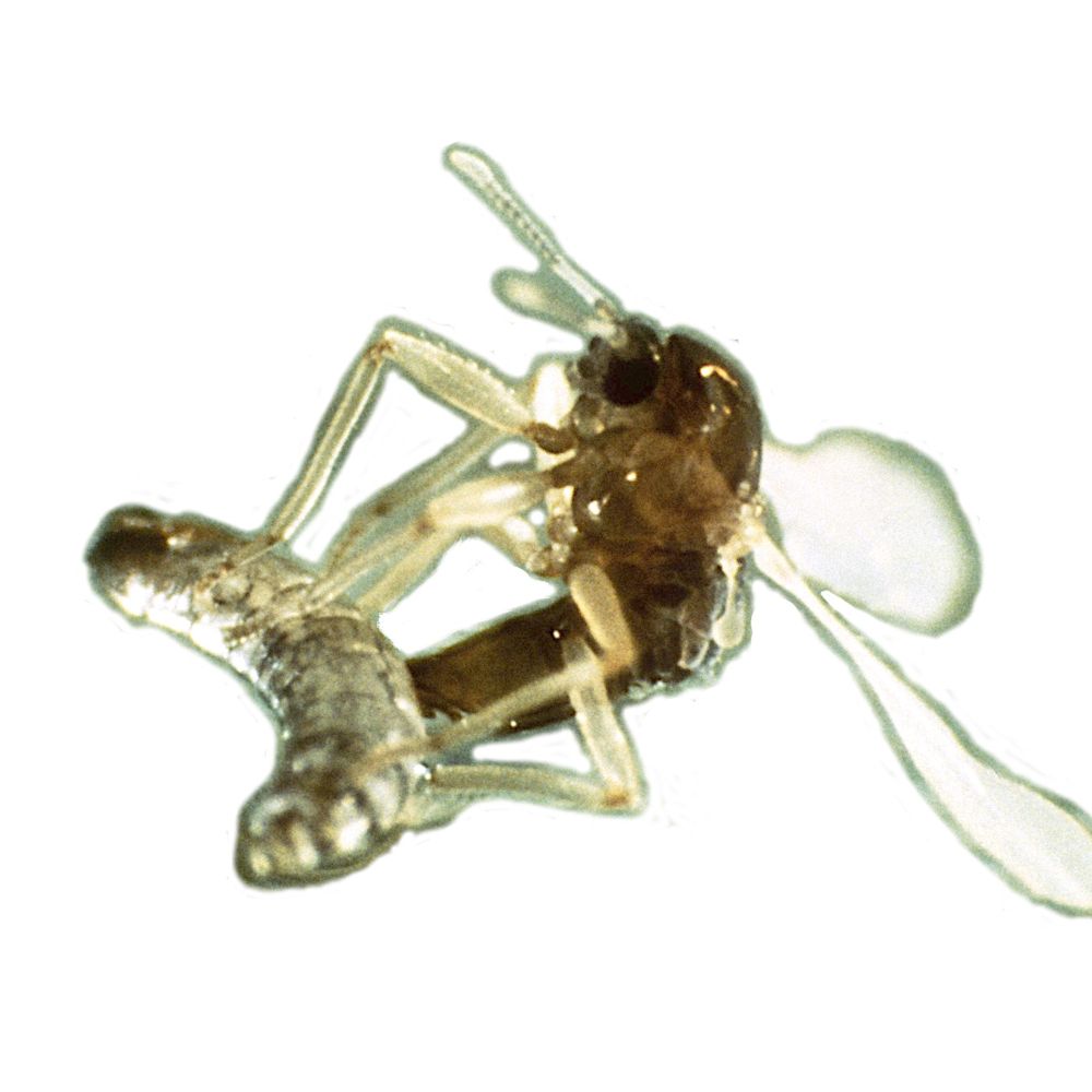
Evolution, ecology & biological clocks - with a focus on lunar rhythms
in development & reproduction of the marine insect Clunio.
+ Genomics | Biodiversity | Behaviour | NeuroBio | MolBio | SciCom
bit.ly/KaiserLab
@mpi-evolbio.bsky.social @maxplanck.de
Laboratory of Stem Cell Biology and Vision Research
Faculty of Medicine, Masaryk University, Czech Republic.
Research Fellow in Manchester. Interested in sensory systems & behaviour.
Usher syndrome, deafblind, retinitis pigmentosa, cochlear implants, guide dog 🦮 🌈, AltText and capitals on hashtags,
♿️ she/her,
Equality, Equity and Justice
JAMA Ophthalmology is a member of the JAMA Network, a consortium of peer-reviewed, general medical and specialty publications.
🌐 JAMAOphthalmology.com
Principal Investigator at University College London
Institute of Ophthalmology
Diabetes UK & Moorfields Eye Charity RD Lawrence Fellow
Retina, vasculature, diabetes, AMD, pericytes, LRG1 and more...
Developing advanced imaging techniques to study how the neurons in the retina talk to each other. PhD student at @sln.icfo.eu
Postdoc Researcher - vision, neurobiology & connectomics in aquatic invertebrates 🪸🦀
I am split between: University of Sussex - badenlab.org
& The Crick, London - prietogodinolab.org
Ph.D. Student at IST Austria - Jösch group 🐭👀
Developmental biologist | Professor at Indiana University Department of Biology | eye development, pattern formation, gene regulation
PhD Student@University of Sussex w/ Tom Baden
MSc Neuroscience@University of Tübingen w/ Thomas Euler
Interested in visual neuroscience, neural design, evolution.
PhD candidate at UW Seattle Molecular and Cellular Biology program 🧬 Studying retinal development and disease 👁️