Huge congrats to @manuelthery.bsky.social and all the Cytomorpho lab. The "Living Architectures" performance at @museeorsay.bsky.social was breathtakingly beautiful
25.01.2026 09:22 — 👍 136 🔁 35 💬 5 📌 2

Postdocs wanted - Please spread the word! We are looking for one bioinformatician and 1-2 cell biologists with different expertise! See the job ad below for more information and links to the job descriptions!
20.01.2026 12:48 — 👍 22 🔁 24 💬 0 📌 1

279 postdoctoral fellowships!
Download freely our database of postdoctoral fellowships and grants. For each entry, we provide eligibility criteria, $ amount, deadline, etc.
Good luck!
Here: research.jhu.edu/rdt/funding-...
20.01.2026 13:22 — 👍 33 🔁 32 💬 1 📌 1

Deep Learning for Microscopy Image Analysis
Topics The following will be covered extensively during lectures, exercises, and project work: Image denoising and restoration (fully supervised and self-supervised) Image translation (e.g.,
Want to learn state-of-the-art deep learning techniques for microscopy image analysis? Apply by Jan. 15 to join us for a bootcamp at HHMI’s Janelia Research Campus. Gain hands-on experience with tools & frameworks you can use in your own research: bit.ly/4sqejiG 🧪
13.01.2026 21:33 — 👍 18 🔁 6 💬 0 📌 0

DendroTweaks Graphical User Interface
Our paper on the #DendroTweaks toolbox is now out as a Version of Record in @elife.bsky.social . doi.org/10.7554/eLif...
Find out more and try the toolbox online at dendrotweaks.dendrites.gr
Happy modeling!
#neuroskyence #compneurosky
13.01.2026 14:39 — 👍 12 🔁 4 💬 0 📌 3
#FluorescenceFriday
Proud of our the last paper @cellcommlab.bsky.social & 1st one of @jboixcampos.bsky.social
We continue to study #CellMigration & how cells decide where to go: ↖️↗️⬇️?
(movie of the actin #cytoskeleton)
Full www.science.org/doi/10.1126/...
@focalplane.bsky.social @science.org 🧪🔬
09.01.2026 16:23 — 👍 47 🔁 12 💬 2 📌 0
#HappyNewYear !
Let's start 2026 with a @cellcommlab.bsky.social paper
How many branches is OK during #CellMigration?
It depends on the #cell: for ex. immune cells are just fine.
In collab. w. #NirGov @hfspo.bsky.social
Full www.science.org/doi/10.1126/...
@focalplane.bsky.social @science.org 🧪🔬
01.01.2026 20:59 — 👍 76 🔁 18 💬 4 📌 0
How do migrating cell groups balance force and flexibility?
Our new preprint shows that RhoGAP15B downregulates RhoA–Myosin activity to stabilise protrusions and enable efficient collective migration in vivo.
Happy New Year!
www.biorxiv.org/content/10.6...
31.12.2025 14:19 — 👍 11 🔁 8 💬 1 📌 0

‼️🚨Hi all! The next Bioc Soc Membrane Contact Site Meeting will be in Chepstow, UK 28-30th Sept 2026. Fantastic speakers with talk slots still available. Sign up & abstract submission now open - limited spaces so register early! Hope to see you all there. Please share + RT 🙏
28.12.2025 07:27 — 👍 16 🔁 14 💬 1 📌 0

@biorxivpreprint.bsky.social CellTrap: A Microfluidic Platform Enabling Cell-Cell Interactions at Variable Effector to Target Ratios
www.biorxiv.org/content/10.6...
27.12.2025 17:34 — 👍 13 🔁 2 💬 0 📌 0
Malina Iwanski @mkiwanski.bsky.social, Lukas Kapitein and colleagues discover that stable microtubules reverse their polarity during neuronal development.
journals.biologists.com/jcs/article/...
#OpenAccess #ReadandPublish
08.12.2025 15:51 — 👍 24 🔁 8 💬 1 📌 2
Do your samples move out of view? Tired of manually adjusting the stage? Introducing DySTrack developed by @zimengwu33.bsky.social and Jonas Hartmann from UCL, a tool that can be integrated into modern microscopes to automate the tracking of moving samples. #MicroscopyMonday doi.org/10.64898/202...
08.12.2025 21:31 — 👍 34 🔁 11 💬 1 📌 0
Interested in cell adhesion, evolution of multicellularity, or developing tools for emerging marine models?
My lab at UM is hiring a postdoc, and the application is now open:
🔗 tinyurl.com/28jvu4aa
If you know anyone looking for a postdoc, please pass this along!
08.12.2025 18:37 — 👍 54 🔁 50 💬 0 📌 2
Come work with us! We are looking for a postdoc in #philbio or #philphysics to work on an interdisciplinary project that adopts the lens of self-organization & active matter to explore the boundary between living & nonliving systems www.kuleuven.be/personeel/jo... #academicsky #philjobs #HPS #evosky
08.12.2025 11:21 — 👍 58 🔁 37 💬 2 📌 3

Torque-based immune cell chemotaxis in complex environments
Directed migration in chemical gradients is crucial to the immune response, yet how immune cells navigate complex tissues remains incompletely understood. Using in vitro migration assays and theoretic...
Hot from the #preprint
Leukocyte + #CellMigration + physics = cool
Microtubules enable torque-based steering during dendritic cell migration.
Actomyosin contractility is required for noise-modulation strategy used by neutrophils.
Full @biorxivpreprint.bsky.social www.biorxiv.org/content/10.1...
23.11.2025 14:39 — 👍 22 🔁 4 💬 0 📌 0
Still posting cytoskeleton videos, it seems. Actin this time.
Sample: Lifeact-eGFP in HeLa cells.
Modality: Airyscan confocal
Timestamp is mm:ss and the scale bar is 5 µm.
23.11.2025 03:21 — 👍 229 🔁 35 💬 14 📌 4
It’s time to talk about epithelial geometry and cancer. How can the architecture of an epithelium affect how tumors will grow and spread? In this thread, @jorgealmagro.bsky.social
23.11.2025 08:01 — 👍 50 🔁 19 💬 1 📌 3
Synchronized development of zebrafish embryos immobilized by snake venom (alpha-bungarotoxin). Credit to Dr. Ian Swinburne. #ZebrafishZunday
23.11.2025 12:11 — 👍 63 🔁 8 💬 3 📌 4
Even if "provisionally" accepted, those are the best words/news I've heard for quite a while...
Here some #blebs to "provisionally" cellebrate.
Let's see this #cell dance under the microscope.
@focalplane.bsky.social #fluorescencefriday #microscopy 🧪🔬
21.11.2025 14:55 — 👍 43 🔁 7 💬 3 📌 0
I'm back at regulardly doing some live imaging so I have new little videos of actin in the immune synapse.
21.11.2025 14:17 — 👍 6 🔁 1 💬 1 📌 0
The law of the jungle.
Interactions of cells in a collective lead to global rotation.
In 80% of the case HUVEC cells turn clockwise.
How many cells does it take for this to happen?
21.11.2025 13:07 — 👍 105 🔁 24 💬 8 📌 2
#AsgardArchaea team, led by @archaeal.bsky.social fr @texasscience.bsky.social — sequencing the DNA collected fr mouth of Rio de la Plata to the continental shelf of Uruguay to detect Asgards, a group of single-celled organisms & our closest microbial relatives on the tree of life. bit.ly/3MdniTH
21.11.2025 00:07 — 👍 91 🔁 25 💬 0 📌 3
HFSP Webinar 2025: Research Grants | Human Frontier Science Program
Do you want to do some frontier of knowledge research? There's still time to register for the next webinar of @hfspo.bsky.social
By far one of the best agencies I've worked with for my beloved HFSP project, even if it's a bumpy road...no risk no glory...
www.hfsp.org/hfsp-events/...
19.11.2025 08:36 — 👍 11 🔁 5 💬 0 📌 0
What a crazy cool paper! First author @pierreucla.bsky.social with a large crew knocked it out of the park. (GIF below from @the.3i.social LLS) Quantifying cell traction forces at the single-fiber scale in 3D: An approach based on deformable photopolymerized fiber arrays www.pnas.org/doi/10.1073/...
13.11.2025 18:21 — 👍 24 🔁 8 💬 0 📌 0

I won't talk about NMeth @natmethods.nature.com forever (probably), but I do want to brag a bit about the November focus issue on cell segmentation and tracking, which is many ways is my last hurrah (and final editorial) for the journal. I am so proud of these papers!! www.nature.com/nmeth/volume...
13.11.2025 20:49 — 👍 123 🔁 20 💬 1 📌 4
This week's #FluorescenceFriday, we'll be treated with one of the classical models of #devbio, the neural crest cells. Here is a beautiful video of neural crest cells with GFP-tagged focal adhesion kinase (🔵) and LifeAct-RFP (🟣) migrating on a Fibronectin matrix.
📹: Adam Shellard
14.11.2025 17:45 — 👍 38 🔁 12 💬 0 📌 0
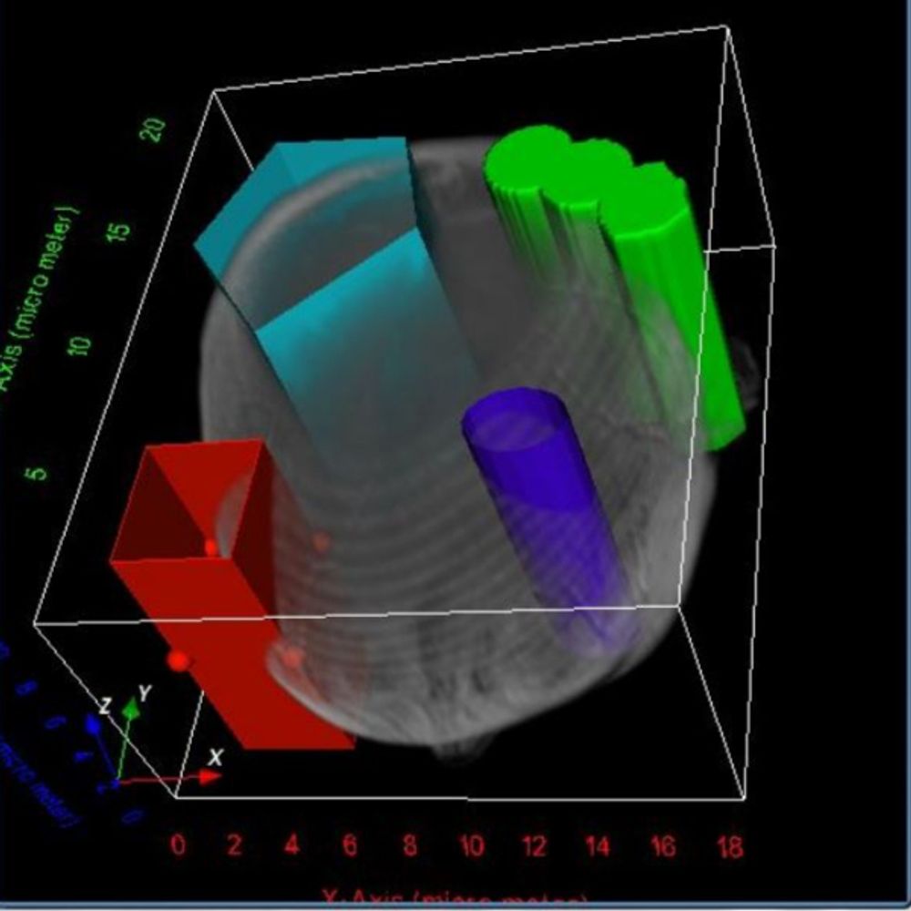
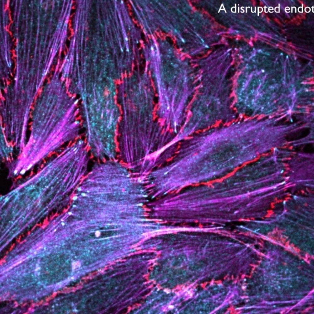
I love to see the endoplasmic reticulum (ER, cyan) hitchhiking over microtubules (MTs, orange).
Also the membrane dynamics are lovely.
It's been a while, but here we are for a late #fluorescencefriday with some #microscopy
#scicomm #scisky @cellcommlab.bsky.social focalplane.bsky.social 🧪🔬
07.11.2025 21:25 — 👍 60 🔁 11 💬 1 📌 1
Neuroscience PhD Student | Purinergic Signaling on Neuron-Glia Interaction | Myelin | 🇨🇱
Mechanobiology, actin, endocytosis @Yale
Shooting electrons, ions and photons (mainly) at plants to study cell-cell communication @mpibiochem.bsky.social & @hhu.de
2025 Schmidt Science Fellow
CDB UCL London and Gurdon Institute Cambridge
Wilson, Simons and Norden labs
Developmental, Systems and Synthetic Biology
Prev: Briscoe, Crick Institute; Zernicka-Goetz and Thomson, Caltech; Banerjee and Goehring, UCL
Molecular cell biology lab exploring the role of mechanical forces in epithelial homeostasis and cancer @ Center for Molecular Medicine UMCU
Postdoc at @MPZ_PhysMed , currently at @IJMonod with Ladoux-Mege group. A material engineer trying to fit into biophysics. Have you ever wondered if cells had memory?
CIRM Postdoc Fellow at UC Irvine: Piezo1, mechanobiology, high-res microscopy. Cinema lover.
We study the molecular and biophysical mechanisms underlying tissue morphogenesis @poldresden.bsky.social @tudresden.bsky.social @erc.europa.eu @embo.org
https://physics-of-life.tu-dresden.de/team/pol-groups/barriga
MPhil @ Cambridge Institute of Medical Research (CIMR)
The BioImage Analysis Unit designs innovative computational methods to address challenging biological questions through rigorous quantitative approaches @Icy_bioimaging.bsky.social
https://research.pasteur.fr/en/team/bioimage-analysis/
@InstitutPasteur
PhD in Dagmar Ibers lab @ ETH Zürich.
How does development really work? Beyond genes and networks towards an holistic mechanistic view
Family & dog - bake, whittle, run, cycle & draw. Nerve repair & biomaterials I teach biology & bioengineering. views=own; he/him. 🇨🇦 🇺🇦 🇬🇧 🇩🇪 🇪🇺 🌍 🌌
It’s way too red!
Blog @ https://bio-mat-sketches-mor.blogspot.com
Director of the Institute of Pharmacology and Vice Dean of the Medical Faculty, Marburg University, Germany. Interested in Epithelial Biology and Pharmacology
Assistant Prof @MBISg, NUS | Interested in mechanobiology | Postdoc, Gardel Lab @UChicago | Schmidt AI in Science and AHA Postdoc | Previously Marie Curie and FRM PhD, Etienne-Manneville Lab @Institut Pasteur
“LaPa-ciencia” es un podcast donde la gente que hace ciencia se muestra tal y como es. Sin batas, sin deadlines, sin experimentos y sin presión. Un sitio para personas, de ciencia, pero personas como tú y como yo.🎙️🧫🦠🔬
LMCB Group Leader at UCL. Cis-regulatory control of cell fate choice in development.
https://delaslab.com/
Schapiro lab at Heidelberg University and Heidelberg University Hospital. We work on #HighlyMultiplexedImaging and #SpatialOmics
PhD at the University of Amsterdam. Microscopy enthusiast 🔬, cytoskeleton admirer 🧫, hopeful sensor builder. Mostly just a biologist finding his way. Opinions my own.