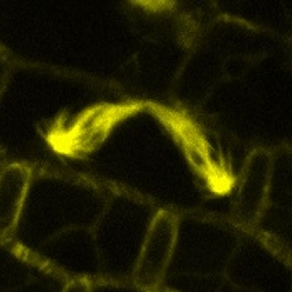
Not something you see in textbooks very often: tripolar mitosis.
07.02.2026 08:00 — 👍 1558 🔁 270 💬 50 📌 22
Yup. This is EB3 fused to mNeonGreen
05.02.2026 22:53 — 👍 2 🔁 0 💬 0 📌 0
I’ll have a look at the raw data tonight but I think you’re in the right ballpark.
05.02.2026 22:29 — 👍 1 🔁 0 💬 0 📌 0
Thanks!
Of course - let me know if you need any additional files.
05.02.2026 22:28 — 👍 0 🔁 0 💬 0 📌 0
An intracellular meteor shower. EB3 comets tracking growing microtubule plus-ends in a cultured cell.
05.02.2026 05:27 — 👍 1706 🔁 245 💬 55 📌 20
Dynamic actin waves in a zebrafish embryo. Credit to @aaandmoore.bsky.social & Dvir Gur. #ZebrafishZunday 🧪
01.02.2026 08:18 — 👍 100 🔁 18 💬 2 📌 0

🧪 We are excited to share this novel open access paper on Phasor Mixing Coefficient to analyze colocalization, developed by folks @i2janelia.bsky.social and @aicjanelia.bsky.social. This is also the first technical paper jointly published with our sister imaging center @malacridalab.bsky.social!
28.01.2026 16:20 — 👍 43 🔁 20 💬 2 📌 2

Image of an animal cell showcases web-like fibers called vimentin — a protein that provides mechanical stability to cells and tissues
Created using fluorescence microscopy, this stunning image of an animal cell showcases thin, web-like fibers called vimentin — a protein that provides mechanical stability to cells and tissues, despite a single vimentin being ~7000x smaller than the width of a human hair. 📸 @aaandmoore.bsky.social
20.01.2026 21:56 — 👍 30 🔁 2 💬 0 📌 0

Direct labeling of microtubule turnover reveals in-lattice repair and stabilization patterns in developing neurons
The microtubule cytoskeleton is the backbone of neuronal morphogenesis, driving the development of the dendrites and axon, and supporting trafficking to distant compartments. How neuronal microtubules...
🚨The Neurocyto lab is branching out in our latest preprint! We used tubulin microinjection to directly visualize microtubule turnover in developing hippocampal neurons, demonstrating the presence of in-lattice repair and a selective stabilization in the nascent axon. Check below, or read on 🧵 1/9
12.01.2026 19:14 — 👍 102 🔁 31 💬 4 📌 2
It typically localizes to the endoplasmic reticulum.
20.12.2025 18:42 — 👍 1 🔁 0 💬 0 📌 0
Call it what you want but it’s technically still just the Golgi Apparatus until you get an act of Congress.
20.12.2025 16:53 — 👍 4 🔁 0 💬 2 📌 0
I don't always get pretty, isolated, AND transfected primary neurons in culture, but when I do I take advantage.... Rat hippocampal neuron overexpressing ThymosinB4-mScarlet and imaged for 16hr on a @zeiss-microscopy.bsky.social LSM880 with Airyscan. #FluorescenceFriday #Microscopy
19.12.2025 14:01 — 👍 120 🔁 27 💬 2 📌 0
This movie shows lysosomes (orange) and keratin (gray) in a cultured cell over 10 minutes.
12.12.2025 06:20 — 👍 268 🔁 42 💬 11 📌 3
I like this movie, but a friend of mine likes to complain about the obvious stitching artifacts. I'll try harder next time, Michael. Vimentin (orange) and ER (blue) in an overnight acquisition.
25.11.2025 06:06 — 👍 52 🔁 6 💬 1 📌 0
What's up Bluesky....here's some live-cell imaging taken in my lab
24.11.2025 12:11 — 👍 45 🔁 5 💬 2 📌 0

is there a direct crosstalk between actin and vimentin intermediate filaments? Our work shows that vimentin promotes actin assembly by stabilizing ATP-subunits at the barbed end.
Fantastic work done by @lilianpaty.bsky.social with @romet-jegou-lab.bsky.social
www.biorxiv.org/content/10.1...
24.11.2025 10:15 — 👍 36 🔁 15 💬 0 📌 0
it’s a little transparent, but that’s the scale bar in the bottom left corner. 5 micrometers.
23.11.2025 18:55 — 👍 1 🔁 0 💬 1 📌 0
Still posting cytoskeleton videos, it seems. Actin this time.
Sample: Lifeact-eGFP in HeLa cells.
Modality: Airyscan confocal
Timestamp is mm:ss and the scale bar is 5 µm.
23.11.2025 03:21 — 👍 231 🔁 36 💬 14 📌 4

24FPS_VimentinCos7GIF_4320p_ultraHQ
This is "24FPS_VimentinCos7GIF_4320p_ultraHQ" by Andy Moore on Vimeo, the home for high quality videos and the people who love them.
vimeo.com/1139537563?s...
I uploaded a version of that vimentin IF movie to my vimeo page. It's still compressed, but looks a lot better. Best results when you set the quality to 4k.
22.11.2025 04:25 — 👍 18 🔁 2 💬 1 📌 0
Yup - imaged it yesterday on a laser scanning confocal.
21.11.2025 17:12 — 👍 12 🔁 0 💬 2 📌 0
Yup!
21.11.2025 16:12 — 👍 0 🔁 0 💬 1 📌 0
Fun fact - this protein belongs to the same family as the hard keratins found in all hair (including the hair you might find in a shower drain).
21.11.2025 16:12 — 👍 1 🔁 0 💬 0 📌 0
We should promote IFs and septins to position 1 and 2. Actin filaments and microtubules have had their day in the sun. We can just start calling them the third and fourth components 😂
21.11.2025 16:08 — 👍 2 🔁 0 💬 0 📌 0
Sure - these are vimentin intermediate filaments which are part of the cell’s internal scaffolding. They confer mechanical resilience to cells and , among other things, help to anchor and position organelles like mitochondria. These filaments are part of the same family that includes keratins.
21.11.2025 16:06 — 👍 8 🔁 1 💬 1 📌 0
Not rude at all - appreciate the suggestion. I’ll give it a shot.
21.11.2025 16:02 — 👍 1 🔁 0 💬 1 📌 0
Ah sorry for the lack of context. Cos7 cells are a cultured cell line originally derived from African green monkey kidney cells. They are exceptionally flat at the cell periphery so imagers like to use them. This one is a bit of a monster - it’s about 5x larger than normal.
21.11.2025 15:56 — 👍 24 🔁 0 💬 2 📌 0
Love it. I acquired the movie at about 10 seconds per frame and the whole thing is just over 15 minutes.
21.11.2025 04:34 — 👍 22 🔁 0 💬 1 📌 0
I mean…does this work? I’m at my wits end.
21.11.2025 04:01 — 👍 68 🔁 0 💬 8 📌 0
Attempt number seven at uploading this video of intermediate filaments in an enormous COS7 cell. I have a feeling the BlueSky compression will not do it any favors.
21.11.2025 03:54 — 👍 940 🔁 83 💬 97 📌 11
iPS cell-derived cardiac myocytes (heart muscle cells) typically beat about once per second, so I usually speed up the movies I post; otherwise, scrollers might miss the action. But every now and then, a cell looks like this in real time. #CellBiology
10.11.2025 01:56 — 👍 45 🔁 10 💬 3 📌 0
PhD student at Wellcome Sanger Institute associated with Haniffa, Parts and Saez-Rodriguez Labs
PhD student | CLEXM | UCD | Cell Bilogist | I like to read
UBC Professor of Zoology; student of birds, genomes, evolution. Pro-democracy. Pro-sanity. Pro-humanity.
Assistant professor of Botany at the University of British Columbia. My lab works on cell division and beyond.
Lab website: https://www.ashraflab.com/
PhD candidate in molecular evolution @UChicago
manyuanlonglab.uchicago.edu
Post-doctoral scholar in Cosgrove lab at Penn State. Interested in Plant Cell Wall and Expansins 🌿
#NewPI @UIC
Studying Host-Symbiont-Virus Interactions in 🪰 and 🦟
#Wolbachia #Spermatogenesis #Embryogenesis #ChromatinBiology #Epigenetics #Microscopy
She/her
Postdoc @Indiana University I flower & fruit development l Microscopy I ➡️De Folter Lab🇲🇽➡️Kierzkowski Lab🇨🇦 ➡️ Nikolov Lab🇺🇲 l 🌱💮 Microscopy lover🔬| AFE The Plant Cell Journal 💻
DevBio PhD student at EMBL, Heidelberg
Former undergrad student at UNAM, Mexico City
Cancer biologist, environment and cancer, guitarist, would-be evolutionary molecular biologist, bread baker, sometimes coder, amateur photographer, professional microscopist, & gardener. Lab site: https://blogs.dal.ca/dellairelab/
📌🇨🇭 / 🗣️🇫🇷 🇬🇧 🇩🇪
⚠️Skeptical inside🧐Rational Rebel
👨🔬 #PhD in the making (neuro)
❤️#sciences , toute curiosité, gros plus pour l'imagerie🔭 📸 🔬 🖼️ 🗺️!
🎮 : KSA/P🚀 GW2 🧙♂️ MLBB 🌈
Géo-localisations sur: @geoloc-holmes.bsky.social
#whereisthis #Thenandnow
Chemotaxis. Math. Computers. Cells. Machine learning.
Prof Universitário, ativista pela legalização da maconha e outras drogas
Author & Senior Editor at Nature. DISCLAIMER: This is a personal account. Reposts aren’t Endorsements. Views needn't reflect the official view of Springer Nature, as it doesn't know where they've been. Glad we've cleared that one up.
Studying zebrafish intestinal biology at the University of Bath with an interest in sex and mucosal immunity. Trying to import the oxford comma to Britain. she/her 🐠 🏳️🌈 🏳️⚧️ 🎶
Assistant Professor in Trinity College Dublin. A mitochondria and immunometabolism enthusiast. Views my own. 🇮🇪
I’m Gant. My lab studies how the structure of cells affects disease progression. Allen Distinguished Investigator in Molecular and Celular Biology at @UCDavis.
https://luxton.faculty.ucdavis.edu