The Lab of Stephan Grill at MPI-CBG has finally made it to Bluesky and we also have a brand new homepage: grill-lab.org
Check it out.
@neipel.bsky.social
Theoretical biophysicists in Grill and Jülicher labs. (MPI-CBG, MPI-PKS Dresden) Interested in anything from genome regulation to developmental biology, currently focusing on left-right symmetry breaking and shape/curvature/ geometry sensing.
The Lab of Stephan Grill at MPI-CBG has finally made it to Bluesky and we also have a brand new homepage: grill-lab.org
Check it out.
bsky.app/profile/neip...
20.07.2025 15:26 — 👍 3 🔁 0 💬 0 📌 0(4/10)
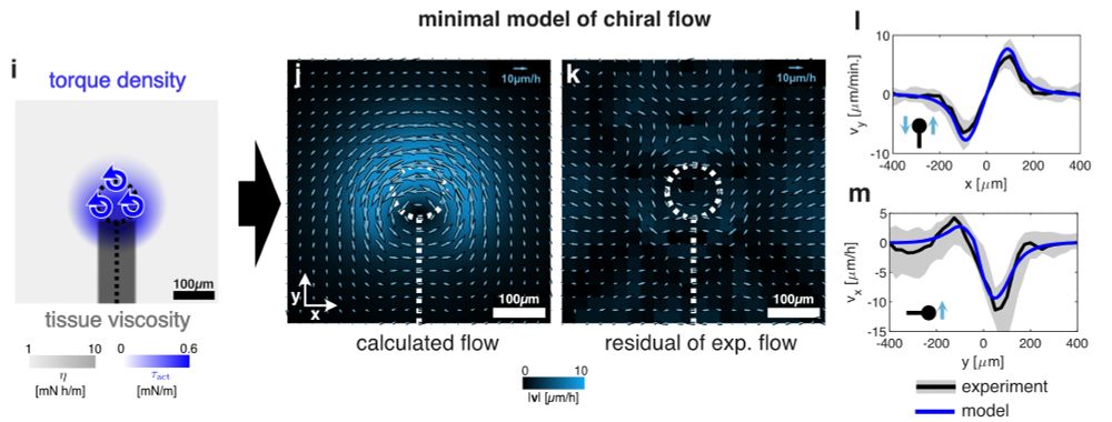
We quantified these chiral tissue flows in 25 quail embryos and inferred forces using a fluid model, suggesting a torque of 6μNμm is generated locally at the node.
(3/10)
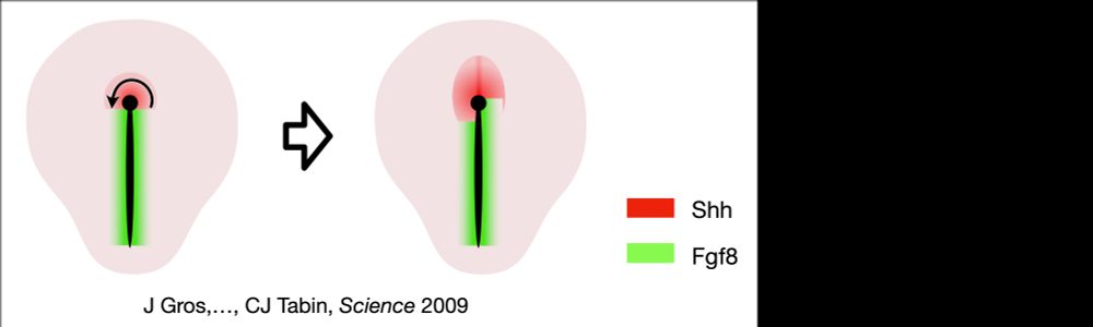
Jerome found cells moving to the left around the node, repositioning a Shh domain to the left, but what makes them do that?
(10/10) Finally, a big thank you to all the people involved, in particular to our PIs @stephangrill.bsky.social and Frank Jülicher as well as the @jeromegros.bsky.social lab for all their support throughout this >5year project!!
20.07.2025 13:42 — 👍 0 🔁 0 💬 1 📌 0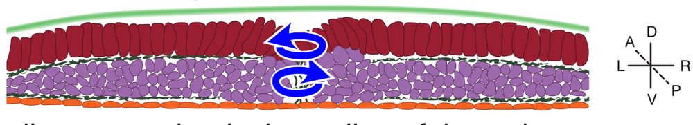
(9/10) In conclusion, we give a mechanical perspective on how the node organises avian development: mechanical coupling of tissue layers allows for the generation of an active chiral torque dipole to break embryonic L/R symmetry. How is this achieved on a cell scale? Stay tuned!
20.07.2025 13:42 — 👍 1 🔁 0 💬 1 📌 0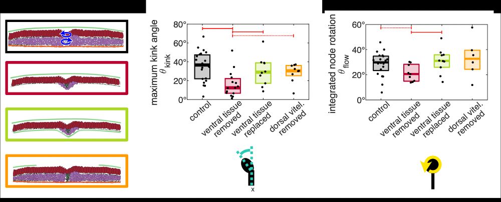
(8/10)
But now comes the really striking result: She found that replacing the meso-/endo tissue with a vitel. membrane is sufficient to restore node rotation. So, you simply need some mech. substrate. sustaining a counter-torque such that the node can drive its own rotation.
(7/10) But, what is the substrate? Is it the underlying meso-/endoderm? Here come the crazy experiments by Julia: She used an eye-brow to remove the meso-/endo showing that it’s required for node rotation. Notably, meso-/endo grows back yielding a delayed and reduced node rotation.
20.07.2025 13:42 — 👍 1 🔁 0 💬 1 📌 0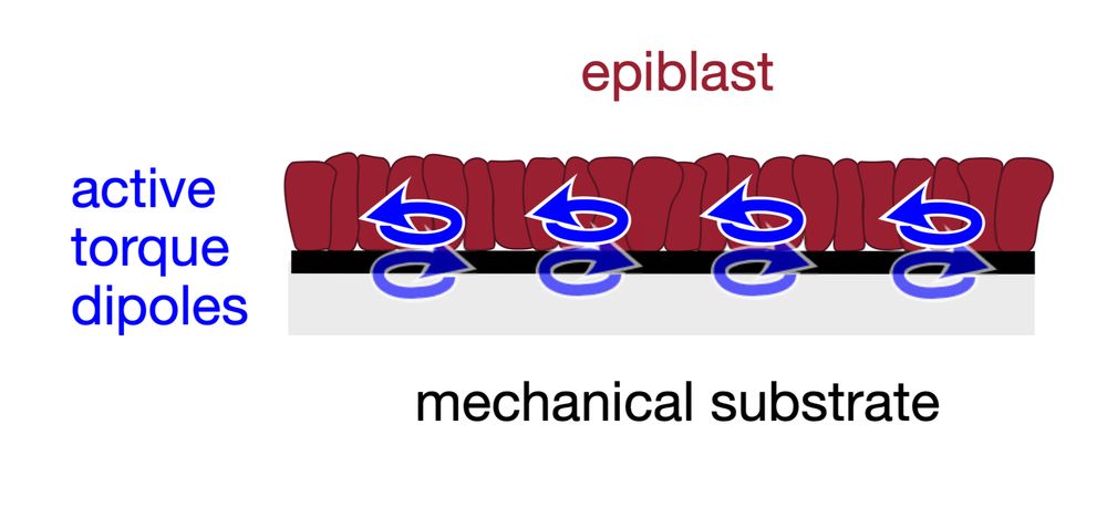
(6/10)
So, the following picture following mechanical picture of avian L/R sym. break. emerges: actomyosin-activity generates cell-scale torque dipoles that propel the rotation of node and surrounding epiblast with respect to some rigid substrate.
(5/10)
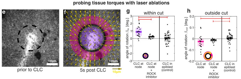
But is the node really capable of making itself rotate? Lasercuts by Julia show yes, providing maybe the first direct evidence for a tissue-scale active chiral torque. Also, we show that the apparent torque is actomyosin-dependent, consistent with previous observations by Jerome.
(4/10)
We quantified these chiral tissue flows in 25 quail embryos and inferred forces using a fluid model, suggesting a torque of 6μNμm is generated locally at the node.
(2/10)
Why is the heart on the left? Somehow, embryos mange to translate mol. chirality to tissue-scale handedness. In various vertebrates, that’s the job of cilia. But birds, chameleons, pigs do it differently, as shown before by the fabulous @jeromegros.bsky.social and @shylonatasha.bsky.social
(1/10)
I am excited to finally share our work on avian left/right symmetry breaking with you. We reveal a tissue-scale active torque dipole generated at the Hensen’s node, thanks to crazy experiments by Julia @juliapfanzelter.bsky.social and some analysis by me.
doi.org/10.1101/2025...