We are the International Academy of Cytology. A scientific, non-profit organization of professionals in the field of Clinical Cytology. Founded in 1957. Our official journal is Acta Cytologica https://www.cytology-iac.org/
General Pathologist working in Cambridge, MA
Trained at Massachusetts General Hospital & Boston Children's Hospital
I daydream about skating ⛸
Pathologist, UCSD, mom, yogi, #UCSD, #GIPath #Pathology #Path
Pathologist | Surgpath, GI and Liver Path, Microbiology | 🇧🇬🇺🇸addopted Appalachian | bcsm BC is my foe #BGSky #Pathsky #IDsky #MedSky
Pathologist & Teacher 😉🇮🇪🏳️🌈🇪🇺🕊️
Northwestern, placenta pathology, machine learning, infections in pregnancy, not giving medical advice, available for media
College of American Pathologists: The leading organization of board-certified pathologists. #pathologists #Pathsky 🔬
General surgical pathologist with specialization in gynecologic pathology
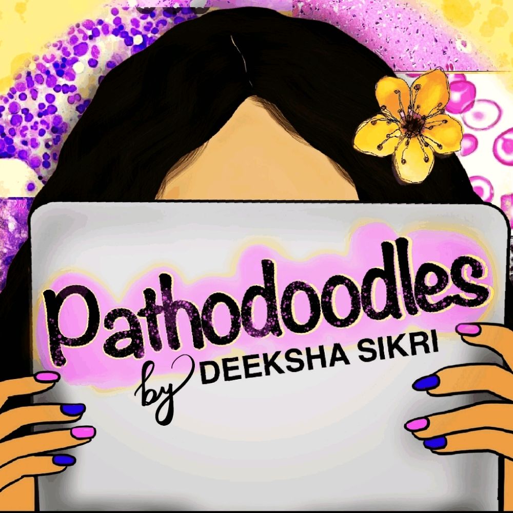
A MedEd enthusiast and doodler powered by caffeine, curiosity and constant-catching up
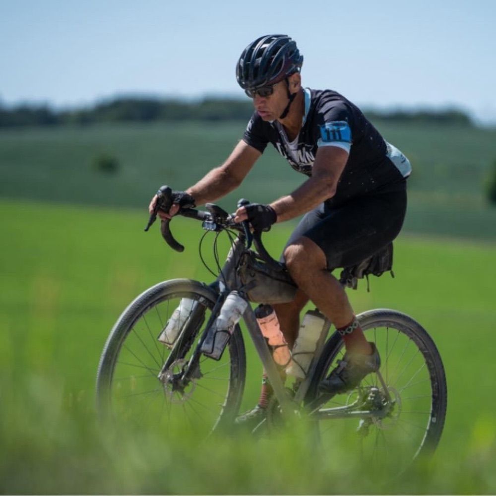
I like my wife and family, bikes, music, dogs, baking bread and soft tissue tumors. Only the last here though.
Visual survey of surgical pathology with more than 13,700 high-quality images of benign and malignant neoplasms & related entities.
Physician (Pathology 🔬) / 🏳️🌈 🕉️ 4️⃣1️⃣
Leftist / BLM / Pro-Choice / LGBTQIA+
Gamer / Geek / Memes and Dad Jokes
#MedSky #PathSky #Pathology #Hemepath #Hematopathology
Pathologist🔬(General Surgical Pathology)
Recording Guitarist (Smooth Jazz, Rock, Gospel)
https://www.linkedin.com/in/folaranmi-olaleke-96a91513
Patólogo General #pathologist #pathology
Interés en todo lo relacionado con la patología y además, los cuerpos extraños en medicina y la histología comparada de animales y plantas.
https://www.flickr.com/photos/foreignbodies/
Tinkering with vLLM @RedHat