Come and join our team!
We currently have two PhD scholarships between Danny Wilson and myself
We are a supportive, productive, and inclusive research group studying parasite cell biology @ The University of Adelaide in beautiful South Australia
scholarships.adelaide.edu.au/Scholarships...
16.06.2025 02:03 — 👍 3 🔁 3 💬 0 📌 0
Thanks very much for having me, and coming along!
28.05.2025 07:57 — 👍 0 🔁 0 💬 0 📌 0
Thanks very much to the wonderful folks in the new Flinders Health & Medical Research Building for hosting me on Tuesday!
Cheers to Nick Eyre and @evahesping.bsky.social for the invitation to speak and for showing me around the beautiful new facility!
28.05.2025 07:56 — 👍 0 🔁 0 💬 0 📌 0

Go Long Huynh! Fancy cell biology and fancy microscopy on artemisinin resistance in Plasmodium parasites at the BioMalPar conference #EMBLMalaria @benliffner.bsky.social @sabsalon.bsky.social @events.embl.org
22.05.2025 08:06 — 👍 13 🔁 2 💬 0 📌 0

The new OrthoMCL-7 with OrthoFinder clustering, Similar Groups, and Phylogenetic Trees is 🌟 now live 🌟at orthomcl.org, part of the @veupathdb.bsky.social
family of resources. The previous version of OrthoMCL-6.21 is still available as a Legacy site.
22.04.2025 14:48 — 👍 4 🔁 3 💬 0 📌 0
Cell biology is producing mountains of microscopy data, usually a given paper got that data for some purpose, but what if you could find relevant data that you could use to answer your own questions? in her new preprint, @marymirvis.bsky.social develops Systematic Review methods to do just that
20.04.2025 15:29 — 👍 37 🔁 4 💬 1 📌 1
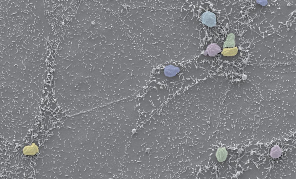
Unlocking new understanding of Plasmodium sporozoite biology with expansion microscopy https://www.biorxiv.org/content/10.1101/2025.04.09.648058v1
12.04.2025 04:18 — 👍 1 🔁 1 💬 0 📌 0
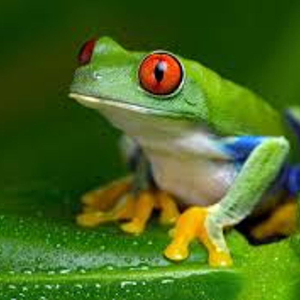
How i feel when I get to look at expanded midguts or salivary glands
12.04.2025 14:55 — 👍 1 🔁 0 💬 0 📌 0
Unlocking new understanding of Plasmodium sporozoite biology with expansion microscopy https://www.biorxiv.org/content/10.1101/2025.04.09.648058v1
12.04.2025 04:18 — 👍 2 🔁 1 💬 0 📌 0
A shoutout to @freddyfrischknecht.bsky.social who has helped me out with my many sporozoite biology queries over the last few years, and likely unknowingly inspired me to find out how many rhoptries a sporozoite has from an email about 6 years ago
12.04.2025 11:27 — 👍 3 🔁 0 💬 1 📌 0
A huge thanks to all my co-authors in this study and a special thanks to @sabsalon.bsky.social for letting me loose on a project that initially had nothing to do with what we were previously working on, but has now greatly shaped our research directions!
12.04.2025 11:27 — 👍 2 🔁 0 💬 1 📌 0
There are plenty of other interesting tidbits and findings in this study, and after peer review we will make all the imaging data available on an online database so anybody can look through the images themselves!
12.04.2025 11:27 — 👍 0 🔁 0 💬 1 📌 0

For the small number of RON11 knockdown parasites that make it to the salivary gland, we found they accumulate in the space between epithelial cells but almost never actually invade or get into the secretory cavity. This interesting observation explains why these parasites are not transmissible!
12.04.2025 11:27 — 👍 0 🔁 0 💬 1 📌 0

More important though, these RON11 knockdown sporozoites only made half the number of rhoptries. And when we looked at the small number of sporozoites that made it to the salivary gland, many of them had no rhoptries at all!
12.04.2025 11:27 — 👍 0 🔁 0 💬 1 📌 0

Looking at these RON11 knockdown parasites, we saw that late in their rhoptry biogenesis, most of them had aberrant looking rhoptries.
12.04.2025 11:27 — 👍 0 🔁 0 💬 1 📌 0
We wanted to put this new knowledge of rhoptry biology to the test by looking at what happens when we knockdown a rhoptry protein, but which to choose? Recently, a study of the protein RON11 showed that knockdown of this protein led to merozoites with 1 rhoptry
journals.plos.org/plosbiology/...
12.04.2025 11:27 — 👍 1 🔁 0 💬 1 📌 0
This led us to the hypothesis that sporozoites have two pairs of rhoptries that are specialised for each of their invasion events: the salivary gland and hepatocytes
12.04.2025 11:27 — 👍 0 🔁 0 💬 1 📌 0
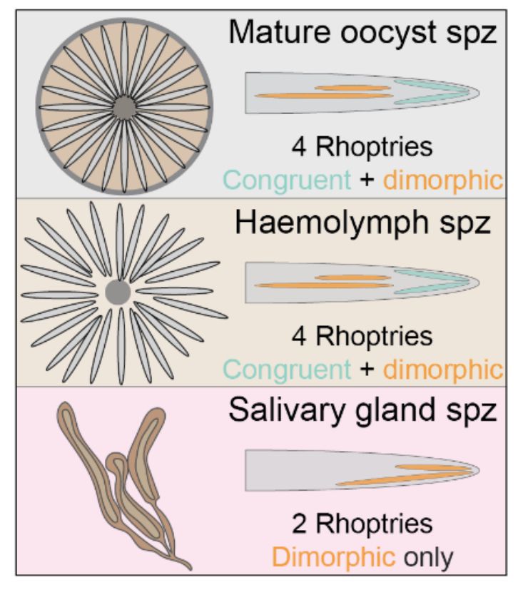
One unexpected observation was that of the usually 2 pairs of rhoptries in oocyst sporozoites, they were morphologically distinguishable. We called these the dimorphic (different size) and congruent (same size) rhoptry pairs, with the congruent rhoptries specifically used up during SG invasion.
12.04.2025 11:27 — 👍 1 🔁 0 💬 1 📌 0
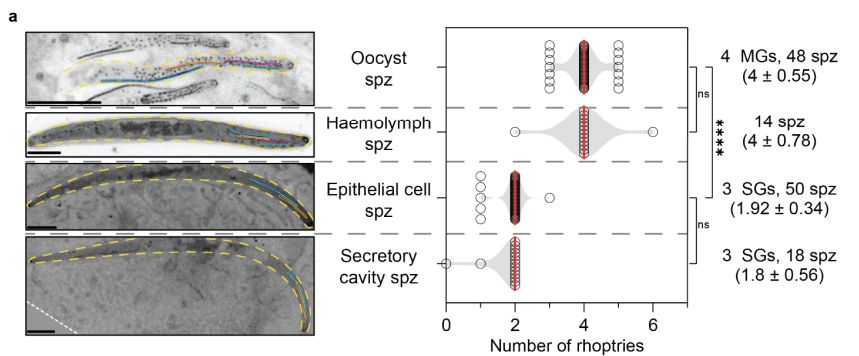
Wanting to follow the fate of rhoptries through the invasion of sporozoites into the mosquito salivary gland, we could differentiate sporozoites at different stages of this process. Excitingly, we could see that sporozoites use up two rhoptries during salivary gland epithelial cell invasion!
12.04.2025 11:27 — 👍 1 🔁 0 💬 1 📌 0

We then put the segmentation score to the test, using it to develop a timeline for the biogenesis of rhoptries in forming sporozoites!
12.04.2025 11:27 — 👍 1 🔁 0 💬 1 📌 0

In the past, oocyst size was used a proxy for development, but one sporozoite formation started, we saw no real correlation with developmental stage and oocyst size. Instead we developed what we call the segmentation score, using progress through cytokinesis as a way to assess sporozoite development
12.04.2025 11:27 — 👍 1 🔁 0 💬 1 📌 0
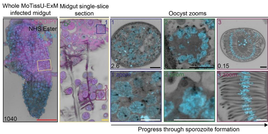
Next, we wanted to look at how sporozoites form within the oocyst. But we ran into a problem, oocysts from the same midgut varied massively in their progress through sporozoite development. So how can we compare different oocysts with each other?
12.04.2025 11:27 — 👍 1 🔁 0 💬 1 📌 0

We could see plenty of other organelles/structures in oocysts too, like the basal complex, apicoplast, apical polar rings, rhoptries, centriolar plaque and more!
12.04.2025 11:27 — 👍 0 🔁 0 💬 1 📌 0
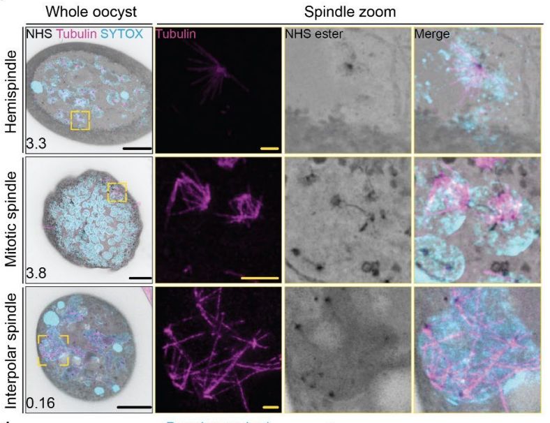
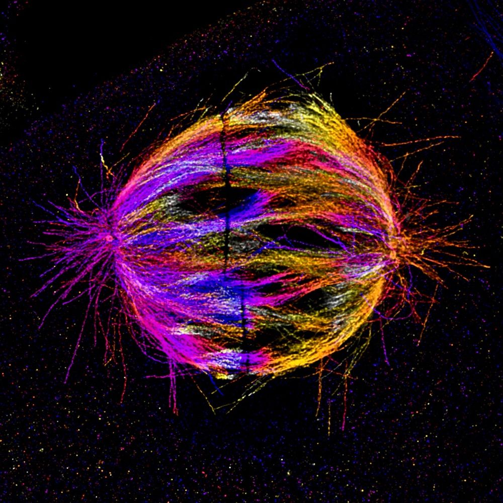
Having seen how powerful expansion microscopy was for studying other aspects of parasite biology, we were really keen to see what this technique could allow us to see in mosquito-stage parasites. Being good cell biologists, we first looked at the cool microtubule spindle structures of oocysts!
12.04.2025 11:27 — 👍 2 🔁 1 💬 1 📌 0
Through this, mosquito tissue ultrastructure expansion microscopy (MoTissU-ExM) came to life! A special shoutout to the IUSM PharmTox postdoc professional development grant for funding this travel
bmcmethods.biomedcentral.com/articles/10....
12.04.2025 11:27 — 👍 0 🔁 0 💬 1 📌 0
In 2022, the idea to have a look at sporozoites using expansion microscopy came up after Alexis gave a seminar at IUSM! Early attempts were hit and miss for different reasons, but we really got the technique running smoothly after a month working it up with Thiago @jvr-lab.bsky.social
12.04.2025 11:27 — 👍 0 🔁 0 💬 1 📌 0
PhD: sand fly/ mosquito 🦟 biologist . Interested in everything parasite-microbiota-vector interactions. Full time Chelsea fan/ LH44 🐐
PhD Biochemistry candidate in BIOPEP Peptide Group and Birkholtz lab (https://b-lab.health/) at Stellenbosch University, South Africa 📍🥼 | Molecular Biologist aspiring to become a Biochemist ⚗️
PhD student at UniGE @Dudin lab
Finishing PhD candidate (Blaskovich Lab, IMB @ UQ)
Synthetic chemistry & microbiology (let's tackle biofilms!)
Can be found dancing, surfing, & cycling 💃🏼🍊🌻🧪🌊🧬⌬
Working and living on unceded lands
Professor of Evolutionary Cell Biology, Director of Cambridge Biosciences BBSRC DTP, Department of Biochemistry, University of Cambridge
I do science for living. Opinions my own.
X: @AliHShaib, my lab: https://www.neuro-physiol.med.uni-goettingen.de/dr-ali-shaib/
Postdoctoral scientist at ICFO. Super Resolution 🔬, Expansion 🔬, Citoskeleton, Retina, Biophotonics, Neuroscience.
Researcher in MeLiS - Lyon. Likes centrioles, cilia and anything graviting around those
Associate Professor of Medicine & Cell Biology, Washington University in St Louis.
We study centrosomes, cilia, kidney and lung ciliopathies 🔬
👉 https://mahjoublab.wustl.edu
NeuroCyto lab 🤹🏻 Institute of NeuroPhysiopathology at CNRS & Aix-Marseille Université 🧠 Neuronal cell biology 🦠 Super-resolution microscopy 🔬 Come for science 👨🏻🔬 Stay for some cooking, sneakers and bike adventures 🥘 👟🚴
(she/her) Scientist, Berlin-based, PhD Student in @ewerslab.bsky.social
Here for Microscopy, Cytoskeleton, Cellbio, Cytokinesis. Black in STEM
🇧🇷🇺🇸Evolutionary Cell Mechanobiology of Archaea.
Associate Professor at Indiana University.
Standing tall on the shoulders of tiny (salty) bugs.
Lab Website: bissonlab.com
PhD Student @ MRC LMB working towards long term imaging of organoid models with James Manton and Madeline Lancaster
Postdoc at the CSSB Hamburg, studying mycobacterial secretion with #CryoEM and #CryoET. Generally something of a nerd. He/him.
Evolutionary biologist exploring the molecular mechanisms behind life's complexity. Currently @International University of Catalonia (UIC), Barcelona.
postdoc@Kosinski EMBL HH
visiting postdoc at giardia lab @FasoLab Uni Bern 🚀
giardia/chc/autophagy/tomo/clem
There are no perks. Permanently with my head in the clouds. Pessimist and lover of chaos, raccoons, celestial bodies and waves.
Associate Professor at the University of Illinois at Urbana-Champaign studying the protozoan parasite Cryptosporidium
Post-doc in malaria parasite molecular biology at the University of Melbourne