Study links (8/8) ME Research UK report (conference analysis): www.meresearch.org.uk/altered-neur...
30.01.2026 13:52 — 👍 0 🔁 0 💬 0 📌 0
Study links (7/8) Godlewska 2022 (Psychopharmacology): doi.org/10.1007/s002...
30.01.2026 13:52 — 👍 0 🔁 0 💬 1 📌 0
Study links (6/8) Hennemann 2025 (J Med Virol): doi.org/10.1002/jmv....
30.01.2026 13:52 — 👍 0 🔁 0 💬 1 📌 0
Study links (5/8) Chaganti 2024 (Front Neurol): doi.org/10.3389/fneu...
30.01.2026 13:52 — 👍 0 🔁 0 💬 1 📌 0
Study links (4/8) Manganotti 2023 (Clin Neurophysiol): doi.org/10.1016/j.cl...
30.01.2026 13:52 — 👍 0 🔁 0 💬 1 📌 0
Study links (3/8) Marinkovic 2023 (Brain Sci): doi.org/10.3390/brai...
30.01.2026 13:52 — 👍 0 🔁 0 💬 1 📌 0
Study links (2/8) Sklinda 2021 (Pol J Radiol):
doi.org/10.5114/pjr....
30.01.2026 13:52 — 👍 0 🔁 0 💬 1 📌 0
Study links (1/8) Thapaliya 2025 (Am J Med): doi.org/10.1016/j.am...
30.01.2026 13:52 — 👍 0 🔁 0 💬 1 📌 0
Overall: results vary.
Some studies report higher glutamate/Glx, others lower Glu/Gln, and some lower GABA.
Different brain areas and timing may explain it.
This supports an idea of altered excitation vs inhibition in some people.
30.01.2026 13:52 — 👍 0 🔁 0 💬 1 📌 0
ME Research UK reported an MRS re-analysis: 48 ME/CFS vs 52 controls.
Glutamine/glutamate higher; choline lower (linked with cell membranes).
They suggested excitotoxicity and neuroinflammation (brain inflammation) as options. Report post, not a full peer-reviewed paper.
30.01.2026 13:52 — 👍 0 🔁 0 💬 1 📌 0
Godlewska et al (Psychopharmacology, 2022): 7T MRS in anterior cingulate cortex. 22 CFS vs 13 controls.
Lower glutathione (antioxidant), creatine (energy buffer), and myo-inositol (linked with glial cells). Using NAA as reference, only myo-inositol stayed lower.
30.01.2026 13:52 — 👍 0 🔁 0 💬 1 📌 0
Hennemann et al (J Med Virol, 2025): whole-brain MRSI in 30 post-COVID syndrome patients vs 30 controls.
Myo-inositol was lower across several regions; creatine higher left frontal; combined Glu+Gln peak lower right parietal. NAA and choline showed no group difference.
30.01.2026 13:52 — 👍 0 🔁 0 💬 1 📌 0
BBB is a filter between blood and brain.
K-trans is an MRI estimate of leakiness.
Myo-inositol is often linked with neuroinflammation.
In this study, cognitive scores did not track K-trans; K-trans and MRS metabolites did not change clearly by 12 months.
30.01.2026 13:52 — 👍 0 🔁 0 💬 1 📌 0
Chaganti et al (Front Neurol, 2024): 14 PASC with cognitive impairment vs 10 controls, scanned ~3 months after infection.
Higher BBB permeability, higher myo-inositol, and lower glutamate/glutamine in frontal white matter and brainstem. 10 were rescanned at ~12 months.
30.01.2026 13:52 — 👍 0 🔁 0 💬 1 📌 0
Manganotti et al (Clin Neurophysiol, 2023): TMS (magnetic pulse test) in long COVID with cognitive complaints (n=18) vs controls (n=16).
Less GABA-B related inhibition and less glutamate-related facilitation. An acetylcholine-linked measure was not different.
30.01.2026 13:52 — 👍 0 🔁 0 💬 1 📌 0
Marinkovic et al (Brain Sci, 2023): mostly young adults with PASC about 6 months after mild COVID. Occipital cortex MRS (back of brain). PASC n=18, controls n=20.
GABA was lower in PASC and linked with worse sleep and more depression. Glx did not differ; NAA tended lower.
30.01.2026 13:52 — 👍 0 🔁 0 💬 1 📌 0
Lactate is a chemical that can rise when cells are short on oxygen or shift how they make energy.
The authors suggested a possible “ischaemic” contribution (reduced blood flow/oxygen), but they called the results preliminary.
30.01.2026 13:52 — 👍 0 🔁 0 💬 1 📌 0
Sklinda et al (Pol J Radiol, 2021): 11 people hospitalized with severe “brain fog” after COVID vs 14 controls. Routine MRI looked normal.
On MRS, NAA, choline and creatine were stable, but Glx (glutamate+glutamine) and lactate changed.
30.01.2026 13:52 — 👍 0 🔁 0 💬 1 📌 0
Same study: long COVID had higher N-acetylaspartate (NAA).
NAA is often used as a rough marker of neuron health or density. The authors also reported that brain chemical levels were associated with self-reported severity scores (correlation, not proof).
30.01.2026 13:52 — 👍 0 🔁 0 💬 1 📌 0
Thapaliya et al (Am J Med, 2025): MRS in the posterior cingulate cortex (midline brain area linked with attention and “inner” thinking). long COVID n=17, ME/CFS n=17, controls n=10.
Glutamate was higher in both patient groups and was similar between them.
30.01.2026 13:52 — 👍 0 🔁 0 💬 1 📌 0
Most results below use 1H-MRS (magnetic resonance spectroscopy):
An MRI add-on that estimates levels of certain chemicals in a small brain area. It does NOT directly measure synapse activity or “extra” glutamate, so interpret carefully.
30.01.2026 13:52 — 👍 0 🔁 0 💬 1 📌 0
Glutamine is closely linked to glutamate (cells convert them back and forth).
Some scans report “Glx” which usually means glutamate + glutamine together.
“Excitotoxicity” means cell damage that may happen with too much activation, but these studies are indirect.
30.01.2026 13:52 — 👍 0 🔁 0 💬 1 📌 0
Glutamate is a main brain signal that increases nerve cell activity.
GABA is a main signal that reduces activity.
Some researchers think symptoms in ME/CFS or long COVID could involve this balance, but evidence is early.
30.01.2026 13:52 — 👍 0 🔁 0 💬 1 📌 0

Brain scans in ME/CFS and Long COVID have found changes in glutamate and GABA, the chemicals that control brain activity.
Multiple studies now suggest disrupted excitation and inhibition may be linked to brain fog, fatigue, and sensory symptoms. Let’s breakdown in simple terms.
30.01.2026 13:52 — 👍 3 🔁 1 💬 1 📌 0
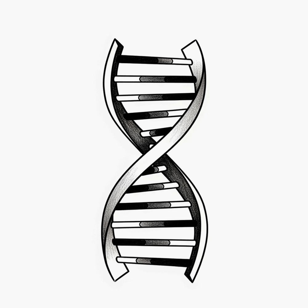
Paper (Nature, 2026):
doi.org/10.1038/s415...
29.01.2026 16:22 — 👍 0 🔁 0 💬 0 📌 0
Practical “what could change”:
This may help sort which non-coding DNA changes matter most, give better guesses for rare disease ‘unknown’ variants, guide which lab tests to run next, and support careful design of DNA or RNA-targeted edits.
29.01.2026 16:22 — 👍 0 🔁 0 💬 1 📌 0
Cancer example (TAL1):
Some leukemia mutations create new DNA switches that raise TAL1 activity. AlphaGenome predicted multi-signal changes (accessibility, histone marks, expression) consistent with known mechanisms.
29.01.2026 16:22 — 👍 0 🔁 0 💬 1 📌 0
TF binding:
Transcription factors are proteins that stick to DNA to help control genes. They tested DNA changes that affect this binding and found the model’s predicted direction and size of change often matched lab results.
29.01.2026 16:22 — 👍 0 🔁 0 💬 1 📌 0
Chromatin accessibility:
When DNA is more ‘open’, proteins can bind to it more easily and genes are often more active. They tested DNA changes known to affect openness and found the model’s predictions closely matched real lab data.
29.01.2026 16:22 — 👍 0 🔁 0 💬 1 📌 0
Poly(A) basics:
Many RNA messages have a stop signal near the end. DNA changes near this signal can change how long the RNA is and how stable it is. AlphaGenome picked up these effects best when the change was close to the stop site.
29.01.2026 16:22 — 👍 0 🔁 0 💬 1 📌 0