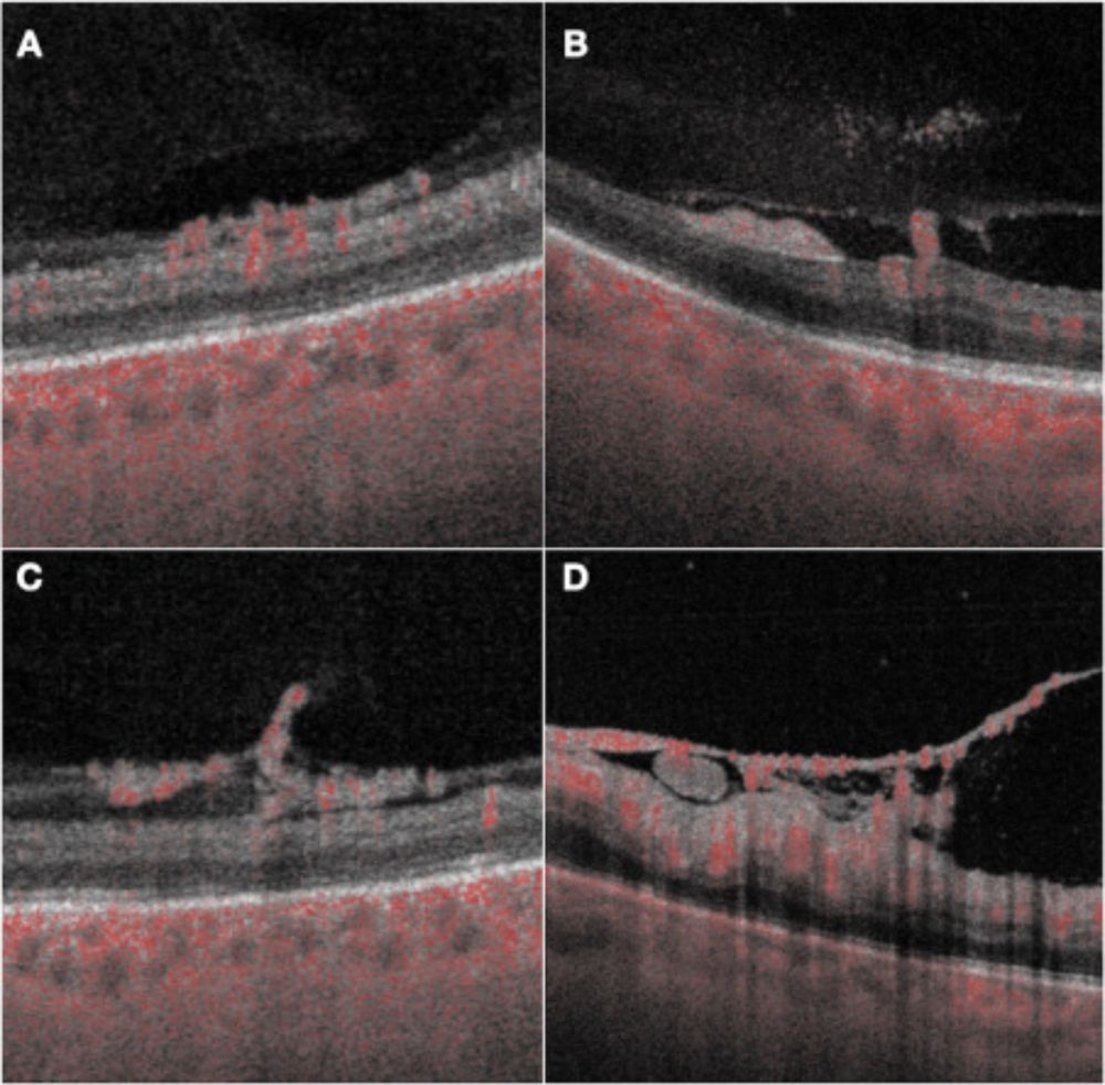
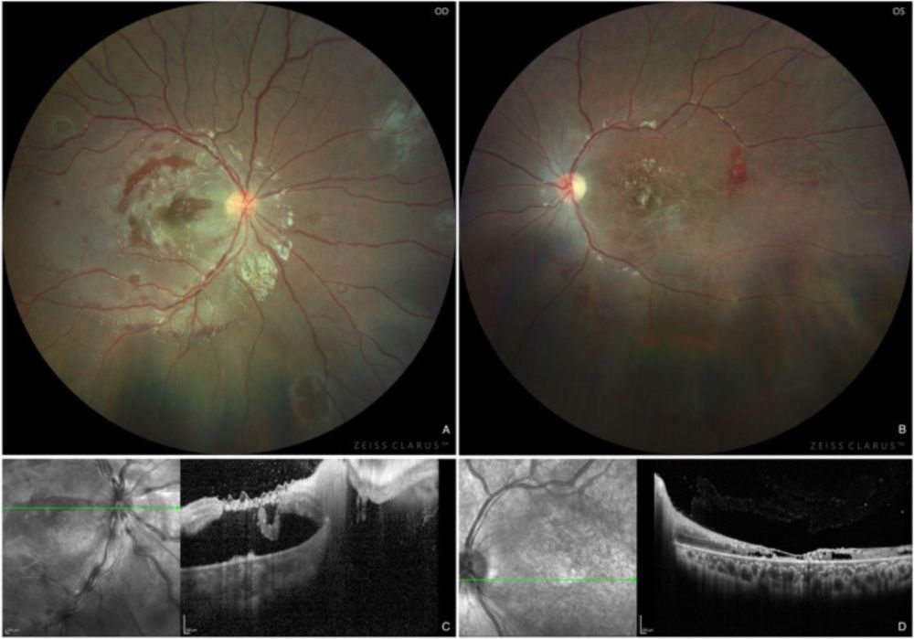
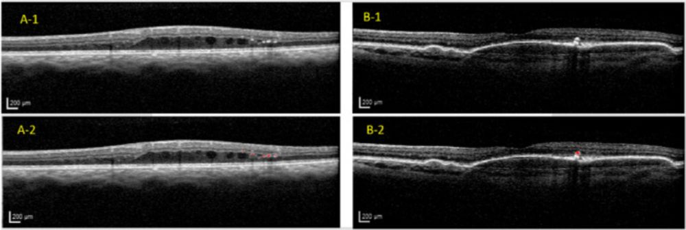
From a team in Italy, this study used multimodal imaging to analyze longitudinal changes of retinal neovessels in eyes with proliferative diabetic retinopathy after 3 monthly intravitreal injections of ranibizumab.
bit.ly/cjo_retinal-...
@cjo-jco.bsky.social
CJO is the official journal of the Canadian Ophthalmological Society. Publishing original, peer-reviewed articles on ophthalmology and vision science. linktr.ee/cjo_jco
From a team in Italy, this study used multimodal imaging to analyze longitudinal changes of retinal neovessels in eyes with proliferative diabetic retinopathy after 3 monthly intravitreal injections of ranibizumab.
bit.ly/cjo_retinal-...
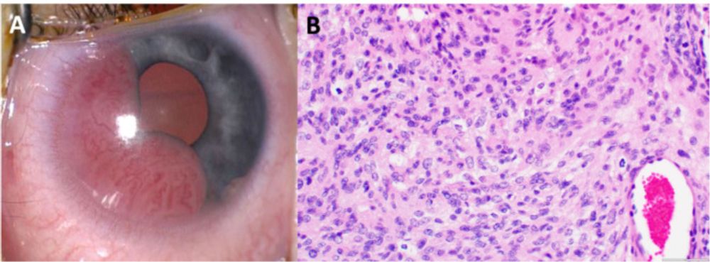
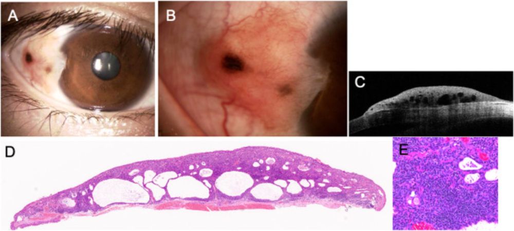
This unique case describes an iris mesenchymal tumour in an infant.
bit.ly/cjo_iris-mes...
#Ophthalmology
Cover of the August 2025 issue of the Canadian Journal of Ophthalmology (CJO). Cover is white with red and black text, and it features two reddish-orange fundus images on a black background in the centre-top.
The August CJO is now online! It features articles on translating ophthalmic medical jargon with AI, 24-hour intraocular pressure fluctuation in glaucoma patients, G-ROP versus WINROP for ROP screening, photo essays, research letters, the F.Y. Eye column, and much more.
bit.ly/cjo_60-4_Aug...
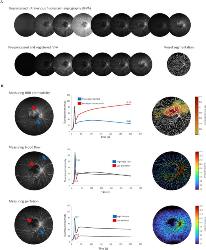
In this study, authors examined the association between quantitative vascular parameters extracted from intravenous fluorescein angiography and baseline clinical characteristics of patients with retinal vein occlusion
bit.ly/cjo_AI-extra...
#Ophthalmology #ClinicalResearch #ArtificialIntelligence
Subacute sclerosing panencephalitis (SSPE) is a chronic infection of the central nervous system by an altered form the measles virus, often presenting in young patients with a history of measles before the age of 2 years old.
bit.ly/cjo_subacute...
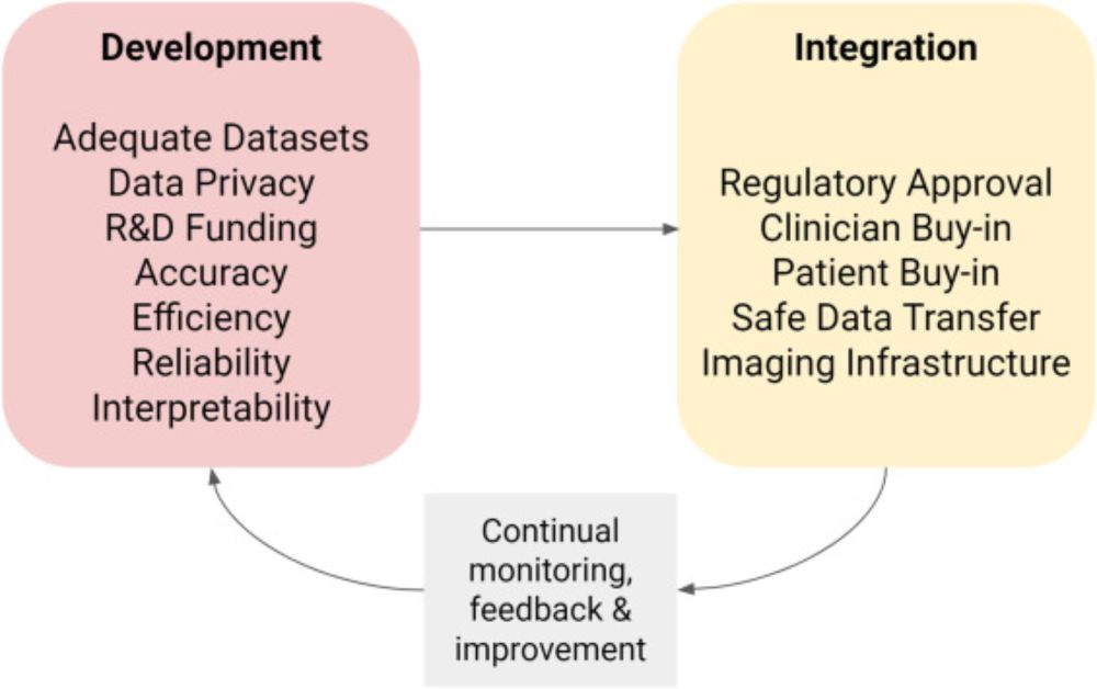
This review describes the current state of Canadian tele-ophthalmology services and AI developments in ophthalmology, and a pathway to integrate these technology-driven strategies in Canada.
bit.ly/cjo_AI-tele-...
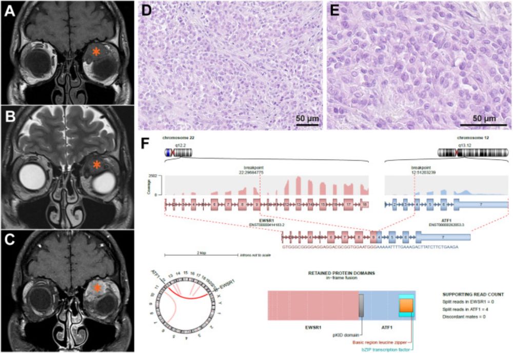
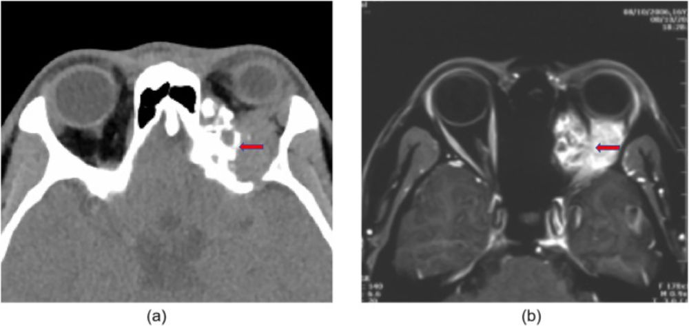
From a team in France, this study assessed the feasibility, accuracy, and safety of ultrasound-guided coaxial core-needle biopsy for histomolecular diagnosis of extra-ocular orbital soft tissue tumours as a minimally invasive alternative to surgical biopsy.
bit.ly/cjo_US-guide...
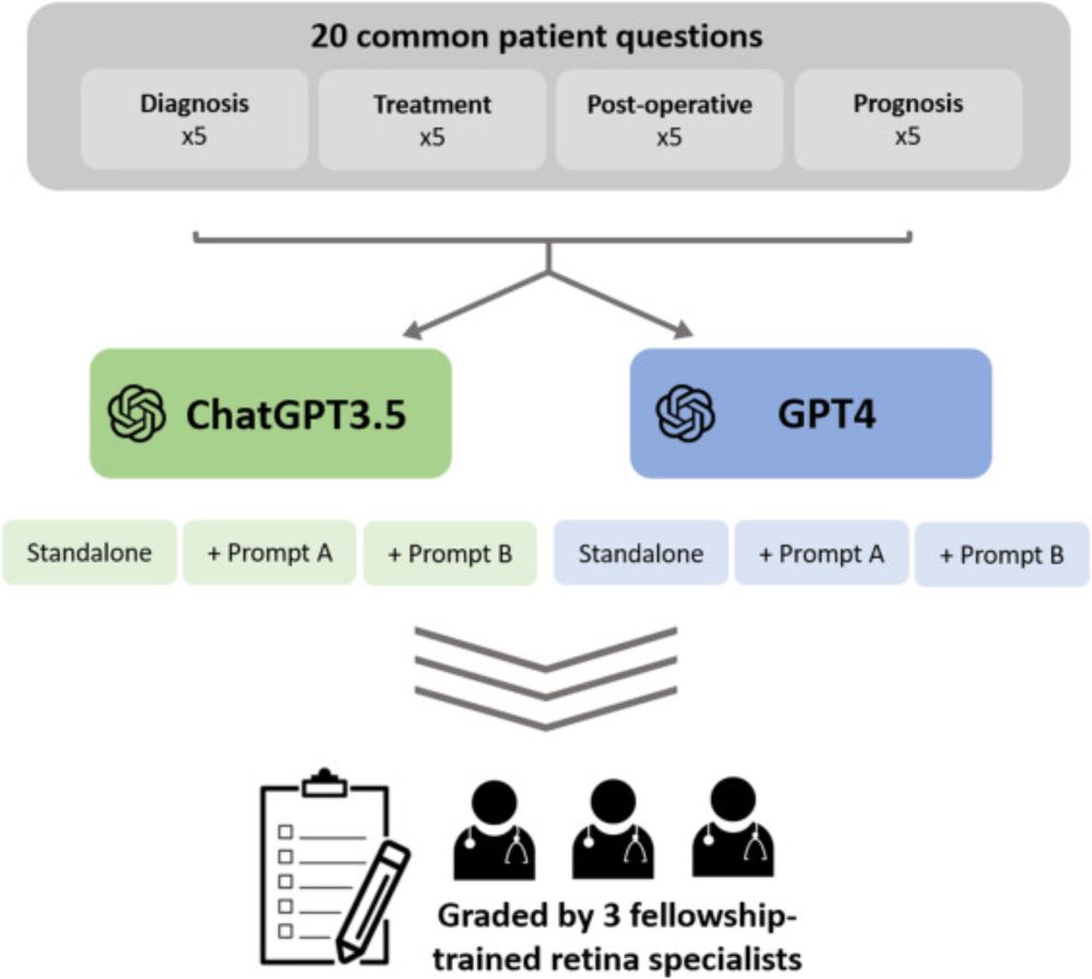
From a team at the University of British Columbia, this study assessed the effect of prompt engineering on the accuracy, comprehensiveness, readability, and empathy of ChatGPT-generated responses to patient questions about retinal diseases.
bit.ly/cjo_ChatGPT-...
VEXAS syndrome is a recently defined multiorgan autoinflammatory disease caused by somatic mutations in hematopoietic progenitor cells. It can present with a variety of symptoms, including inflammatory ocular changes, as seen in this case.
bit.ly/cjo_VEXAS-sy...
This study evaluated the prevalence of tamoxifen retinopathy in a major metropolitan area based on multimodal retinal imaging and determined whether the additional peripheral retina captured in ultra-widefield imaging aids in the diagnosis of tamoxifen retinopathy.
bit.ly/cjo_tamoxife...
From a team in China, this study analyzed the clinicopathological characteristics of primary orbital mesenchymal chondrosarcoma, a rare malignant tumour, and identified the risk factors influencing its prognosis.
bit.ly/cjo_orbital-...
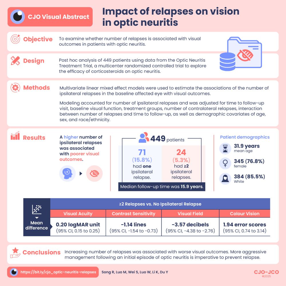
Visual abstract infographic presents a summary of the study, including the objective, study design, methods, results, and conclusions.
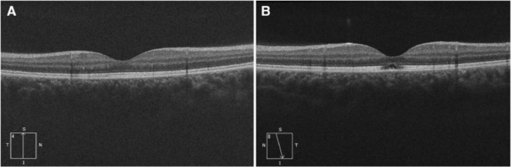
Each relapse in acute optic neuritis results in an approximate one-line decrease in Snellen chart vision. This study found that a higher number of ipsilateral relapses is independently associated with poorer visual outcomes.
bit.ly/cjo_optic-ne...
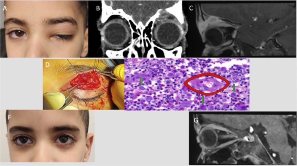
Pediatric orbital Langerhans cell histiocytosis is a benign disorder of dendritic cell origin with an annual incidence of 4−9 cases per million in children under the age of 15. This article describes the clinical, imaging characteristics, and treatment of this disorder.
bit.ly/cjo_Langerha...
Although immunotherapeutic agents show promise in treating recalcitrant malignancies, their adverse effects can be sight threatening. This article describes a unique case of bilateral corneal perforation secondary to immunotherapy for metastatic melanoma.
bit.ly/cjo_corneal-...
#OcularOncology

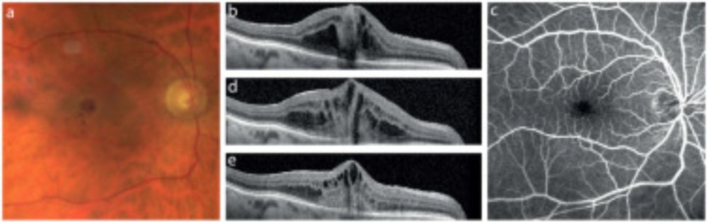
From a team in Austria, this study investigated the localization, distribution, and type of central microaneurysms and their relationship with retinal vascular alterations in patients with retinal vein occlusion.
bit.ly/cjo_microane...
#Ophthalmology #VisionScience #OcularImaging
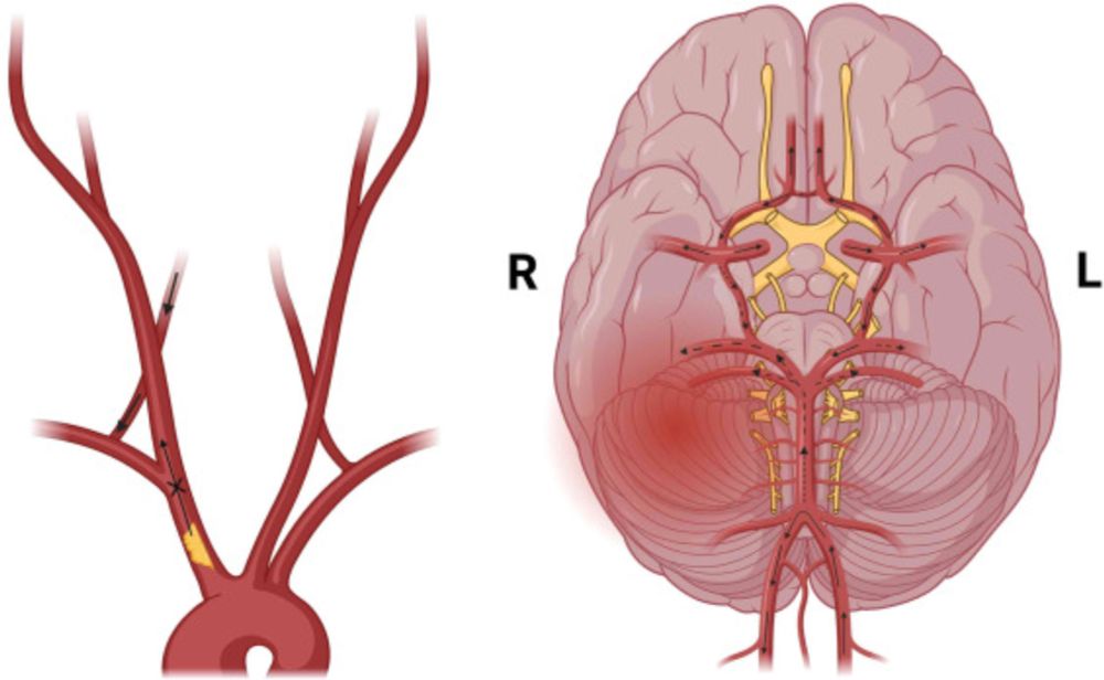
This unique case reports a reversal of cortical blindness following an urgent retrograde stenting procedure for complete innominate artery stenosis.
bit.ly/cjo_cortical...
In this study, authors assessed the prevalence of unintended placement of IOL haptics in the sulcus and its association with posterior capsular opacification in pseudophakic post-mortem eyes.
bit.ly/cjo_IOL-hapt...
#Ophthalmology #ClinicalResearch #VisionScience
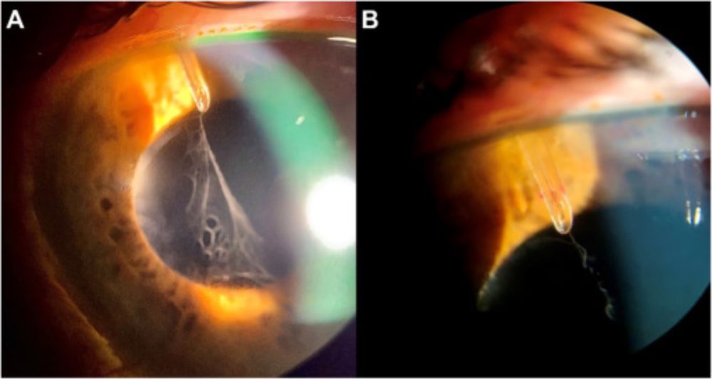
This article describes the first case of an early postoperative complication of PreserFlo microshunt, which was occlusion of the shunt with fibrin-like material, and the novel use of intraluminal Nd:YAG laser to treat it.
bit.ly/cjo_fibrin-o...

A woman in a white karate gi with a green belt strikes a punching stance in profile. One fist is held next to her face and the other fist is extended straight out in front of her. A grey, industrial-looking wall serves as the background.
In the United States alone, over 6 million people participate in martial arts, which are associated with injuries to the head, neck, face, and eyes. This study characterized the nature and incidence of martial arts-related eye injuries in the United States from 2012 to 2021.
bit.ly/cjo_seeing-t...
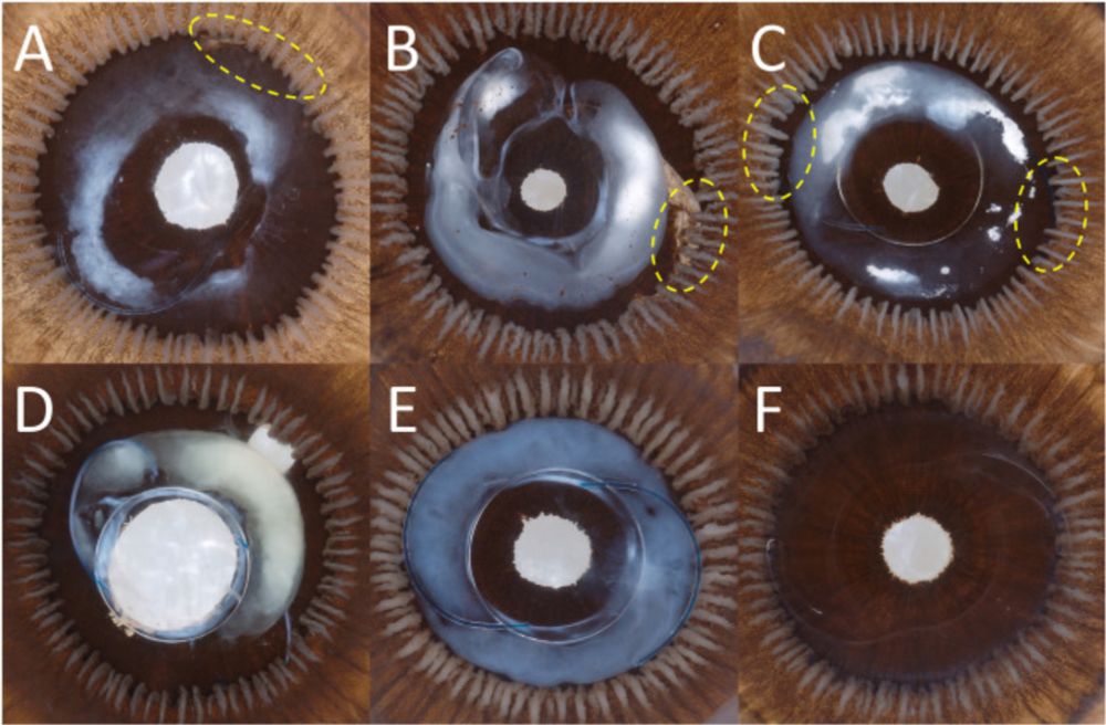
This article presents two unique cases of pterygium-like conjunctival nevus with multiple large pseudocysts within the lesion and analyzes the clinicopathological correlation.
bit.ly/cjo_pterygiu...
#Ophthalmology #OcularImaging
Cover of the June 2025 issue of the CJO. The background is white. The journal name, issue, and volume appear in red and black print at the top. An picture featuring two fundus images is in the centre, and the names of three articles are featured below the image.
The June CJO is now online! It features a review on integrating AI with tele-ophthalmology, articles on ocular injuries in martial arts, Langerhans cell histiocytosis of the orbit, visual recovery following pituitary adenoma surgery, the F.Y. Eye column, and more.
bit.ly/cjo_60-3_Jun...
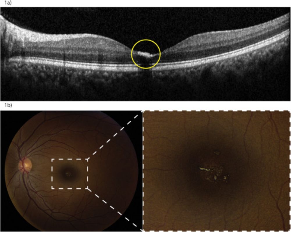
From a team at the Université de Montréal, this article reports on two cases of solar retinopathy following a total solar eclipse on April 8, 2024, which were identified and confirmed by retinal specialists using OCT scans.
bit.ly/cjo_solar-re...
#Ophthalmology
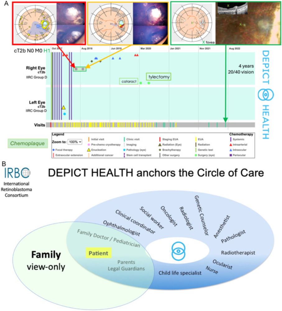
From a team at SickKids, this article describes novel methods to evaluate retinoblastoma research outcomes on the basis of treatment event and date, presently determined by retrospective eCancerCareRB data, and prospectively through DEPICT HEALTH on the cloud.
bit.ly/cjo_digital-...
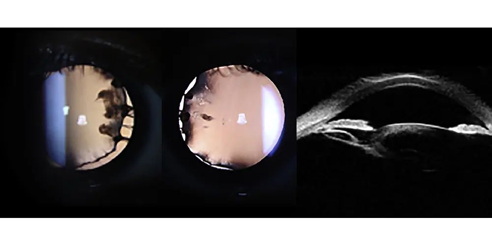
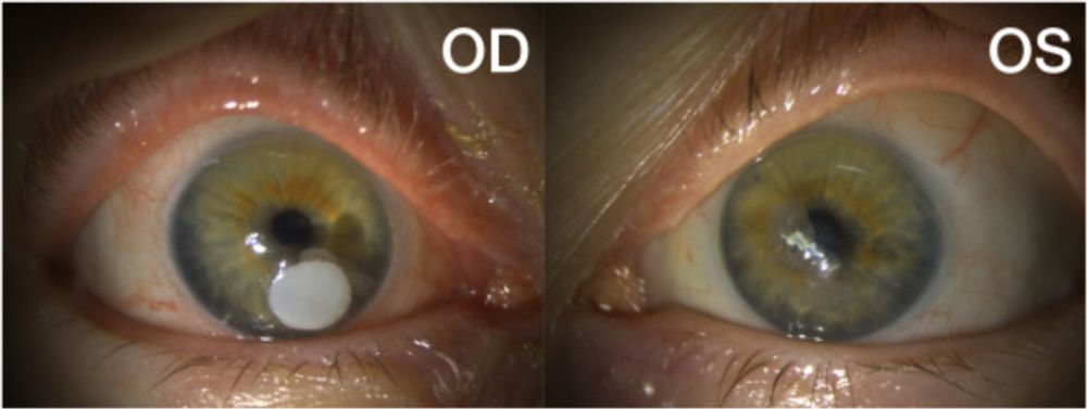
Photo essay from a teenager without surgical history who presented with decreased vision. Gene panel testing identified a heterozygous COL11A1 variant.
https://bit.ly/cjo_heterozygous-COL11A1-mutation
#Ophthalmology #COL11A1Mutation
Sympathetic ophthalmia is a rare bilateral granulomatous uveitis, usually from penetrating ocular trauma or intraocular surgery. This case highlights its rare development after plaque brachytherapy for uveal melanoma with extraocular extension.
bit.ly/cjo_sympathe...
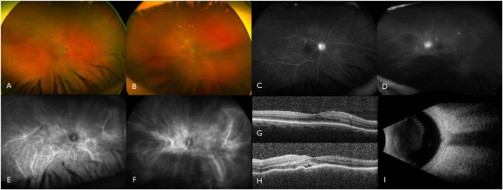
This study analyzed and compared spectral-domain OCT characteristics of intraretinal hyper-reflective foci in eyes with diabetic retinopathy versus eyes with age-related macular degeneration.
bit.ly/cjo_SD-OCT-c...
#Ophthalmology #OcularImaging
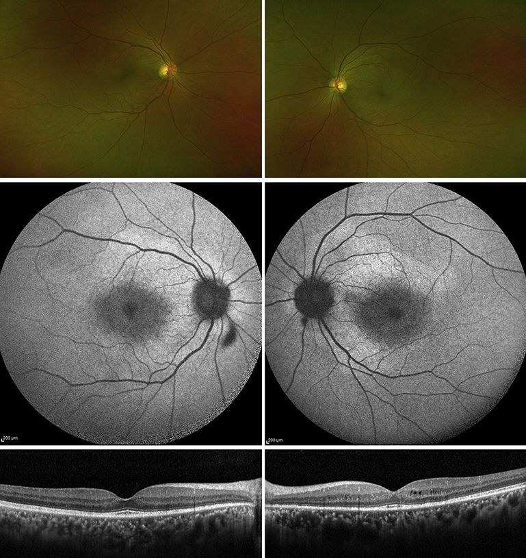
Multimodal imaging from male patient. Top images show wide-field colour fundus photos. Middle images show black and white fundus autofluorescence images. Bottom images show black and white spectral-domain optical coherent tomography.
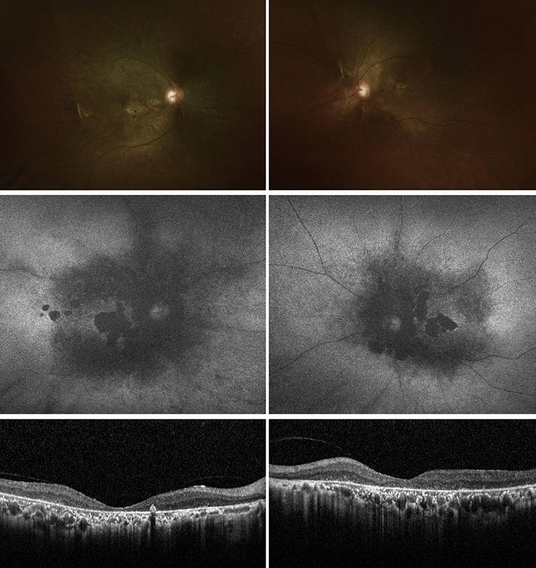
Multimodal imaging from female patient. Top images show wide-field colour fundus photos. Middle images show black and white fundus autofluorescence images. Bottom images show black and white spectral-domain optical coherent tomography.
Neuronal intranuclear inclusion disease is a complex neurodegenerative disorder characterized by eosinophilic intranuclear inclusions in the nervous system and visceral organ cells. This article highlights 2 cases from the same family.
bit.ly/cjo_neuronal...
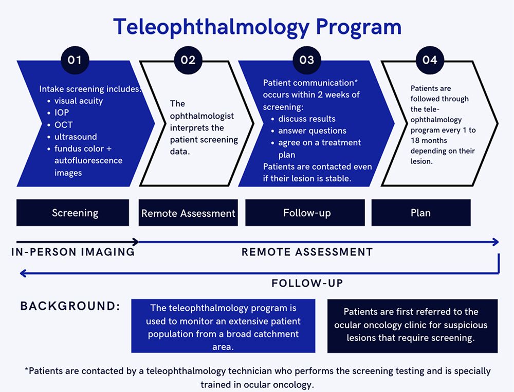
From a team in Alberta, this study evaluated patient satisfaction with a teleophthalmology program for ocular oncology that provides screening, remote assessment, care planning, and follow-up.
https://bit.ly/cjo_oncology-teleophthalmology
#Ophthalmology #ClinicalResearch
Slit lamp images showing chronic recurrent conjunctival erosions secondary to cabozantinib.
Cabozantinib is an oral tyrosine kinase receptor inhibitor used in the management of clear cell renal carcinoma. This case presents the first report of chronic recurrent conjunctival erosions secondary to cabozantinib.
bit.ly/cjo_conjunct...
#Ophthalmology
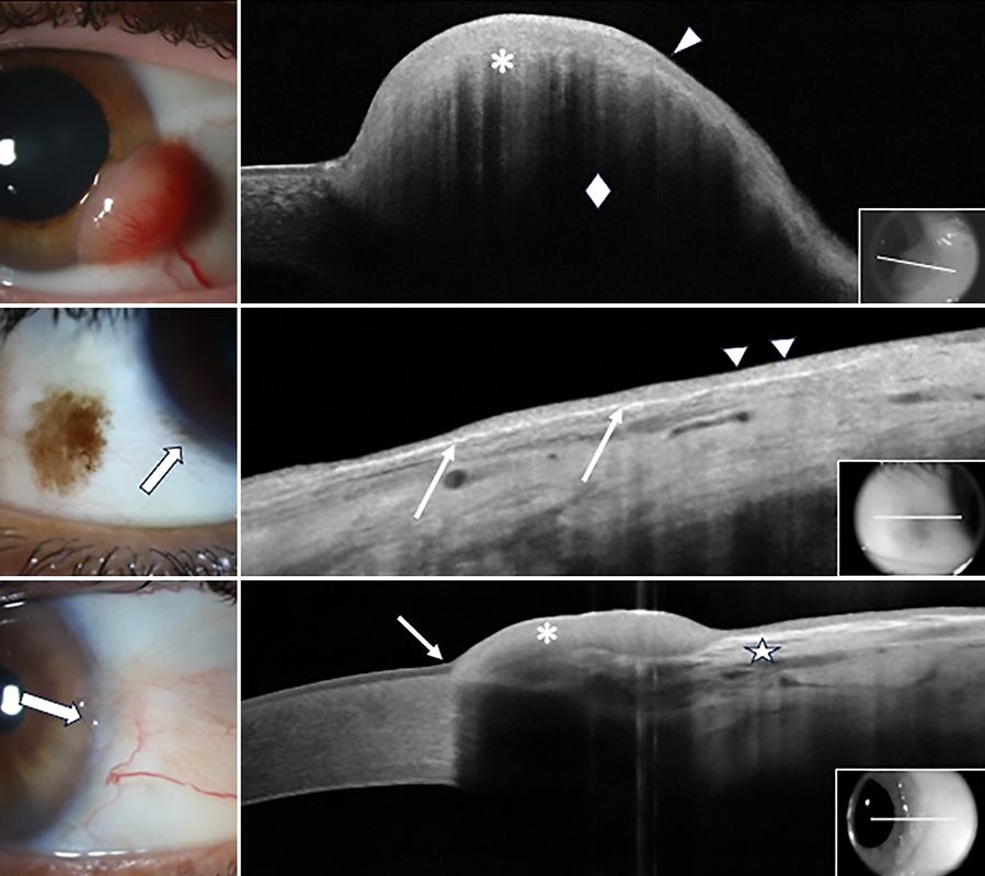
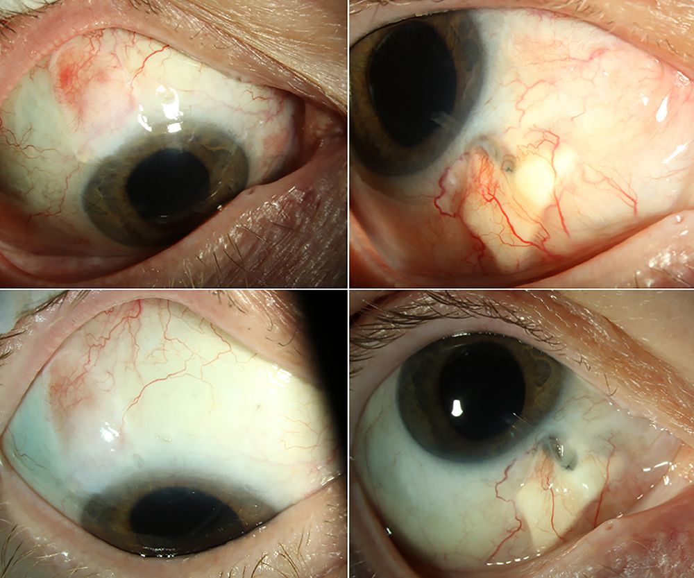
Three colour images showing eyes with different ocular surface lesions appear in a column on the left side of the image. Next to each is a corresponding black and white high-resolution optical coherence tomography image show the location of each lesion.
This review describes how high resolution OCT can be used to take “optical biopsies” that aid in the diagnosis, differentiation, and management of several benign and malignant ocular surface lesions.
bit.ly/cjo_OCT-ocul...
#Ophthalmology #ClinicalResearch