Axonal pathfinding of zebrafish retinal ganglion cells forms the optic nerve. Credit to Dr. Matthew Bostock @houartlab.bsky.social. #ZebrafishZunday 🧪
16.11.2025 20:30 — 👍 178 🔁 58 💬 4 📌 2
What a crazy cool paper! First author @pierreucla.bsky.social with a large crew knocked it out of the park. (GIF below from @the.3i.social LLS) Quantifying cell traction forces at the single-fiber scale in 3D: An approach based on deformable photopolymerized fiber arrays www.pnas.org/doi/10.1073/...
13.11.2025 18:21 — 👍 24 🔁 8 💬 0 📌 0

When the brain talks back to the eye
The state of our brain shapes what we see, but how early in the visual system does this start? This Primer explores a new PLOS Biology study which shows that brain state-dependent release of histamine...
When brain talks back to the eye "The state of our brain shapes what we see, but how early in the visual system does this start? A new study in PLOS Biology shows that brain state-dependent release of histamine modulates the very first stage of vision in the retina" journals.plos.org/plosbiology/...
05.11.2025 21:30 — 👍 14 🔁 7 💬 0 📌 1
This is such a cool paper! Congrats!
04.11.2025 11:22 — 👍 2 🔁 0 💬 0 📌 0

Retinal glia regulate development of the circadian photoentrainment circuit
Circadian photoentrainment depends on intrinsically photosensitive retinal ganglion cells (ipRGCs), which convey environmental light information to th…
Our latest paper is out! While the circadian photoentrainment circuit has been extensively studied, the mechanisms regulating its development remain poorly understood. Here we show that retinal Müller glia play a key role in this process. Check it out! www.sciencedirect.com/science/arti...
29.10.2025 14:27 — 👍 42 🔁 14 💬 4 📌 2
This is really cool work! Congrats, Olivier, and the whole team!
26.10.2025 06:59 — 👍 4 🔁 1 💬 0 📌 0
Extremely important K+ and Ca2+ indicator development work in these papers.
22.10.2025 17:35 — 👍 4 🔁 4 💬 0 📌 0

Subretinal Photovoltaic Implant to Restore Vision in Geographic Atrophy Due to AMD | NEJM
Geographic atrophy due to age-related macular degeneration (AMD) is the leading cause of irreversible blindness and affects more than 5 million persons worldwide. No therapies to restore vision in ...
Bigtime publication on retinal prosthesis
@science.xyz implanted 38 patients with macular degeneration with the PRIMA subretinal implant. Paired with camera-equipped glasses, the system improved central vision in 80% of participants, allowing them to read again
@maxhodak.bsky.social #neuroskyence
21.10.2025 19:03 — 👍 6 🔁 1 💬 0 📌 0
Science and our wider society is advanced through the practice of basic science.
Basic science is the engine that makes it all work.
Janni changed the world through her work in fruit fly embryonic development that taught us about evolution, transcription and cell fate during development.
20.10.2025 16:36 — 👍 21 🔁 5 💬 0 📌 0
YouTube video by arvoinfo
ARVO 2026 Annual Meeting
#ARVO2026 registration is open! Join @arvoinfo.bsky.social in Denver (May 3-7) for an immersion of cutting-edge new eye/#VisionScience under the theme, Achieving precision ophthalmology through innovative #VisionResearch. Sign up early and save: https://youtu.be/OSpr4hlp1OM
17.10.2025 13:00 — 👍 3 🔁 2 💬 0 📌 0
Brian, we are thrilled to have you here! Amazing, that you and other colleagues came all the way from the States😍
26.09.2025 21:29 — 👍 2 🔁 0 💬 1 📌 0

An image showing each of the short listed nominees for the Nature Inspiring Women in Science Award with the award logo at the top. "Meet the shortlists for the Inspiring Women in Science Award" is in the middle. The Estee Lauder logo is at the bottom.
We are excited to present the shortlists for the 2025 Nature Awards for Inspiring Women in Science. Congratulations to the thirteen excellent candidates who made it to this year’s shortlist. Read more about the candidates and learn more about the award: go.nature.com/46nlAWy #WomeninStem
22.09.2025 18:14 — 👍 31 🔁 15 💬 1 📌 1
Webvision – The Organization of the Retina and Visual System
We are pleased to announce that Webvision has been fully migrated to our new home at the University of Pittsburgh. www.webvision.pitt.edu
All traffic should not automatically be redirected from the old URLs, but please update your links.
Heads up: @pittophthalmology.bsky.social
19.09.2025 07:45 — 👍 17 🔁 6 💬 0 📌 1

Vision scientist Alecia Gross at the lectern talking about retinal degeneration
It’s @alecia144g.bsky.social talking about her work in retinal degeneration at #RD2025
19.09.2025 09:49 — 👍 15 🔁 2 💬 0 📌 0
Beautiful image! I feel like I recognize this from our amazing tour in Prague😎
19.09.2025 23:21 — 👍 1 🔁 0 💬 0 📌 0

It’s #NationalPostdocAppreciationWeek! Postdocs are at the heart of discovery and innovation, pushing the boundaries of research and making a lasting impact on science and society. Join us in celebrating their invaluable contributions! #NPAW #Postdocs #FASEB
15.09.2025 19:15 — 👍 3 🔁 1 💬 0 📌 0
Kun on tässä seurannut tutkimusrahoitukseen liittyvää ns. keskustelua, niin ei voi välttyä ajatukselta, että olemme menossa kohti ankeita aikoja. Ymmärrys rahoituksen roolista tutkimukselle, korkeakoulutukselle ja maan tulevaisuudelle on monilta täysin hukassa. Pieni ketju aiheesta. 1/18
07.09.2025 15:59 — 👍 114 🔁 22 💬 1 📌 1
Come to the second edition of the world wide sodium channel conference! Submit an abstract by September 26.
06.09.2025 04:56 — 👍 5 🔁 3 💬 0 📌 0
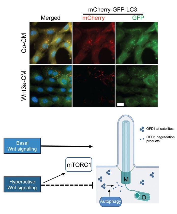
Top: Inhibition of mTOR signaling rescues cilia formation in Wnt activated cells. RPE1 cells stably expressing mCherry-GFP-LC3 were treated with Co-CM and Wnt3a-CM and fixed 16 h after serum starvation for direct fluorescence analysis of mCherry and GFP signals. Representative images show GFP+mCherry+ foci (yellow dots, autophagosomes). Bottom: Model of how Wnt signaling affects ciliogenesis. Basal Wnt signaling promotes cilia formation, whereas Wnt hyperactivation prior to ciliogenesis delays this process by increasing mTORC1 activity and impairing the removal of OFD1 from centriolar satellites. M, mother centriole; D, daughter centriole.
The #PrimaryCilium regulates several signaling pathways, but what role do these pathways play in #cilium formation? This study shows that modulating Wnt & mTOR signaling affects #ciliogenesis in human retinal epithelial cells @plosbiology.org 🧪 plos.io/45Yg5gB
03.09.2025 17:01 — 👍 7 🔁 2 💬 0 📌 0
LinkedIn
This link will take you to a page that’s not on LinkedIn
🌍✨ Call for Nominations: ISER International Prizes ✨🌍
@iser.bsky.social is now accepting nominations for its prestigious international prizes! 🏆
📅 Deadline: September 8, 2025
🔗 lnkd.in/eUQMFGsr
Don’t miss the chance to recognize excellence in our field — submit your nomination today!
21.08.2025 14:49 — 👍 2 🔁 2 💬 0 📌 0

90-vuotias | Seppo Lindblom tietää, miten talous saadaan kasvuun: satsaamalla tieteeseen ja tutkimukseen
”Vahva tiedepolitiikka on ainoa tepsivä tuottavuustemppu, joka lopulta auttaa velka- ja puolustuskuluissa”, Seppo Lindblom sanoo.
Viisautta ja sivistystä huokuu tästä synttärihaastattelusta. Seppo Lindblom puolustaa velanhoidon ja puolustuksen pinteeseen jäänyttä hyvinvointia. Ja tutkimusta!
Paras on silti tämä:
”-Mitä sanoisit 20-vuotiaalle itsellesi?
-En kaksikymppisenä kuuntelisi näin vanhojen miesten neuvoja.”
08.08.2025 16:34 — 👍 47 🔁 11 💬 0 📌 0
Extremely interesting work. Congrats to the team!
05.08.2025 05:50 — 👍 2 🔁 0 💬 0 📌 0
Sounds fabulous indeed! I’m sure that the course will be extraordinarily good. My student was admitted but couldn’t come because of the visa interview freeze, which was really unfortunate. Hopefully the course will be organized next year as well.
03.08.2025 05:31 — 👍 0 🔁 0 💬 0 📌 0

Generated image of cross section of a yellow channel in a plasma membrane
You still got time!
🚨Call for papers - Ion channels and channelopathies🚨
Deadline: 27 October 2025 #BMCBiology
Guest Editors:
Zhuo Huang, Peking University
Soile Nymark @snymark.bsky.social, Tampere University
www.biomedcentral.com/collections/...
#ImmunoSky #NeuroSky #calcium 🧪
17.07.2025 19:38 — 👍 1 🔁 1 💬 0 📌 0
Projection Targeting with Phototagging to Study the Structure and Function of Retinal Ganglion Cells
Visual information from the retina is sent to diverse targets throughout the brain by different retinal ganglion cells (RGCs). Much of our knowledge about the different RGC types and how they are routed to these brain targets is based on mice, largely due to the extensive library of genetically modified mouse lines. To alleviate the need for using genetically modified animal models for studying retinal projections, we developed a high-throughput approach called projection targeting with phototagging that combines retrograde viral labeling, optogenetic identification, functional characterization using multi-electrode arrays, and morphological analysis. This method enables the simultaneous investigation of projections, physiology, and structure-function relationships across dozens to hundreds of cells in a single experiment. We validated this method in rats by targeting RGCs projecting to the superior colliculus, revealing multiple functionally defined cell types that align with prior studies in mice. By integrating established techniques into a scalable workflow, this framework enables comparative investigations of visual circuits across species, expanding beyond genetically tractable models. Motivation Visual information from the retina is distributed to diverse targets throughout the brain. Much of our knowledge about how visual information is processed and routed to these brain targets is based on mice because of the large library of genetically modified mouse lines. For most other species, such libraries are not available. Therefore, we were motivated to develop an approach for characterizing diverse retinal projections into the brain that can be applied to other species. We aimed to achieve projection targeting with retrograde viral vectors, followed by identification of circuit-specific retinal ganglion cells (RGCs) with optogenetics, functional characterization with multi-electrode array (MEA) recordings, and morphological description with in situ and confocal microscopy. The resulting high-throughput approach permits functional and morphological investigation of dozens to hundreds of cells in individual experiments. By replacing the need for genetically modified animals with a suite of standard techniques, this approach should improve cross-species comparisons of the initial stages of visual processing. Summary Understanding the structure-function relationships across neurons is challenging, particularly when circuits are composed of dozens of distinct cell types. We refined an approach, called projection targeting with phototagging, that allows simultaneous elucidation of the projections, morphology, and visual response properties of diverse RGC types in the mammalian retina. The approach combines retrograde virally mediated phototagging of RGCs, microscopy, and large-scale MEA measurements. Importantly, the approach does not rely on transgenic animals and thus is generalizable across species. We validated this approach in rats by targeting retinal projections to the superior colliculus (SC). We showed that multiple RGC types project to the SC and that these results in rats align well with prior findings from transgenic mouse studies. ![Figure][1]</img> Highlights ### Competing Interest Statement K.R. Is a coauthor on a patent for AAV2retro (Application No. 62/350,361 filed June 15, 2016, and U.S. Application No. 62/404,585 filed October 5, 2016). * AAV : adeno-associated virus cMRF : central mesencephalic reticular formation CRF : contrast response functions DpG : deep gray layer of the superior colliculus DpWh : deep white layer of the superior colliculus EI : electrical image GCL : ganglion cell layer IC : inferior colliculus INL : inner nuclear layer InG : intermediate gray layer of the superior colliculus IPL : Inner Plexiform Layer InWh : intermediate white layer of the superior colliculus ISI : inter-spike interval distribution MEA : multi-electrode array MGN : medial geniculate nucleus of the thalamus OT : nucleus of the optic tract Op : optic nerve layer of the superior colliculus OSI : orientation-selective index PAG : periaqueductal gray PC : posterior commissure POD : post-operative day PT : pretectum ReaChR : red-shifted channelrhodopsin RF : receptive field RGC : retinal ganglion cell SRF : spatial receptive field SuG : superficial gray layer of the superior colliculus TRF : temporal receptive field Zo : zonal layer of the superior colliculus Duke Institute for Brain Sciences (DIBS) Incubator Award NIH R01 EY034004 K99 EY032119 NIH P30 EY005722 Core Grant for Vision Research at Duke University NIH P30 EY000331 for Vision Research and an Unrestricted Grant from Research to Prevent Blindness at UCLA [1]: pending:yes
Awesome work by @gregdfield.bsky.social et al. www.biorxiv.org/content/10.1... Projection Targeting with Phototagging to Study the Structure and Function of Retinal Ganglion Cells | bioRxiv
08.07.2025 06:47 — 👍 5 🔁 1 💬 0 📌 0
Tributes paid to pioneering eye researcher Professor Pete Coffey
Tributes have been paid to one of the world’s top eye researchers, Professor Pete Coffey, who has sadly passed away after a long illness.
It’s incredibly hard to express what Professor Pete Coffey meant to so many of us. The Macular Society has written a beautiful tribute to Pete and I'm honoured to have contributed a few words.
Thank you, my friend, for so many happy days.
www.macularsociety.org/about/media/...
07.07.2025 15:51 — 👍 11 🔁 6 💬 3 📌 1
Science Events helps you organise a scientific conference or workshop. Use our platform to organise your next event! Visit our platform at https://www.sci.events
Associate Professor | UNC Chapel Hill, Pharmacology Department | Lineberger Comprehensive Cancer Center | Research in Endocrinology, Epigenetics, & Wnt signaling| Views my own. https://www.med.unc.edu/pharm/pruittlab/team/
Global Services Manager @ Hyland | Passionate about services, healthcare, digital health, AI & cloud | BSc, PRINCE2, MSP, L6σ Green Belt | Opinions are my own | https://linktr.ee/paulcochrane
Papers is an award-winning reference manager that improves how you discover, annotate, collaborate and cite research.
Sign up for a 30-day free trial: linktr.ee/get_papers
NIH T32 Postdoctoral Fellow in Weissleder Lab at CSB, MGH, @harvardmed.bsky.social, PhD from Rotello Group, UMass Amherst
Developing advanced imaging techniques to study how the neurons in the retina talk to each other. PhD student at @sln.icfo.eu
Cell aims to publish the most exciting and provocative research in biology. Posts by Scientific Editors on the Cell editorial team. See the latest papers at https://www.cell.com/cell/
An open-access @natureportfolio.bsky.social journal publishing high quality primary research, reviews and commentary in all areas of the biological sciences since 2018.
https://www.nature.com/commsbio/
An open access @NaturePortfolio journal publishing high-quality primary research articles, reviews and commentary in all areas of the chemical sciences.
nature.com/commschem/
An open access @natureportfolio.bsky.social journal publishing high quality primary research, reviews, and commentary in Earth, environmental, and planetary science.
nature.com/commsenv/
The multi-disciplinary engineering journal from the Nature Portfolio. Fully open access. You can read all our content here for free: https://www.nature.com/commseng/
An open access journal from Nature Portfolio publishing important advances in all areas of materials science.
A selective open access journal from @NaturePortfolio publishing research, reviews & commentary across clinical, translational & public health research fields. nature.com/commsmed
Communications Physics is an open access journal from Nature Portfolio publishing high-quality research, reviews and commentary in all areas of the physical sciences.
https://www.nature.com/commsphys/
Communications Psychology is a selective, peer reviewed, open access journal in the @natureportfolio.bsky.social, publishing high-quality research, reviews and commentary across psychology.
https://www.nature.com/commspsychol/
A Nature journal dedicated to presenting the very best research across the disciplines of astronomy, astrophysics, cosmology and planetary science.📡
www.nature.com/natastron
Publishing the best of biotech science and business. Find us on Twitter, Facebook & Instagram. Part of @natureportfolio.nature.com.
Publishing the latest advances across all areas of cancer research and oncology. Part of @natureportfolio.nature.com
🌐 www.nature.com/natcancer/
📍New York, London, Berlin and Heidelberg
Nature Catalysis publishes high quality work across all areas of catalysis, including both fundamental and applied studies.
https://www.nature.com/natcatal/
A journal dedicated to publishing the latest advances across all areas of cell biology. Part of @natureportfolio.nature.com
nature.com/ncb/index.html