sure
05.10.2025 16:30 — 👍 0 🔁 0 💬 0 📌 0


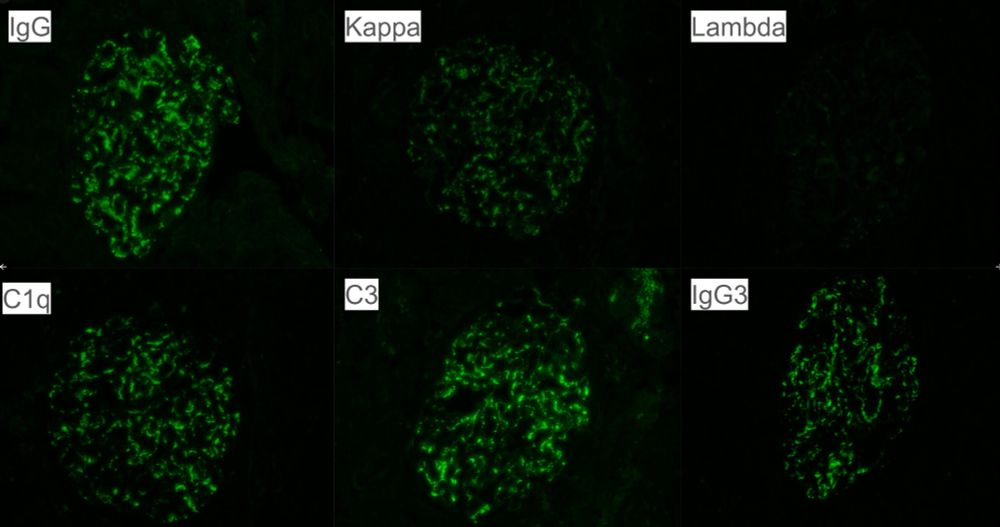
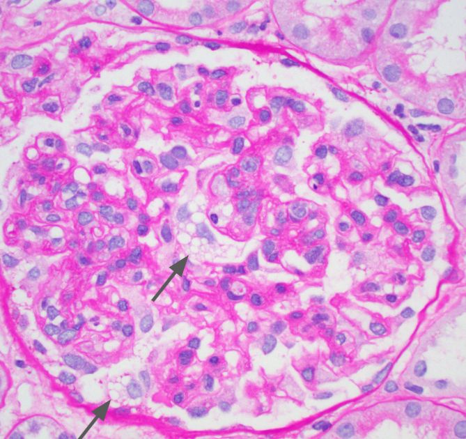
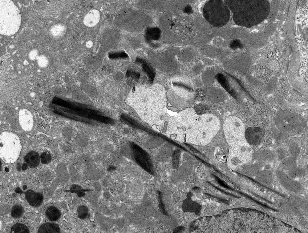
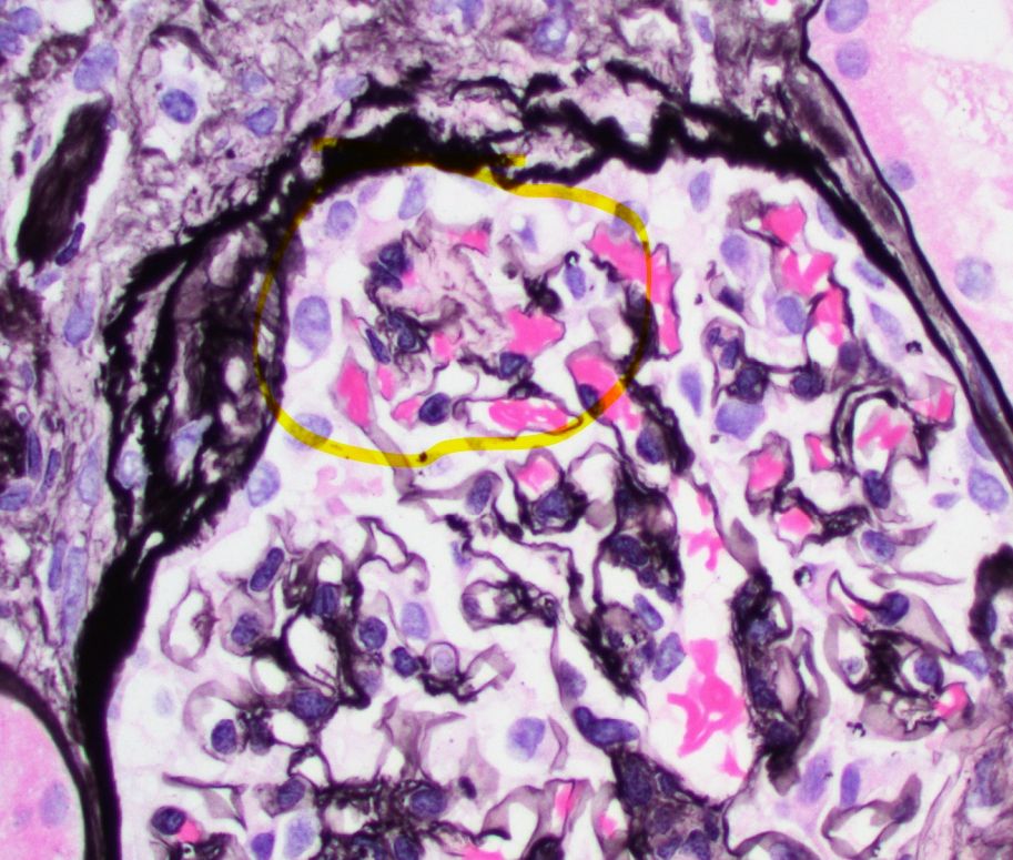
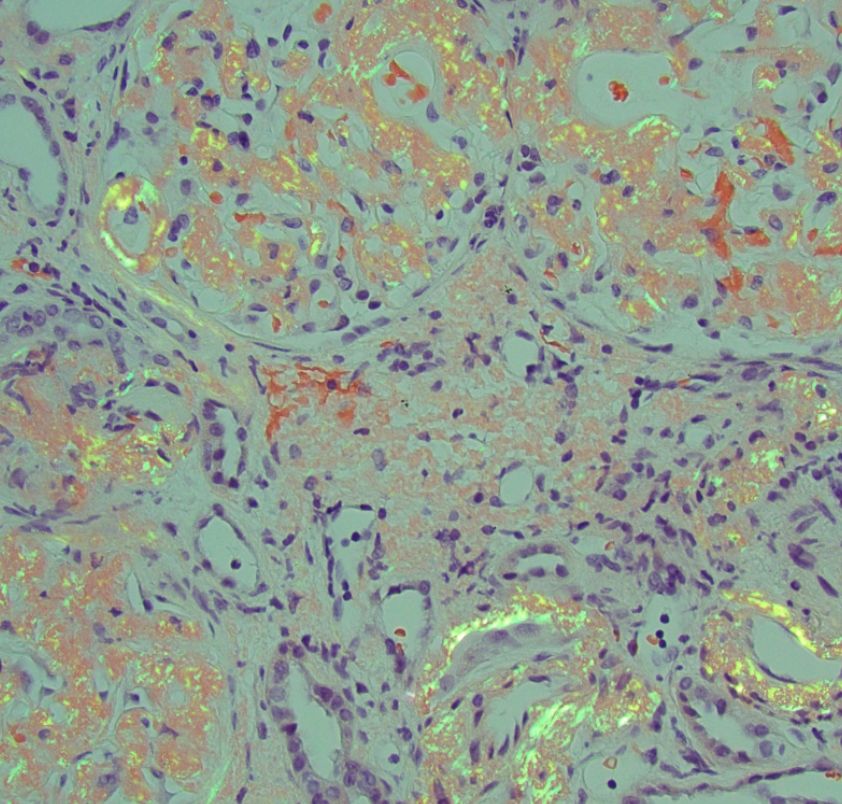
Heavy Chains, Heavy Consequences: A Case of Concomitant Heavy Chain Amyloidosis and Heavy Chain Deposition Disease. Fibrils and powdery deposits. #renalpath #nephsky #pathsky www.sciencedirect.com/science/arti...
17.09.2025 18:13 — 👍 7 🔁 5 💬 1 📌 0
🔬 We are now recruiting a Renal Pathologist! #pathology #path2path #PathSky #renalpath
Join our team today! ➨ bit.ly/46CqsrU
17.09.2025 16:33 — 👍 2 🔁 1 💬 0 📌 0

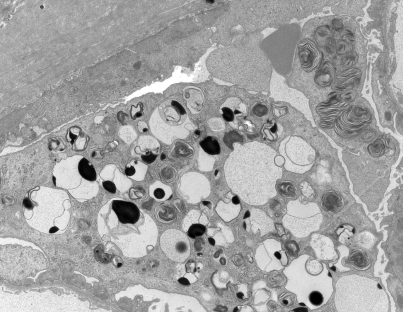
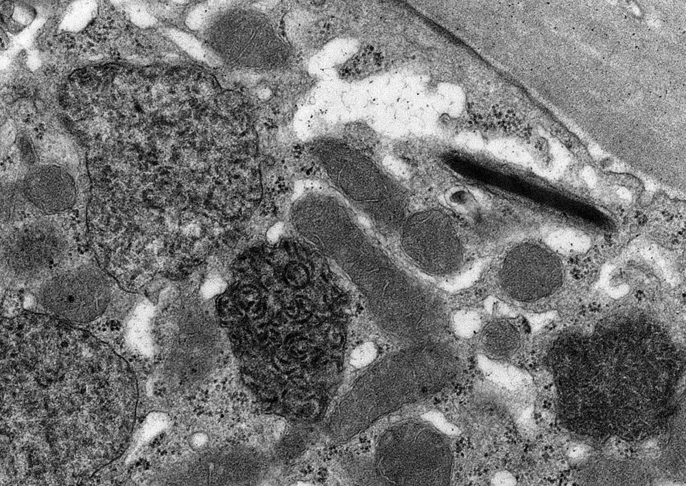
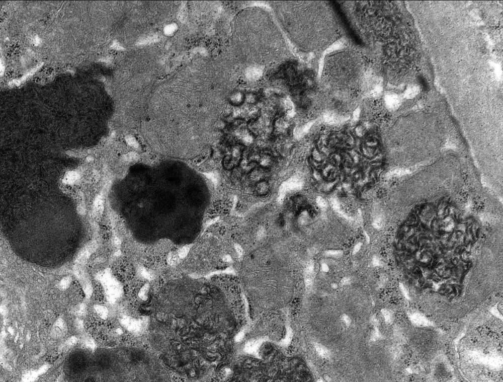
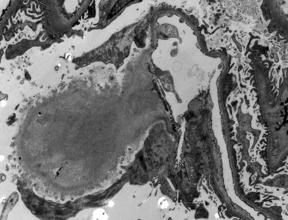
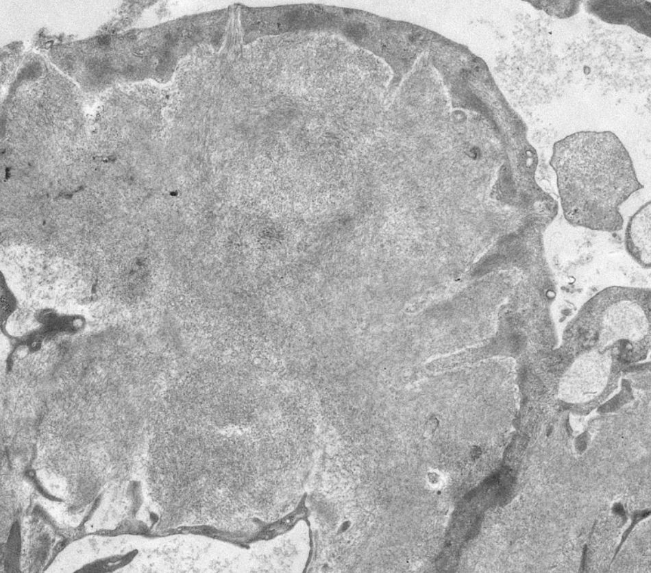
Also some rare myelin figures in a podocyte, because why not. #renalpath #nephsky #pathsky
11.09.2025 17:44 — 👍 6 🔁 0 💬 0 📌 0

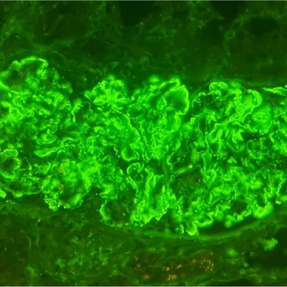
toluidine blue stained section was also quite pleasing. #renalpath #pathsky #nephsky
28.08.2025 22:06 — 👍 3 🔁 0 💬 0 📌 0


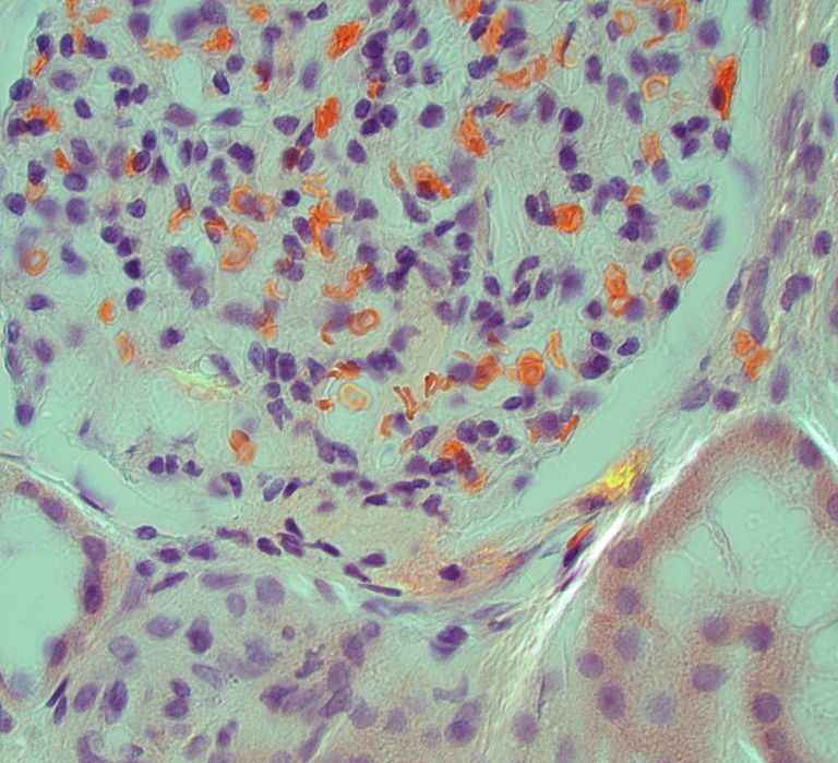
Nice example of urate crystals in gouty tophi involving the medulla. Polarizable crystals can be see using unstained sections from the IF tissue. #renalpath #pathsky #nephsky
28.08.2025 19:04 — 👍 10 🔁 3 💬 1 📌 0

ANCA GN Working Group Meeting
More info for EUVAS Florence Working Group - Symposium on ANCA GN Classification-Scoring Systems, click link
👉https://vasculitis.org/
Register: app.donorfy.com/form/ZU81NH9...
@brixsilke.bsky.social & Ingeborg Bajema #renalpath #vasculitis #pathsky #nephsky
18.08.2025 22:12 — 👍 4 🔁 4 💬 0 📌 0
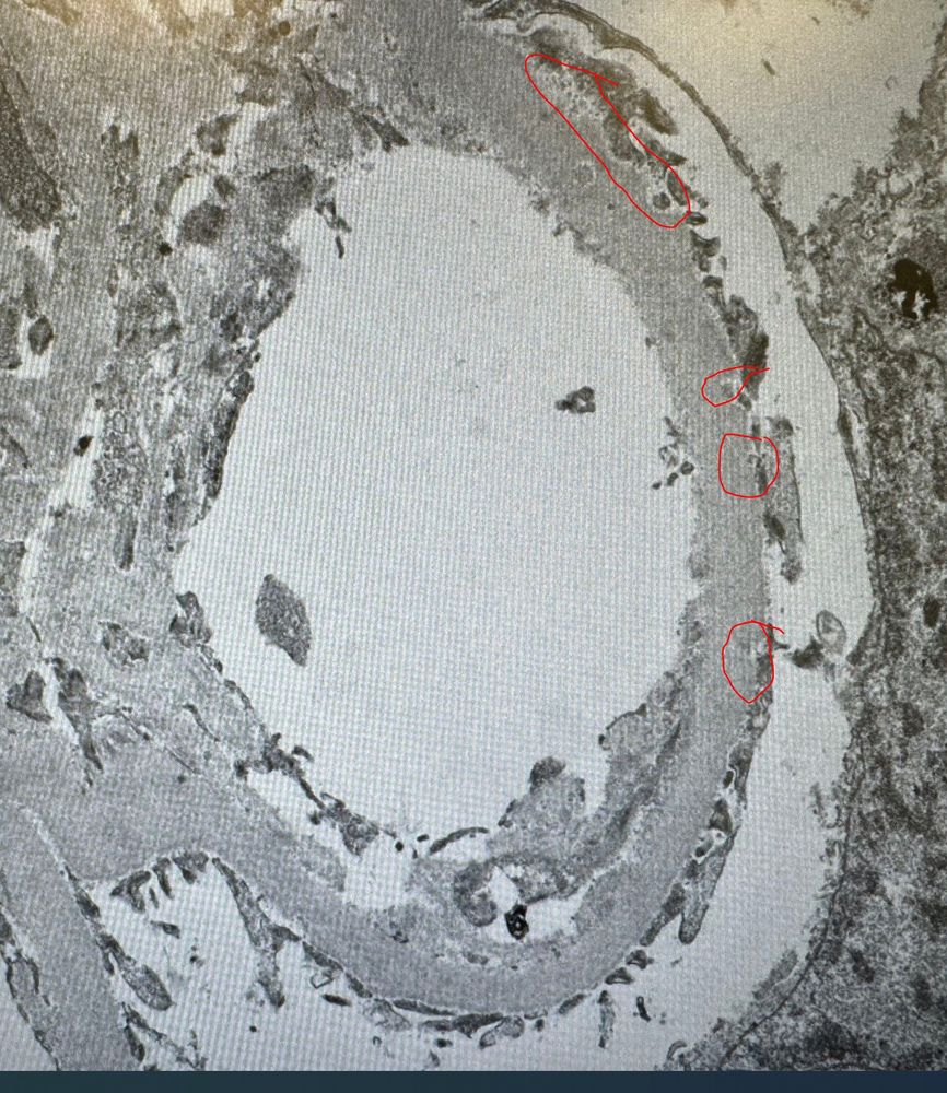
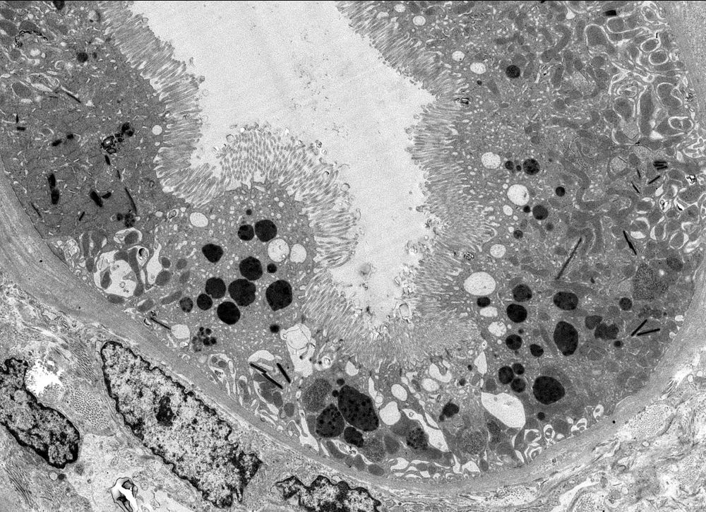
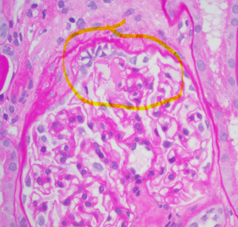
these are the areas I am referring to in the EM in one image. Not super specific, but could be the correlate for your IF staining.
10.08.2025 20:15 — 👍 2 🔁 0 💬 0 📌 0
or perhaps secondary overload injury from an original membranous nephropathy that has long resolved with just some IC staining. This is the best way I can put this together, but just my opinion. I agree this is not a typical case.
10.08.2025 20:13 — 👍 1 🔁 0 💬 1 📌 0
Given the GBM thickening and maladpative glomerular injury(hypertrophy), the IC staining may be incidental at this time and the patient's dx is mostly overload injury. Is the patient obese, have diabetes, HTN, smoking, MPN, low bithweight, etc.?
10.08.2025 20:10 — 👍 1 🔁 0 💬 1 📌 0
despite being much weak in reality. The GBM in the EM images do seem to show some undulation of the subepithelial surface which may be a subtle clue to some small mostly resorbed subepithelial deposits. So we may be seeing minimal residual subepithelial deposits from prior disease.
10.08.2025 20:10 — 👍 1 🔁 0 💬 1 📌 0
GBMs looks thick, you also mention glomerular hypertrophy. Wonder if some of the staining you see in the capillary loops is just "pseudolinear" IgG as we see in diabetic glomerulopathy and other metabolic dx associated glomerulopathy. Perhaps the small granular deposits are being enhanced here.
10.08.2025 20:10 — 👍 1 🔁 0 💬 1 📌 0
Can you post some of the EM images. Did you have conventional EM not just re-processing on the frozen?
09.08.2025 18:54 — 👍 0 🔁 0 💬 1 📌 0
"stage 0" membranous nephopathy can occur. Typically this seen early in recurrent MN in transplants. but I am very surprised by the EM discrepancy in this case. Can you clarify the clinical situation for the biopsy?
09.08.2025 18:52 — 👍 3 🔁 0 💬 0 📌 0
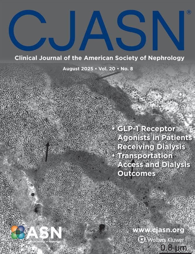
The August issue of CJASN is now available online! Topics covered this month include:
- GLP-1 receptor antagonists in patients receiving dialysis
- Transportation access and dialysis outcomes
- and more
Check out the issue here: journals.lww.com/cja...
#ASNCJASN
Cover by @JZRenalpath et al.
08.08.2025 15:00 — 👍 1 🔁 1 💬 0 📌 0

🔬 We are now recruiting a Renal Pathologist! #pathology #path2path #PathSky #renalpath
Join our team today! ➨ bit.ly/4ll8K09
07.08.2025 19:16 — 👍 3 🔁 3 💬 0 📌 0
Thank you. I am fortunate to have an excellent histology lab. The staining is mostly done on the grid (Uranyl acetate/ lead citrate). Osmium tetroxide is used in block preparation which also provides some contrast.
26.07.2025 14:59 — 👍 1 🔁 0 💬 0 📌 0
College of American Pathologists: The leading organization of board-certified pathologists. #pathologists #Pathsky 🔬
BlueSky account of the Renal Pathology Society (RPS) #renalpath
https://www.renalpathsoc.org/
Reader in Renal Pathology Imperial College #renalpath#kidneypath
#Nephrology Division, University of California, San Francisco | At the forefront of research, education, and patient care in #Kidneydisease
Renal pathologist @CedarsSinai
#renalpath #kidneydisease
opinions are my own
Chief UTSW Nephrology | Harvard, VUMedicine & BIDMC alum | ASNKidney Secretary & Exec Councilor | Chair NIH PBKD
#NephSky #MedSky
https://profiles.utsouthwestern.edu/profile/202716/samir-m-parikh.html
Creating a 🌎 without #kidneydiseases. #KidneyWk
Oregon Health and Science University Division of Nephrology and Hypertension. Providing comprehensive care and educating nephrologists for the future in PDX.
https://bit.ly/2mhCY9y
Nephrologist @StanfordNeph, Chair @HDAE_Official, Director Social Media @HemodialysisInt, Defender of the 4 nephrons.
Academic Nephrologist|Director @GlomCon fellowship|Director👩🏼⚕️Glomerular Disease & PKD Clinics👠📟
Nephrology Dialysis Transplantation (NDT) is the leading nephrology journal in Europe. NDT is an official journal of the European Renal Association and is published by Oxford University Press.
Global Science: Local Change. Advancing kidney health worldwide through evidence-based guidelines, collaboration, and innovation. Follow us for the latest updates and resources from KDIGO.
The Indian Society of Nephrology was born when 10 like-minded and strong-willed individuals sat together on a balmy day in Mumbai (then Bombay) in 1970.
ISNCON 2025 is in Lucknow. Register NOW
Website: www.isn-india.org
Northwestern's Division of Nephrology & the Northwestern University George M. O'Brien Kidney Resource Center
#NephMadness 2025 is live on the #AJKDBlog: https://bit.ly/WelcomeNM25 | Organized by @ajkd.bsky.social
Publishing reviews and commentaries across the field of nephrology. Posts are from the editors.
www.nature.com/nrneph/
Podocyte Talk is your go-to channel for in-depth discussions on cutting-edge kidney disease research and clinical innovations. Join us weekly as we explore groundbreaking studies, novel therapies, and practical insights into nephrology. #Nephsky
Original clinical research and reviews on all aspects of acute and chronic diseases affecting kidney function in children. Official journal of IPNA.
Nephrologist
#cardiorenal #CKM syndrome
Postdoc @georgeinstitute.bsky.social @UNSWMedicine Sydney, Australia
@ajkd.bsky.social intern 22/23