Figure showing the comparison between spinning disk (SD) confocal, SIM and ExM when imaging Drosophila cemtrioles.
Emma Burns, Anastasia Amoiroglou, Gregory Rogers, Nasser Rusan and colleagues develop an expansion microscopy protocol for Drosophila cultured cells and tissues, which they use to dissect centriole biology.
journals.biologists.com/jcs/article/...
22.01.2026 12:09 — 👍 17 🔁 9 💬 1 📌 0
Prime numbers 2-4999, animated with ggplot2+gganimate.
#Rstats code: gist.github.com/stephenturne...
20.01.2026 10:13 — 👍 67 🔁 21 💬 6 📌 2

Don't miss out. Apply to our 2026 Advanced Research Training Courses today!
15.01.2026 13:46 — 👍 18 🔁 16 💬 1 📌 1

Laboratory Engineer (Senior), in Advanced Imaging, 1.5.2026-31.12.2028
🚨 Job Alert! 🚨
We have an opening at our Microscopy Core Facility in Turku 🇫🇮.
If you love imaging 🔬 and helping scientists succeed, we want you! 👇
abo.rekrytointi.com/paikat/index...
#Microscopy #ScienceJobs #CoreFacility #Imaging
@turkubioscience.bsky.social
15.01.2026 14:04 — 👍 48 🔁 72 💬 0 📌 1

My lab in the NIH Intramural Program (Bethesda, MD) will be recruiting postdoctoral fellows over the next year with flexible start dates. We work on nanoscale cellular imaging of the plasma membrane and related organelles. Please reach out if you’re interested. www.training.nih.gov/jobs/pdp-053...
22.12.2025 14:57 — 👍 59 🔁 44 💬 1 📌 3
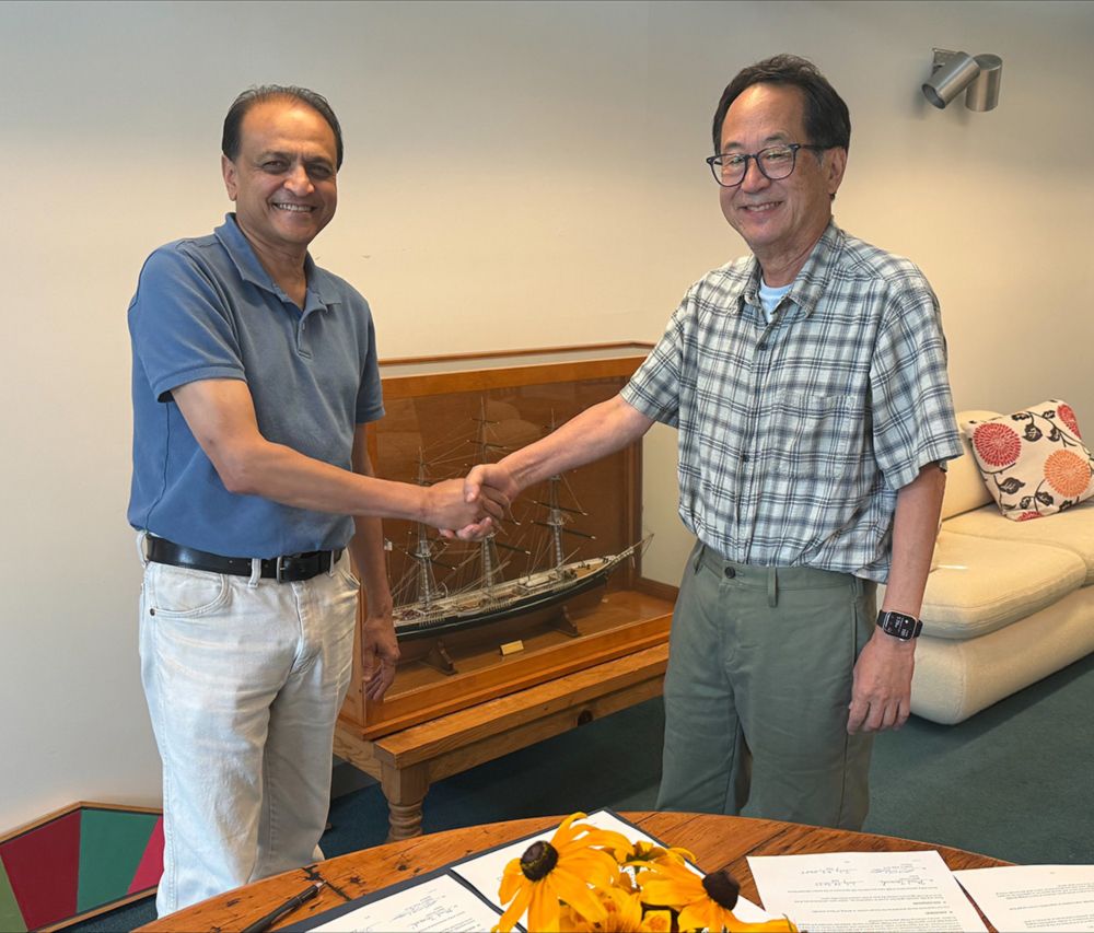
Addgene: Justin Taraska Lab Materials
Yes! Many of our plasmids are here www.addgene.org/Justin_Taras... and both NPY-GFP (exocytic vesicles) and clathrin light chain-GFP (endocytic sites) make excellent sub-diffraction biological targets.
22.12.2025 14:31 — 👍 2 🔁 0 💬 0 📌 0

Hoya bloom
30.06.2025 23:03 — 👍 6 🔁 0 💬 0 📌 0

Metro
10.05.2025 14:35 — 👍 10 🔁 0 💬 0 📌 0
Japanese Garden, Powell’s Bookstore downtown, Forest Park, SE Division or SE Hawthorne St, Coava coffee, OHSU gondola.
07.04.2025 20:24 — 👍 3 🔁 0 💬 1 📌 0
 31.03.2025 02:30 — 👍 15 🔁 0 💬 0 📌 0
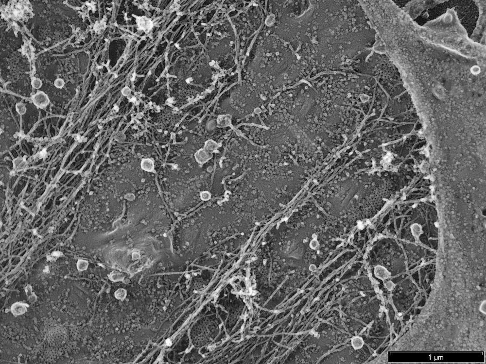
I am excited to share our first Pt replica EM images. It took us a little while, but now we have establish the unroofing, drying and Pt coating workflow 🎉 Great work by our postdoc Luis Wong Dilworth! The image below shows the cytosolic membrane leaflet of a fibroblast 👇
13.02.2025 14:33 — 👍 38 🔁 7 💬 2 📌 0
 12.02.2025 15:39 — 👍 8 🔁 0 💬 0 📌 0
Super cool. Septin/actin rings maybe.
12.02.2025 15:14 — 👍 2 🔁 0 💬 1 📌 1
 06.02.2025 14:31 — 👍 5 🔁 0 💬 0 📌 0
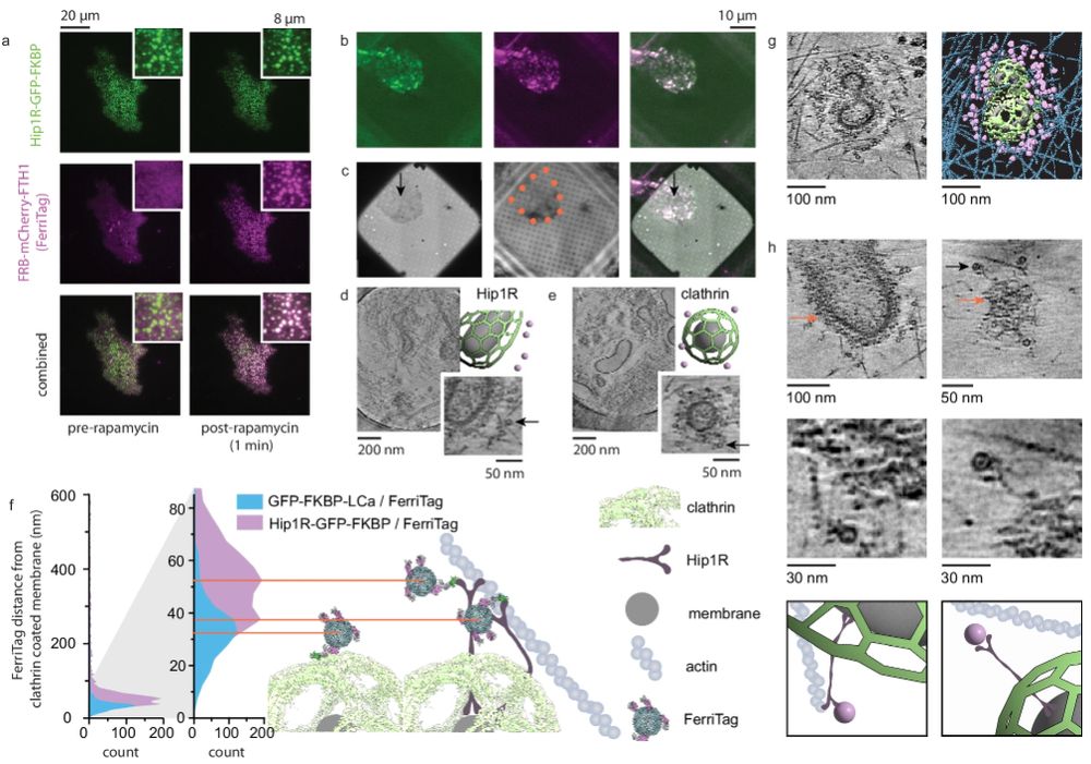
This is a scientific figure from the paper mentioned in the post. It shows fluorescence and electron microscopy images and associated schematics for using the ferritag to image clathrin coated pit proteins.
I'd like to draw your attention to this truly excellent paper from the Taraska lab on cryoET of plasma membrane associated proteins. Everyone who is thinking about probes in the cryoET space should also see what they could do with ferritag (fig 6) www.nature.com/articles/s41...
22.01.2025 16:40 — 👍 404 🔁 58 💬 11 📌 7
 22.01.2025 02:53 — 👍 6 🔁 0 💬 0 📌 0
Not necessarily unhappy. Muscle cells have extensive flat clathrin lattices. Some cells stimulated with EGF also have them. Highly metastatic cells can accumulate them. They are used as adhesion and receptor organization sites.
14.01.2025 21:08 — 👍 3 🔁 0 💬 0 📌 0
Is the third image at the bottom surface of the cell? Those are likely large flat clathrin lattices that can accumulate in some cell types at the bottom of the cell.
14.01.2025 20:29 — 👍 3 🔁 0 💬 1 📌 0
 02.01.2025 22:49 — 👍 6 🔁 0 💬 0 📌 0
CD44 and Ezrin restrict EGF receptor mobility to generate a novel spatial arrangement of cytoskeletal signaling modules driving bleb-based migration https://www.biorxiv.org/content/10.1101/2024.12.31.630838v1
01.01.2025 20:30 — 👍 2 🔁 1 💬 0 📌 0
It’s incredibly exciting when a completely different approach supports an unexpected finding from your group. Recently, we published that LDLR is captured with EGFR and FGFR1 at clathrin sites after EGF stimulation. I love science.
www.molbiolcell.org/doi/full/10....
bsky.app/profile/scie...
13.12.2024 18:21 — 👍 10 🔁 1 💬 0 📌 0

Extracellular proximal interaction profiling by cell surface–targeted TurboID reveals LDLR as a partner of liganded EGFR
A modified form of TurboID identifies extracellular interactions between transmembrane proteins.
In a new #ScienceSignaling study, researchers introduce extracellular #TurboID, a refinement of the popular proteomics tool that interrogates #ProteinProteinInteractions on the exterior cell membrane, and characterize interactions between #EGFR and #LDLR. scim.ag/3DgfJr9
13.12.2024 17:22 — 👍 53 🔁 10 💬 0 📌 1
Condolences Rita, I’m so sorry.
11.12.2024 14:26 — 👍 2 🔁 0 💬 0 📌 0
Your Only Source For Professional Dog Ratings
nonprofit: @15outof10.org ❤️🩹
links.weratedogs.com
Molecular logic of complex cell behaviors.
Orion Weiner's lab at UCSF. Account run by lab members.
#CellMigration #CellPolarity #Mechanics #OptoTools
weinerlab.com
MPG group leader and AvH fellow with interests in development, physiology and CLSM. I need to know how 🌱 communicate with environment. privately: huge cat-fan
Official Bluesky account for NOAA's National Weather Service.
Bryan William Jones
Neuroscientist
Vision Neuroscientist
Connectomics / VolumeEM
Photographer / #Leica
Professing at Pitt
In the data
bryanwjones.com
ORCID 0000-0001-5527-6643
It's me, the sea lady.
I study Sexual Selection, Host Sex/Parasite Interactions, and Behavior (also interested in Bio Ed!)
Currently looking for a postdoc position!
Biologist/Photographer/Artist/Jedi/Autistic
https://bfitzwater.wixsite.com/bfitzwater
Postdoc at UC Irvine | Functional morphologist & evolutionary biologist | PhD from @GWBiology | NSF GRFP fellow | Full time ichthyologist 🐠 & part time herpetologist🦎| He/Him
jonathanhuie.com
Postdoctoral Researcher in the Mitchell Lab at UNC Chapel Hill | Genomics, symbiosis, & modeling | Corals -> fungi | she/her
scottgenomics.com
Postdoc at University of Michigan
My research focuses on the morphological evolution of living and fossil fishes.
I mainly post about fish and weird things I enjoy
https://emilymtroyer.weebly.com/
Quantitative ecologist working mostly on modeling populations and communities of plants and critters. Queer. He/him
Trophy husband, PhD vertebrate paleontologist. 0 time rank 1 wow PVP player. I also write about the history of fishes: https://fishhistory.substack.com/. He/him
Evolutionary and behavioural ecologist | PhD from Australian National University | '24 and '25 field manager for @phenoweb.bsky.social | BTO bird ringer 🐥🐣 | Remote sensing | Long-term studies | https://scholar.google.com/citations?user=l9Wiy-IAAAAJ&hl=en
🦇 echolocation researcher.
postdoc in New Hampshire; NSF PRFB
bioacoustics, cognitive science, photography, fútbol, & a healthy dose of political shitposting
he/him/his 🇦🇷
amarotuninetti.me
Evolutionary biologist specialising in trait evolution
Big nerd
studying the changing world 🐟🐚🧬🌊 | #PopGen, rapid evolution, transcriptomics, range shifts | he/they 🌈
李慕安, GiljeGiljau Makakaruwang, son of Paiwan and Ketagalan on the beautiful island 🌸
[hire me to be your postdoc!!]
andymuanlee.com
Former PhD Student @ieesparis.bsky.social studying small population dynamics and environmental changes. Focused on extinction risks and phenotypic plasticity in ectotherms.
Open to work for a postdoc position 🌱
PhD Candidate: Plant community ecology & evolutionary ecology; monkey flowers, bumblebees, pollinator decline.
https://scholar.google.ca/citations?user=BlN5SkwAAAAJ&hl=en
PhD Candidate UW-Madison🦡
Alaska Stickleback Project🐟
Adaptation, plasticity, genomics, & connecting ecology with evolution🧬
B.S. Biology Univ of North Florida🐦
On the postdoc market
https://emilykerns.github.io/
She/her
Neuroscientist in Oslo. Former Editor. Mucking about with Voltage Imaging and patching. Aiming for a mix of fun, science, and funny science. Trying to be kind. He/him. https://dorst-lab.org/
 03.02.2026 05:06 — 👍 52 🔁 14 💬 10 📌 0