YouTube video by TU Dresden entdecken
Wie wird Insulin produziert? 🩸 | Kurze Frage über Diabetesforschung & Betazellen
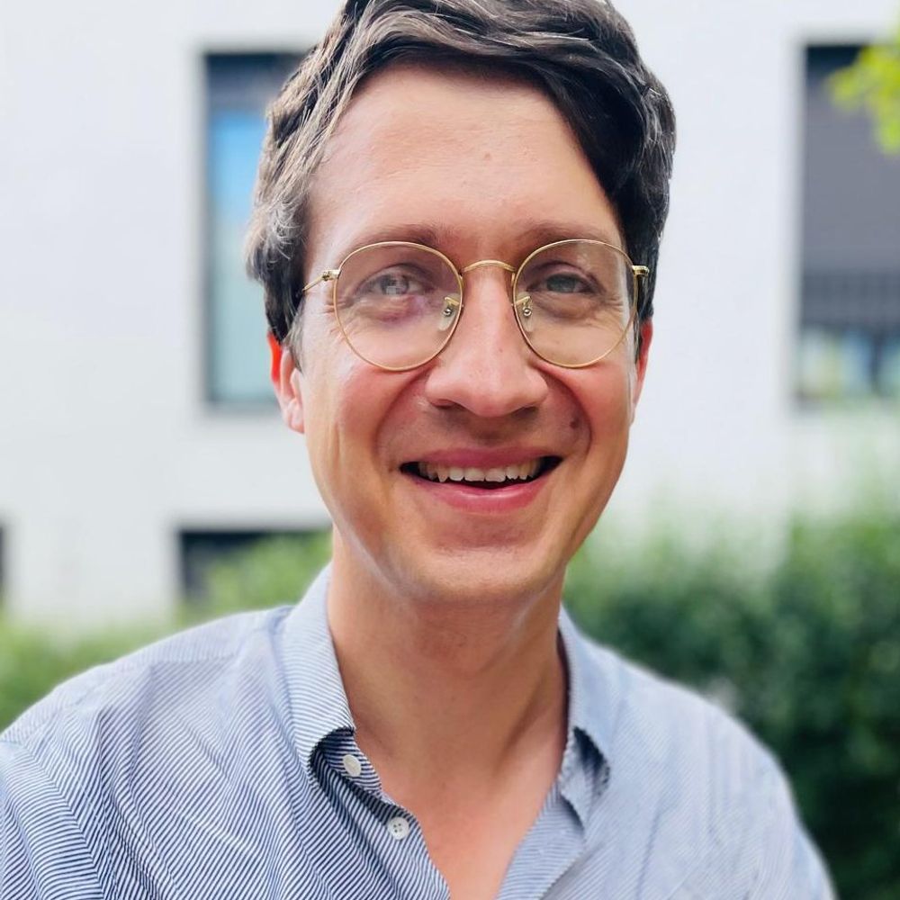
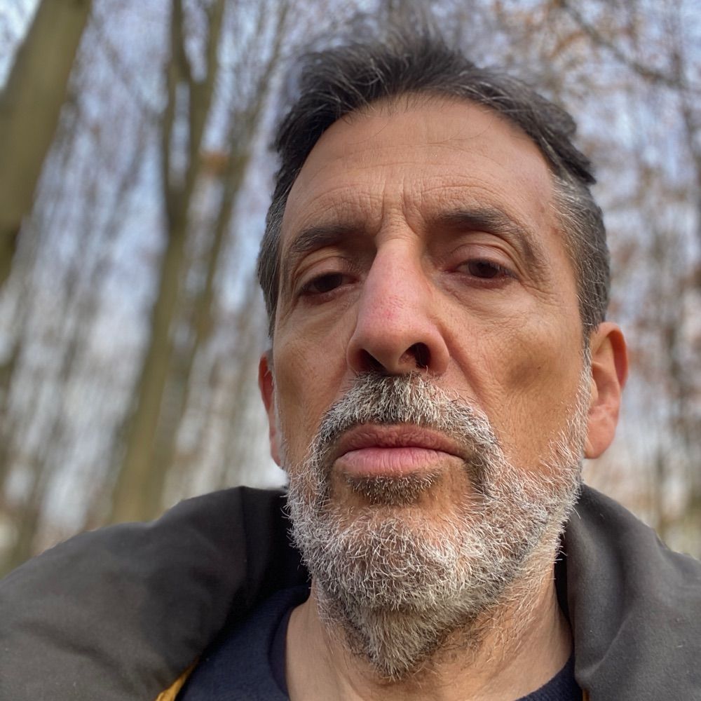
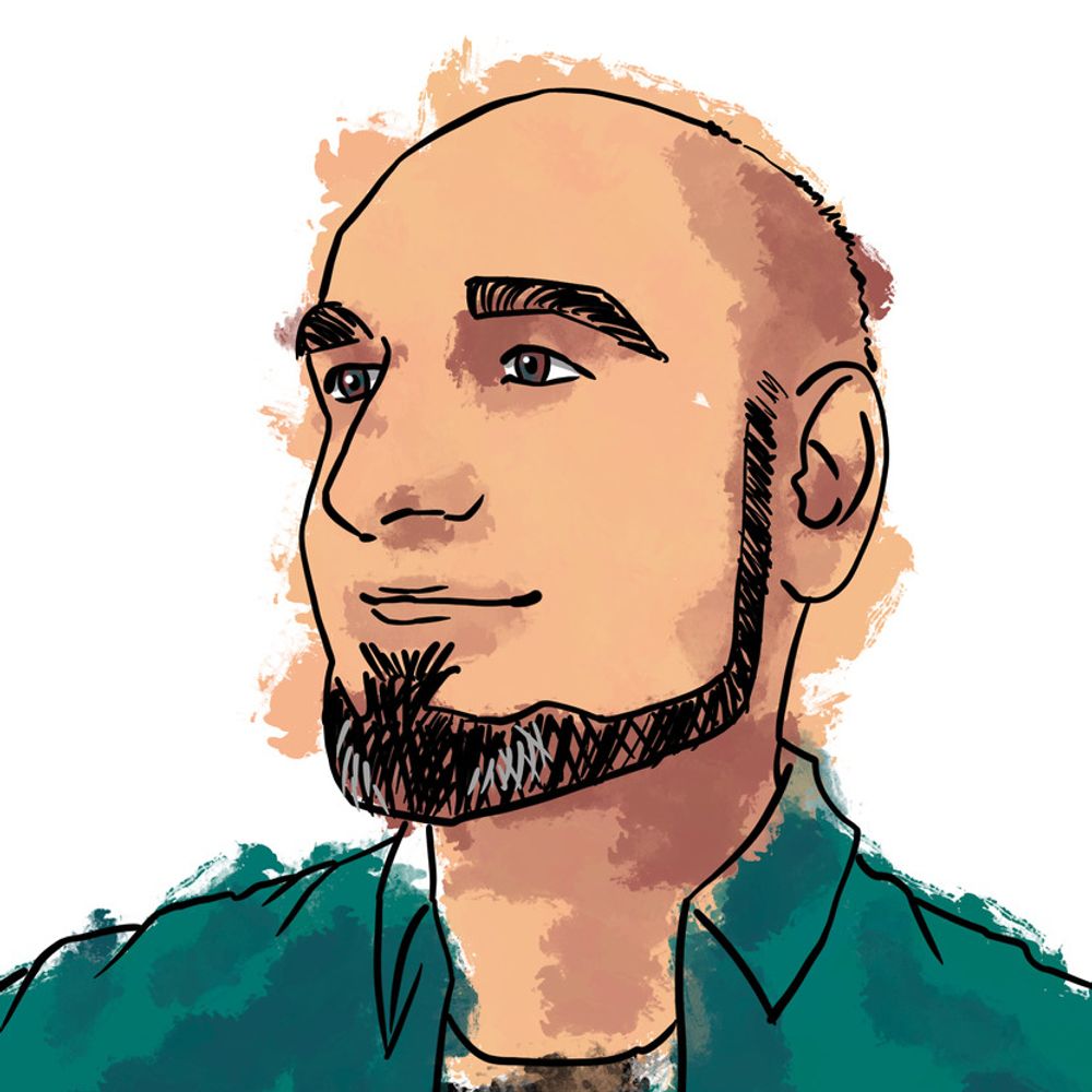
The TUD has made a very nice video in their "Kurze Frage" series featuring PLID member Andreas Müller where he explains how insulin is produced by the beta cells of the pancreas and why this is important in diabetes (German only)
#TUDresden #DiabetesResearch #PLID #HelmholztzMunich
03.12.2025 12:24 — 👍 1 🔁 1 💬 0 📌 0
Here is a short video by @tudresden.bsky.social (in German) of me explaining what beta cells do and talking about our research on investigating the structural features of their function and dysfunction.
03.12.2025 11:28 — 👍 12 🔁 1 💬 0 📌 0
Interested in applying U-ExM to cells, tissues, organoids, or cryo-sections? Our EMBO workshop is open for applications!
🧫🔬 Deadline: 12 Jan 2026 — don’t miss it!
02.12.2025 15:13 — 👍 17 🔁 9 💬 0 📌 0
Our new paper showcasing molecular connectomics with pan-expansion microscopy is out in @natbiotech.nature.com! www.nature.com/articles/s41...
This wonderful collaboration with @bewersdorflab.bsky.social was led by Ons M'Saad (now founder/CEO of Panluminate) and @allisonphysics.bsky.social. (1/5)
27.11.2025 04:39 — 👍 57 🔁 22 💬 1 📌 0
#Cilia and #Centrosome friends- tomorrow is the big day!! 🎉 🎉
Organoids for #PCD, #CryoET of the base of motile cilia, TZ organization in #IRDs, macrophages & cystic niches in #PKD, how pro-inflammatory lipids & #adipogenesis AND cilia as signal-sending organelles shedding #EVs! 🤩 😍 Join us!!
24.11.2025 11:04 — 👍 13 🔁 5 💬 0 📌 1
Scientific journals: we don’t want you using generative AI because it makes shit up.
Same journal: here is an AI summary or evaluation of this paper. This might be more useful than the actual abstract or paper that the authors freaking wrote
Same journal: AI cover art which makes no sense? Oooh!
23.11.2025 01:51 — 👍 196 🔁 42 💬 5 📌 2

Calling on my #science network! TeamNDF is #hiring a postdoc to start in 2026!
Are you fascinated by metabolism and the brain? Do you want to dig into life long impacts and the molecular cause?
Learn more about the team and our research here: www.dife.de/en/research/...
Please share! 🧠 🍔 💉
22.11.2025 12:15 — 👍 8 🔁 11 💬 0 📌 1
New on bioRxiv! 🚨
“Pericyte and Endothelial Primary Cilia and Centrioles have Disparate Organization Across the Brain Microvasculature”
This project studying #microvascular cells took an unexpected turn when we discovered that #pericytes have primary #cilia!
🔗 www.biorxiv.org/content/10.1...
🧪 🧵1/8
20.11.2025 21:03 — 👍 37 🔁 11 💬 1 📌 2

We just released a new major version of TrackMate (v8), the cell and organelle tracking plugin of Fiji.
It ships many new features, detailed below, but that are articulated around the following:
20.11.2025 11:42 — 👍 176 🔁 74 💬 4 📌 6
Das ist ein ganz kleiner Teil einer Langerhans'schen Insel, die unseren Blutzuckerspiegel reguliert. Eine Insel besteht aus etwa 1000 hormonproduzierenden Zellen. Man sieht in der Animation 2 Betazellen (mit Insulin) einen Teil eines Blutgefäßes und einen Teil einer Nervenzelle (Axon).
17.11.2025 18:57 — 👍 3 🔁 1 💬 1 📌 0

My new ImageJ / Fiji toolkit is out 🔥! The goal is to make image handling & visualization easy, with an intuitive interface! Install it on Fiji with the "Image Viewer" update site
#microscopy #ImageJ #FluorescenceFriday #microscopyMonday
imagej.net/plugins/imag...
04.11.2025 22:40 — 👍 184 🔁 56 💬 4 📌 4
As a neuroscientist, I’d suggest there is a profound disconnect between what *some* computer scientists think is representative of “intelligence”, cognitive ability, or descriptions of consciousness from some in AI work.
LLMs are not how neural systems process information, nor how brains function.
06.11.2025 17:19 — 👍 244 🔁 84 💬 20 📌 8
Dodonova Group – Organisational principles and 3D architecture of archaeal chromatin
We are looking for motivated #postdocs to join the lab! Biochemistry/microbiology/cryo-EM experience? Excited by #cryo-EM/ET, #chromatin? ❄️🧬🧫
Interdisciplinary projects, top EMBL facilities!
Email CV + short cover letter → svetlana.dodonova@embl.de
Info about the group: www.embl.org/groups/dodon...
15.10.2025 10:38 — 👍 9 🔁 7 💬 0 📌 1
Everything you wanted to know about #cilia & #signaling in one big beautiful poster format from field thought leaders @wachtenlab.bsky.social & @sorentc.bsky.social- from our just live @jcellsci.bsky.social Special Issue… 🤩🤩🤩
30.10.2025 13:11 — 👍 14 🔁 8 💬 0 📌 0
Volume 138 Issue 20 | Journal of Cell Science | The Company of Biologists
Our Special Issue on Cilia and Flagella: from Basic Biology to Disease, guest edited by @cilialab.bsky.social and @lottepedersen.bsky.social, is building. We’ll be highlighting all the articles over the next couple of weeks. Follow along at #JCSciliaSI.
journals.biologists.com/jcs/issue/13...
27.10.2025 10:30 — 👍 14 🔁 10 💬 0 📌 3

SynapseNet is a deep learning based software tool that automates the segmentation of vesicles, mitochondria, synaptic compartments, and the active zone. This is visualized in three panels. On the left, a section of an electron tomogram with a synaptic compartment densely filled with vesicles, which appear as round structures with dark boundary and light body in the image, is shwon. The top right panel shows the segmentation result of vesicles, visualized by masks with an individual color per vesicle and outlines for the segmented compartment (red) and active zone (blue). The bottom right panels shows a 3D rendering of the segmentation with vesicles shown as yellow spheres.
Are you studying synapses in electron microscopy? Tired of annotating vesicles? We have the tool for you! SynapseNet implements automatic segmentation and analysis of vesicles and other synaptic structures and has now been published:
www.molbiolcell.org/doi/full/10....
17.10.2025 11:09 — 👍 39 🔁 13 💬 2 📌 0

Expanded mouse olfactory bulb imaged with fluorescence at 20-50 nm resolution!
Ingenious concept - light sheet on serial sections by photo-etching the expanded hydrogel, layer by layer.
Photo-sectioning is quite slow, and the volumetric images are insanely large and complex, but the data look wonderful.
www.science.org/doi/10.1126/...
17.10.2025 07:00 — 👍 21 🔁 5 💬 0 📌 0
16.10.2025 13:49 — 👍 46 🔁 16 💬 2 📌 1

Poster of the 100-year history of The Company of Biologists.
1925: On this date, 100 years ago, The Company of Biologists is founded by George Parker Bidder III. Throughout today, we will be posting some of our key milestones. Read more about the past 100 years at bit.ly/3WnTVA1.
15.10.2025 08:04 — 👍 77 🔁 32 💬 0 📌 3
I found these immune cells just finished mitosis but their midbody is still yet to be resolved. How cool? If you zoom in to the midbody, you can see its classical shape just like in the textbooks. I love these little details.
14.10.2025 10:55 — 👍 113 🔁 28 💬 4 📌 1
Join us *next Monday*!
#CrickXrays25
X-ray #nanoimaging of biological tissues
🇬🇧🌐 @crick.ac.uk & online
tinyurl.com/crickxrays25
Sponsored by @dectris.bsky.social, @webknossos.org and @biologists.bsky.social and backed by @brukercorporation.bsky.social and Histomography
07.10.2025 09:20 — 👍 7 🔁 5 💬 1 📌 1

We present multi-immersion Oblique Plane microscope (miOPM), a light-sheet platform that can be adapted to a wide range of applications, from sensitive live cell imaging to imaging organs and cleared tissues.
www.biorxiv.org/content/10.1...
06.10.2025 17:52 — 👍 122 🔁 44 💬 5 📌 2
🪰 A team of researchers has unveiled the complete connectome of a male fruit fly central nervous system—a seamless map of all the neurons in the brain and nerve cord of a single male fruit fly and the millions of connections between them.
🔗 https://hhmi.news/4o3EJnk
06.10.2025 14:16 — 👍 45 🔁 20 💬 1 📌 6

I’m happy to share some plugins I’ve been developping this summer: "Channels and Contrast" and LUTs Manager!
I can’t find new bugs and ideas by now so I need your help to please test them in your machines and report bugs, feedbacks and ideas! forum.image.sc/t/looking-fo...
04.10.2025 11:50 — 👍 99 🔁 35 💬 6 📌 3
Check the live stream of the Göttingen Expansion Forum 2025 if you're interested! I think the link changes every day will try to update. Program is here (pdf link): www.rizzoli-lab.de/wp-content/u...
28.09.2025 08:52 — 👍 22 🔁 7 💬 0 📌 0
To me the most exciting part is the exploration. Finding new connections between cells and figuring out what role they play in signaling.
26.09.2025 06:34 — 👍 1 🔁 0 💬 0 📌 0
Thank you. That's a very good question. It definitely opens new lines of research. Next, we need to find out what these connections actually do and if they change in diabetes. Then we look for ways to restore their function.
25.09.2025 19:19 — 👍 1 🔁 0 💬 1 📌 0
Head of Genome Biology Dept @EMBL,
Scientist, Principal investigator, Professor
Exploring genome regulation during development,
and everything to do with enhancers.
3D genome, chromatin topology,
cell_fate, embryonic Development,
Single Cell genomics
Willkommen an der TUD | The Collaborative University
inventive. transformative. engaged.
🗞️ tud.de/newsportal
🎓 tud.de/sins
ℹ️ tud.de/impressum
BioImagingUK is an organic organisation of UK Scientists that develop, use or administer #imaging solutions for #LifeScience Research
Join our Mailing List to get all the updates: http://tinyurl.com/5n6hrhzj
Biophysicist, senior researcher at Institut Jacques Monod, CNRS, université Paris cité
Regulation of cytoskeleton dynamics lab
(she/her) Scientist, Berlin-based, PhD Student in @ewerslab.bsky.social
Here for Microscopy, Cytoskeleton, Cellbio, Cytokinesis. Black in STEM
PhD in Biochemistry and Biotechnology
An archaeologist who loves Eurovision and popular tv. Sharing content on heritage, politics and entertainment.
She/her
My opinions are my own.
Collaborative Research Center investigating the formation and function of dynamic cellular interfaces at the University of Münster, Germany. Funded by the German Research Foundation (DFG).
https://www.uni-muenster.de/SFB1348/en/index.html
I’m a trans 🏳️⚧️ creator and I make Sci-fi, action, adventure, and LGBTQIA+ comics. 1312. 4B. LinkTree: https://linktr.ee/RileysolutionLLC
Naturwissenschaften, Familie und viel Musik 🎻🎵🎶, noAFD, #dieMaskeBleibtAuf, #CovidIsNotOver, #TeamWissenschaft, 7x💉, in Stuttgart geboren, 12 Jahre in Tübingen, jetzt im Rheinland zu Hause, 🧶, K2 mit Diabetes Typ 1, 🌻, Balkonsolar
Pronomen: sie/ihr
love cilantro, physics, psychology, knitting, CNC milling, and anything that could be considered procrastination.
Director, Dept. of Mechanistic Cell Biology, Max Planck Institute of Molecular Physiology, Dortmund (DE). Opinions my own
Durch die Wälder streifend, Windspargel pflanzend.
Historikerin
Wissenschaftliche Mitarbeiterin https://geschichte-statt-mythen.de & Diss @FSU
Redakteurin https://zeitgeschichte-online.de/themen/zeitgeschichte-der-rechten & perspektiven.blog
_________________________________
Behavioural and evolutionary ecologist with a penchant for frogs 🐸 and a love of beer 🍺 | HERper | Brewer | interested in Ethnoherpetology, Art & Music (she/her)
https://sites.google.com/view/carolindittrich/
immer unterwegs für eine #besserewelt
Doctor of the Things of Nature
Learning Technology Officer
at Munich University of Applied Sciences
Makes YouTube videos:
youtube.com/@sumandproduct
youtube.com/@untitleddevlog
He/him. #IchBinHanna
https://www.bernhard-werner.de