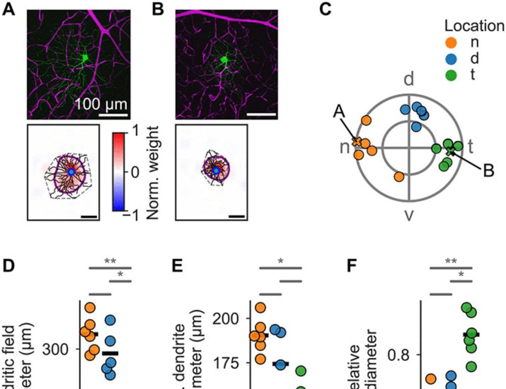
Projection Targeting with Phototagging to Study the Structure and Function of Retinal Ganglion Cells
Visual information from the retina is sent to diverse targets throughout the brain by different retinal ganglion cells (RGCs). Much of our knowledge about the different RGC types and how they are routed to these brain targets is based on mice, largely due to the extensive library of genetically modified mouse lines. To alleviate the need for using genetically modified animal models for studying retinal projections, we developed a high-throughput approach called projection targeting with phototagging that combines retrograde viral labeling, optogenetic identification, functional characterization using multi-electrode arrays, and morphological analysis. This method enables the simultaneous investigation of projections, physiology, and structure-function relationships across dozens to hundreds of cells in a single experiment. We validated this method in rats by targeting RGCs projecting to the superior colliculus, revealing multiple functionally defined cell types that align with prior studies in mice. By integrating established techniques into a scalable workflow, this framework enables comparative investigations of visual circuits across species, expanding beyond genetically tractable models. Motivation Visual information from the retina is distributed to diverse targets throughout the brain. Much of our knowledge about how visual information is processed and routed to these brain targets is based on mice because of the large library of genetically modified mouse lines. For most other species, such libraries are not available. Therefore, we were motivated to develop an approach for characterizing diverse retinal projections into the brain that can be applied to other species. We aimed to achieve projection targeting with retrograde viral vectors, followed by identification of circuit-specific retinal ganglion cells (RGCs) with optogenetics, functional characterization with multi-electrode array (MEA) recordings, and morphological description with in situ and confocal microscopy. The resulting high-throughput approach permits functional and morphological investigation of dozens to hundreds of cells in individual experiments. By replacing the need for genetically modified animals with a suite of standard techniques, this approach should improve cross-species comparisons of the initial stages of visual processing. Summary Understanding the structure-function relationships across neurons is challenging, particularly when circuits are composed of dozens of distinct cell types. We refined an approach, called projection targeting with phototagging, that allows simultaneous elucidation of the projections, morphology, and visual response properties of diverse RGC types in the mammalian retina. The approach combines retrograde virally mediated phototagging of RGCs, microscopy, and large-scale MEA measurements. Importantly, the approach does not rely on transgenic animals and thus is generalizable across species. We validated this approach in rats by targeting retinal projections to the superior colliculus (SC). We showed that multiple RGC types project to the SC and that these results in rats align well with prior findings from transgenic mouse studies. ![Figure][1]</img> Highlights ### Competing Interest Statement K.R. Is a coauthor on a patent for AAV2retro (Application No. 62/350,361 filed June 15, 2016, and U.S. Application No. 62/404,585 filed October 5, 2016). * AAV : adeno-associated virus cMRF : central mesencephalic reticular formation CRF : contrast response functions DpG : deep gray layer of the superior colliculus DpWh : deep white layer of the superior colliculus EI : electrical image GCL : ganglion cell layer IC : inferior colliculus INL : inner nuclear layer InG : intermediate gray layer of the superior colliculus IPL : Inner Plexiform Layer InWh : intermediate white layer of the superior colliculus ISI : inter-spike interval distribution MEA : multi-electrode array MGN : medial geniculate nucleus of the thalamus OT : nucleus of the optic tract Op : optic nerve layer of the superior colliculus OSI : orientation-selective index PAG : periaqueductal gray PC : posterior commissure POD : post-operative day PT : pretectum ReaChR : red-shifted channelrhodopsin RF : receptive field RGC : retinal ganglion cell SRF : spatial receptive field SuG : superficial gray layer of the superior colliculus TRF : temporal receptive field Zo : zonal layer of the superior colliculus Duke Institute for Brain Sciences (DIBS) Incubator Award NIH R01 EY034004 K99 EY032119 NIH P30 EY005722 Core Grant for Vision Research at Duke University NIH P30 EY000331 for Vision Research and an Unrestricted Grant from Research to Prevent Blindness at UCLA [1]: pending:yes
Awesome work by @gregdfield.bsky.social et al. www.biorxiv.org/content/10.1... Projection Targeting with Phototagging to Study the Structure and Function of Retinal Ganglion Cells | bioRxiv
08.07.2025 06:47 — 👍 5 🔁 1 💬 0 📌 0

a picture of a woman with the words this is exciting below her
ALT: a picture of a woman with the words this is exciting below her
Exciting news:
⭐I'm thrilled to receive a grant from the Brain Research Foundation! tinyurl.com/2p9ku4sd
⭐"Atypical retinal ganglion cell function in a mouse model of Fragile X syndrome" was published! tinyurl.com/htk3ekce
⭐my review on retinal receptive fields was published! tinyurl.com/48b9xbs2
02.07.2025 02:06 — 👍 58 🔁 7 💬 4 📌 1
This is a fantastic up-to-date summary of modern questions in retinal circuits, clearly presented and with great figures!. Required reading for anyone interested in the amazing computational capabilities of neural circuits. Congrats and a big thank-you to the authors.
16.05.2025 04:17 — 👍 11 🔁 3 💬 1 📌 0

65 years after Lettvin’s bug detector neurons in the frog retina, we revisit how the retina drives behavior—from reflexes to prey capture to brain-state modulation
New review with @annaintegrated.bsky.social & @serenariccitelli.bsky.social in Annual Review of Vision Science 👇
tinyurl.com/ymp3vs4d
15.05.2025 17:10 — 👍 42 🔁 17 💬 0 📌 3

Happy Birthday Gerald Westheimer. 101 years on May 13 and still publishing papers! #livinglegend of #visionscience. @ucberkeleyofficial.bsky.social
13.05.2025 03:51 — 👍 25 🔁 3 💬 4 📌 2
Frontiers | AAV-mediated transduction of songbird retina
I am delighted to present to all my latest research paper titled " #AAV-mediated transduction of #songbird #retina" which has been published in the journal " #Frontiers in #Physiology", in its special issue "Rising Stars in Avian Physiology: 2024".
#birds #AAV
www.frontiersin.org/journals/phy...
19.03.2025 11:40 — 👍 8 🔁 1 💬 1 📌 0
Thanks! 🙂
13.03.2025 13:29 — 👍 0 🔁 0 💬 0 📌 0
Thank you, Soile 😊
13.03.2025 07:28 — 👍 1 🔁 0 💬 0 📌 0
Thank you 🙂
01.03.2025 15:32 — 👍 0 🔁 0 💬 1 📌 0

Gravelbike leaning against a tree
x/4 😉 and thank you cycling 🚴
28.02.2025 13:34 — 👍 4 🔁 0 💬 0 📌 0
4/4 Many thanks to everyone involved ❤️
28.02.2025 13:34 — 👍 1 🔁 0 💬 1 📌 0
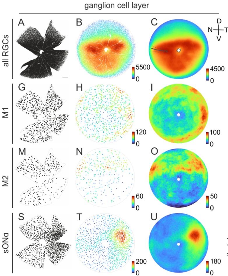
Density distributions of retinal ganglion cells across complete mouse retinas
3/ Fantastic work by Sabrina Duda that beautifully illustrates how cell types of the early visual system are distributed according to their functional role and to meet the demands of the visual environment.
28.02.2025 13:34 — 👍 2 🔁 0 💬 1 📌 0
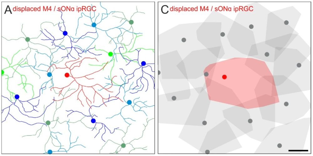
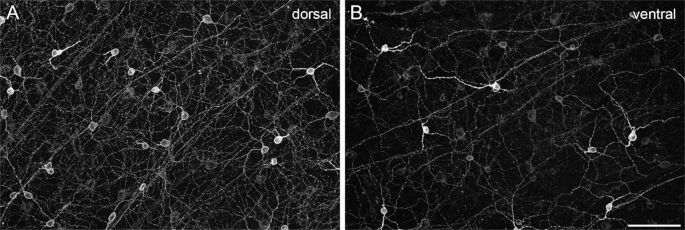
Dendritic trees of regular and displaced retinal ganglion cells and how the align to form a mosaic
2/ Dendrites of 🐭 displaced retinal ganglion cells complete the mosaics of their regular counterparts.
28.02.2025 13:34 — 👍 1 🔁 0 💬 1 📌 0
Apply for your participation in the Systems Vision Science Summer School and Symposium 2025: summerschool.lizhaoping.org/application/ Application deadline is March 31, 2025. Experimental vision researchers, vision theorists, engineers and computer scientists should not miss this unique event!
09.01.2025 12:30 — 👍 22 🔁 10 💬 0 📌 0
Visual Neuroscience | Marine Biological Laboratory
Experience hands-on training in modern visual neuroscience techniques in the new Visual Neuroscience course. Join us for a life-changing experience learning the visual system - and return home with te...
More complete announcement to come, but heads-up about a new summer course on Visual Neuroscience at the MBL, Woods Hole! Hands-on training in imaging, EM, bioinformatics, electrophysiology, visual behavior. From octopus to mouse, from retina to brain! Aug.
1-16, 2025 www.mbl.edu/education/ad...
05.12.2024 18:42 — 👍 28 🔁 16 💬 1 📌 1
Volume 1 of the "Early Vision" pack is now full-up so I started populating Volume II go.bsky.app/5z7mP26
Addition requests, as before, please fill this google form:
forms.gle/4ZdNjejitMQu...
11.12.2024 14:30 — 👍 12 🔁 7 💬 1 📌 1
I'm a bit puzzled why the retina never makes it into these big consensus think pieces. Last I checked, it was just as much a part of the CNS as the brain and spinal cord, and also where a lot of the early single-cell work along these lines was first done.
11.12.2024 17:18 — 👍 28 🔁 10 💬 3 📌 0
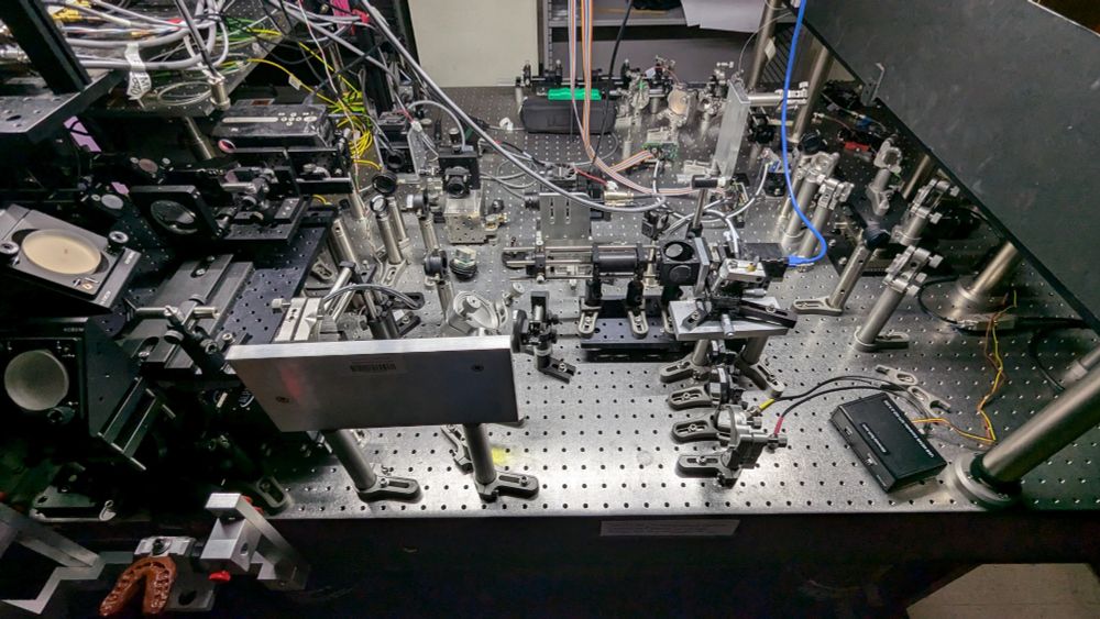
To think that this entire optical system is devoted to looking at a single photoreceptor cell today. In the lower left of this image you'll see the bite-bar where I 'lock in'.
05.12.2024 18:47 — 👍 16 🔁 3 💬 0 📌 0
😅👏
06.12.2024 20:51 — 👍 2 🔁 0 💬 0 📌 0
🧪 E11 Bio is excited to share a major step towards brain mapping at 100x lower cost, making whole-brain connectomics at human & mouse scale feasible (🧠→🔬→💻). Critical for curing brain disorders, building human-like AI systems, and even simulating human brains.
Read more: e11.bio/news/roadmap
03.12.2024 14:58 — 👍 170 🔁 54 💬 6 📌 34
“Weird axonless” is totally true though 😄
30.11.2024 08:09 — 👍 3 🔁 0 💬 1 📌 0
Among the 40ish types of amacrine cells, this AIS-like segment has been found only on few of them. The vGluT3 amacrine cell type hasn’t been reported to have it…
30.11.2024 08:07 — 👍 2 🔁 0 💬 1 📌 0
vGluT3-positive amacrine cells. An interneuron that inhibits via glycine OR excites via ribbon-release of glutamate!? What more can you ask for? 😄
29.11.2024 19:43 — 👍 5 🔁 2 💬 2 📌 0
Congrats 🙌
29.11.2024 12:02 — 👍 1 🔁 0 💬 0 📌 0
Thanks for the effort ❤️
29.11.2024 10:32 — 👍 0 🔁 0 💬 0 📌 0
Early vision Neuroscience community, Vol 1
We are talking eyes, retinas, and some of the more ancestral bits of visual brains. No species restrictions. List is close to full, will start building Vol 2 before too long
Addition requests, please see link in the below thread
go.bsky.app/BotZr1g
29.11.2024 08:16 — 👍 42 🔁 20 💬 4 📌 0
Political economist, sustainability transformation researcher, author, speaker, lecturer - www.maja-goepel.de
The Center for Visual Science (CVS) is an interdepartmental program that brings together vision scientists at the University of Rochester.
Neurobiologist @ MRC LMB in Cambridge, UK
Vision scientist, electrical synapse enthusiast, Zebrafish addict, and maybe a few other things
Visualize, share, segment, and annotate your large 3D images online.
Researcher at San Martino Hospital in Genova, exploring how the retina makes sense of the world! / former IIT & WIS
Glia biologist. Vision scientist. Interested in mechanisms of development and ageing in the retina. We love glia. You should too.
News, commentary and research coverage from the international monthly journal publishing the highest quality of work in all areas of neuroscience.
https://www.nature.com/neuro/
Neuroscience, NeuroAI | https://kfranke.com/ | Senior Scientist at Stanford working at https://enigmaproject.ai/
Vision scientist🐟🦎🔬 Advocator of science in Africa
Assit. Prof. at Washington University in St Louis
Asso. prof. at BioRTC, Yobe State University
Headed by Prof. Dr. Ueffing. Investigation of degenerative, neoplastic and vascular diseases of the eye and the visual pathway. Therapy & treatment strategies.
Co-Founder of scalable minds. Building WEBKNOSSOS, a tool visualizing, annotation, managing and sharing of large 3D images. Contributing to OME-Zarr.
he/him @normanrz@mastodon.social
Co-Founder of scalable minds. Building WEBKNOSSOS, a tool visualizing, annotation, managing and sharing of large 3D images.
Neurobiologist & Drosophilist


