I would be remiss if I did not add that this project started long ago, with draft text and figures so clear in a folder optimistically named "PaperInAWeek20221019"
19.12.2024 20:35 — 👍 0 🔁 0 💬 0 📌 0
Frontal areas are then only split into sides because the constraints of development and anatomy divide the brain into hemispheres. 23/24
19.12.2024 20:34 — 👍 0 🔁 0 💬 1 📌 0
in frontal areas than in sensory or motor ones b/c frontal cortex makes computations about an overall plan/decision for the whole animal. When deciding on what benefits the animal overall, it’s important to be of one mind and execute the plan in a coordinated manner. 22/24
19.12.2024 20:33 — 👍 0 🔁 0 💬 1 📌 0
Do we get to speculate about why this is the case? Separate sensory and motor areas work fine when each is processing info appropriate to their own side of the body. But I conjecture that contralateral corticocortical and corticostriatal projections may play a larger role 21/24
19.12.2024 20:33 — 👍 0 🔁 0 💬 1 📌 0
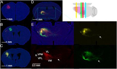
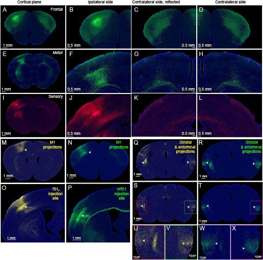
These were not as rich as the IT-type axons because corticothalamic projections only weakly cross the midline. But maybe we will be able to make some useful study of the differences of these two thalamic inputs in the future. 20/24
19.12.2024 20:33 — 👍 0 🔁 0 💬 1 📌 0
We were also asked by the reviewers about the corticothalamic projections. Here we used some other mouse lines in L5B (PT-type projections, Sim1_KJ18_Cre) and L6 (CT-type projections, Ntsr1_GN220_Cre). 19/24
19.12.2024 20:32 — 👍 1 🔁 0 💬 1 📌 0
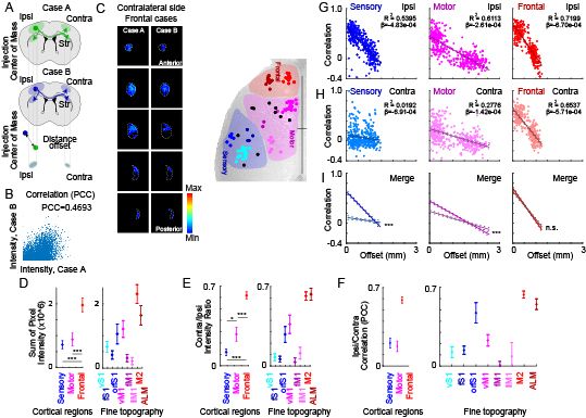
Again, we see that injections close to each other – low offset (in um) – are again pretty similar (7GHI, top). This is especially strong in frontal and sensory. But correlations across the midline were symmetrical/strong for frontal but not motor or sensory (7GHI, middle). 18/24
19.12.2024 20:32 — 👍 0 🔁 0 💬 1 📌 0
The striatal comparison is similar to the result in cortex: frontal projections to the other side are stronger than motor which in turn is stronger than sensory (7E). 17/24
19.12.2024 20:31 — 👍 0 🔁 0 💬 1 📌 0
We made similar comparisons in striatum. These images of ipsi and contralateral frontal corticostriatal projections are quite pleasing to the eye. They are, as you might predict from cortex, the most symmetrical for more anterior projections. 16/24
19.12.2024 20:31 — 👍 0 🔁 0 💬 1 📌 0
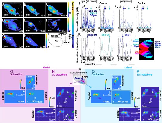
We even subdivided medial and lateral parts of S1 (suggested by a reviewer) and you can see how they differently project to the contralateral entorhinal areas (5MNOPQ). This figure was fun to make. 15/24
19.12.2024 20:31 — 👍 0 🔁 0 💬 1 📌 0
So, we made 3D pictures of each brain, leaving only the detectable axons (suprathreshold voxels to make it sound more scholarly). We could average these together, subtract left from right, and show where projections are stronger or weaker on each side. This is nice (5A-H). 14/24
19.12.2024 20:30 — 👍 0 🔁 0 💬 1 📌 0
But the correlations across the midline followed a different pattern: these were strong for frontal but not motor or sensory (3GHI, middle). This finding (and the similar one in striatum) I think is pretty neat. 13/24
19.12.2024 20:30 — 👍 0 🔁 0 💬 1 📌 0
This shows that injections close to each other – low offset (in um) – are pretty similar in the intensity of their correlation. They project to the same places ipsilaterally. This is especially strong in frontal and sensory. 12/24
19.12.2024 20:29 — 👍 0 🔁 0 💬 1 📌 0
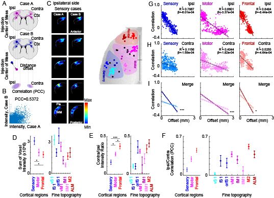
The cortical comparison shows that frontal projections to the other side are stronger than motor which in turn is stronger than sensory (3E). But even more than the strength, we were able to compare each injection to nearby injections from different mice (3GHI, top). 11/24
19.12.2024 20:29 — 👍 0 🔁 0 💬 1 📌 0
Digital anatomy I think will let us play with the data in some fun ways. We can digitally make a mirror image to compare left/right brains in the same mouse. I suspect computational ability is beginning to outpace my ability to come up with what questions we should ask ... 10/24
19.12.2024 20:29 — 👍 0 🔁 0 💬 1 📌 0
Alignment lets us define which voxels are in defined cortical and striatal areas, and using the same definition across mice, make quantitative, objective comparisons. 9/24
19.12.2024 20:29 — 👍 0 🔁 0 💬 1 📌 0
Because we can do neat computational things like align the whole image stack to a standard brain, then we can compare injections across mice, tracking how close the injection sites were and also directly compare the axonal projections in the aligned brain space. 8/24
19.12.2024 20:28 — 👍 0 🔁 0 💬 1 📌 0
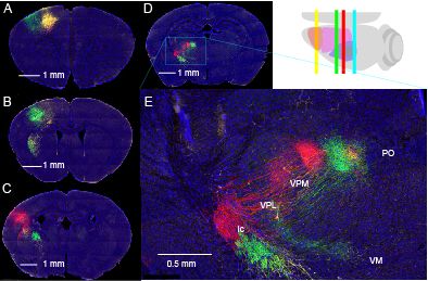
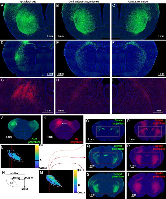
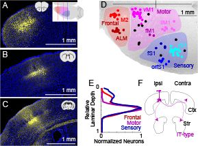
Focus on 3 parts of cortex: anterior mouse brain (frontal), motor, and somatosensory areas. I tried to stick to a red/purple/blue color scheme for these areas through the paper. Here we show where the injection was, with the ipsilateral and contralateral cortex in detail. 7/24
19.12.2024 20:28 — 👍 0 🔁 0 💬 1 📌 0
This line is pretty well restricted to layer 5 and we have a library of injections all over the dorsal cortex. As you might expect in anatomy work, we try to show some good examples of well labeled axons in a few examples. 6/24
19.12.2024 20:28 — 👍 0 🔁 0 💬 1 📌 0
We have a beautiful library of images from defined cell types, including the strong corticostriatal projection in layer 5 (we call these IT-type cells, we label them with a great transgenic mouse line, Tlx3_PL56_Cre). 5/24
19.12.2024 20:27 — 👍 0 🔁 0 💬 1 📌 0
Striatum is generally involved in motivated movements, but many cortical areas, including primary sensory ones, send projections there. So how do these across the midline compare? Are they strong or weak? And do they project to comparable parts of the brain for each side? 4/24
19.12.2024 20:27 — 👍 0 🔁 0 💬 1 📌 0
But some projections cross the midline & connect to the other side. There's lots of anatomical examples in our data. So, we were curious about how cortical projections to contralateral cortex & basal ganglia, esp. striatum, compared across left and right sides of the body. 3/24
19.12.2024 20:26 — 👍 0 🔁 0 💬 1 📌 0
Generally, the human brain is organized in a criss-crossed manner with the left side of the brain seeing/touching the right side of the world & controling movement of the body's right side. This is fascinating when we first learn it in school, but not well understood why. 2/24
19.12.2024 20:26 — 👍 0 🔁 0 💬 1 📌 0
Here’s a brief summary of our new paper with
@andrewpapale.bsky.social on ‘Symmetry in frontal but not motor and somatosensory cortical projections’ now
@sfnjournals.bsky.social www.jneurosci.org/content/44/3... 1/24
19.12.2024 20:26 — 👍 1 🔁 1 💬 1 📌 0
My lab is interested in understanding how the specific cell types in the mammalian motor system, esp. cortex and basal ganglia, are connected, how these specific connections enable the motor system to control movement, and how these connections change during motor learning & neurodegeneration.
19.12.2024 19:20 — 👍 0 🔁 0 💬 0 📌 0
News, commentary and research coverage from the international monthly journal publishing the highest quality of work in all areas of neuroscience.
https://www.nature.com/neuro/
OCD Researcher. Psychiatrist. Nerdy Mom.
Psychiatrist | Neuroscientist | Author | Deputy Editor American Journal of Psychiatry | Deputy Editor Neuropsychopharmacology | AI for Mental Health Initiative | OCD Research | Human Neural Circuitry (HNC) Program
Director, Penn State Neuroscience Institute - University Park; Huck Early Career Chair in Neurobiology and Neural Engineering, Departments of Biology & Biomedical Engineering.
Researching the neural basis of computations that guide economic decision-making at the Center for Neural Science at NYU.
PhD candidate in Neuroscience in the @yttrilab.bsky.social at @CMU_Bio, retired footballer, maker of good food
https://markolas11.github.io
Neuroscientist, runner, mom • Assistant Professor at the Medical College of Wisconsin • pain, itch, and neuropeptides • first gen • she/her • opinions my own • sheahanlab.com • taylersheahan.com
Professor at Cornell BME. Studying the neurobiology of psychiatric #drugs including #ketamine and #psychedelics.
https://alexkwanlab.org
Ketamine Karen, Dopamine Dame
Here via Tehran/KU/Yale/Pitt
author of KETAMINE https://mitpress.mit.edu/9780262542241/
and a lot of good papers https://scholar.google.com/citations?user=WNB2KvkAAAAJ&hl=en
Post-doctoral fellow in the Bruchas lab at the University of Washington.
neuroscientist, psychiatrist, writer
optogenetics.org
karldeisseroth.org
https://www.amazon.com/Projections-Story-Emotions-Karl-Deisseroth/dp/1984853694
My tweets represent my individual opinions and do not reflect upon my professional roles or responsibilities.
Neuroscientist, hockey mom
Principal Investigator Pitt/UPMC - Neuroscience, Genomics, Epigenetics, & Pharmacology (UTAustin, VCU, EKU)
Scientist 👩🔬 & EPFL Prof 🇨🇭 | DeepLabCut.org , 🦓 cebra.ai | neuroscience & ML 🧠 mackenziemathislab.org | ✨CSO at Kinematik.ai | occasionally 🐦⬛birds/🌱outdoors/🍣food/👠fashion
Associate Professor @VanderbiltU. Associate Director @VCARscience. Researching the neural dysfunction that underlies substance use disorder. @TheCalipariLab
The wanderer. The clown. The chairman.
President and CEO, @alleninstitute.org
Opinions are my own.
Y. Eva Tan Professor in Neurotechnology, MIT. Investigator, HHMI. Leader, Synthetic Neurobiology Group, http://synthneuro.org. Scientist, inventor, entrepreneur.
Interested in cognitive processes, neural circuits, philosophical constructs, economic theories and computational models of choice. And in a few other things.
Neuroscientist and psychiatrist. Nash Family Professor of Neuroscience and Director of the Friedman Brain Institute at the Icahn School of Medicine at Mount Sinai