More to come soon, both in continuing to examine the genomic regulation, but also defining the cellular behaviors that are necessary for early heart tube formation. For now, huge thanks to @benoitbruneau.bsky.social for supporting me as we pursued this ambitious and open-ended project!
(8/8)
03.09.2025 22:04 — 👍 6 🔁 1 💬 0 📌 0
In what @benoitbruneau.bsky.social aptly described as our “lets go nuts” experiment, we confirmed this interaction by demonstrating that reduced Nr2f2 dosage partially rescues the Mef2c KO heart tube phenotype.
(7/n)
03.09.2025 22:04 — 👍 1 🔁 0 💬 1 📌 0
Finally, Alex Clark and @jsauce7.bsky.social helped us construct gene regulatory networks for each of the outflow tract, ventricle, and inflow tract segments of the heart tube. Using these networks, we identified a previously unknown genetic interaction between Nr2f2 and Mef2c.
(6/n)
03.09.2025 22:04 — 👍 4 🔁 0 💬 1 📌 0
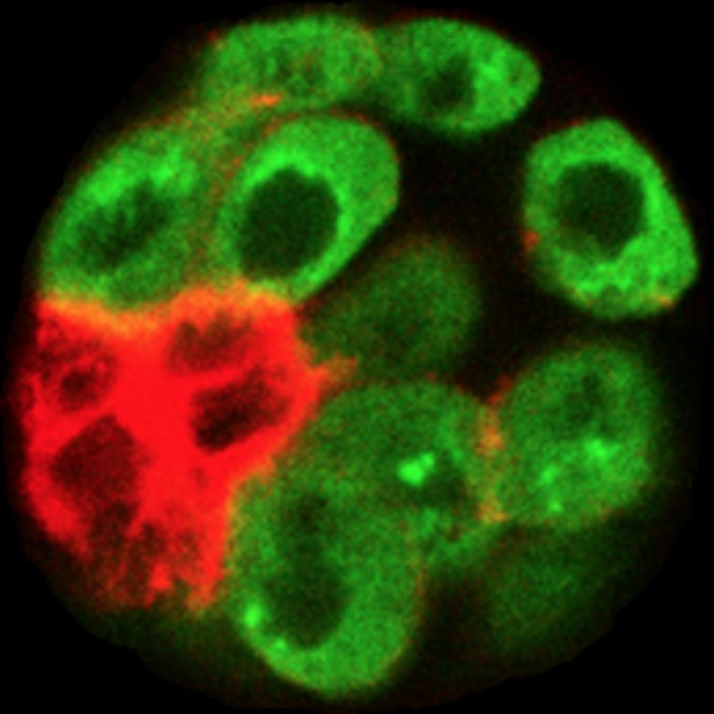
By collaborating with @tanvis.bsky.social and Brian Black at UCSF, we showed via zebrafish transgenesis that many of our predicted elements exhibited clear enhancer activity in the developing heart.
(5/n)
03.09.2025 22:04 — 👍 4 🔁 0 💬 1 📌 0
We hypothesized that MEF2C acts via segment-specific enhancers to drive distinct patterns of gene expression in the early heart and used our multiomics datasets to predict such candidate enhancers.
(4/n)
03.09.2025 22:04 — 👍 2 🔁 0 💬 1 📌 0
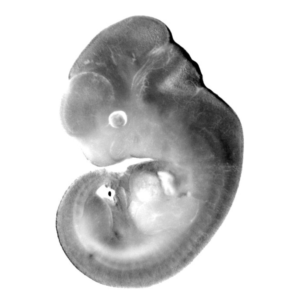
We found that loss of MEF2C causes a broad “posteriorization” of the early heart tube. In both gene expression and chromatin accessibility modalities there were some changes that were universal throughout the heart tube, and others that were specific to distinct segments of the tube.
(3/n)
03.09.2025 22:04 — 👍 2 🔁 0 💬 1 📌 0
Loss of the transcription factor MEF2C leads to embryonic lethal defects in early heart development. In this paper, we used multiomics (snRNA- and snATAC-seq) to understand precisely what goes wrong when MEF2C is absent.
(2/n)
03.09.2025 22:04 — 👍 1 🔁 0 💬 1 📌 0
Was a pleasure to present this piece of my postdoctoral work at #bcvs2025! If you missed it or are looking for more, check out our preprint: www.biorxiv.org/content/10.1...
24.07.2025 17:27 — 👍 10 🔁 1 💬 0 📌 0
A peer support group for postdocs on academic track🔬🔭📡💻🧬🌱⚗️🧪🧫💊🩺 To join us ✉️ future.pi.slack@gmail.com provide Gmail & lab/department profile link.
https://futurepislack.wordpress.com/
Postdoctoral fellow in the Saucerman Lab at UVA studying cardiac development and electrophysiology
Bioinformatics & Genomics scientist, https://github.com/bioinfoDZ. Professor at Albert Einstein College of Medicine, study brain / heart development & diseases, and cancers
RNA biology of embryonic development and disease #heart #ribosome #translational_control #RBP #splicing @UK_Frankfurt Alum:@salkinstitute @UcSandiego
https://kurianlab.com/
Group Leader. Australian Regenerative Medicine Institute. Associate Professor Monash University. Melbourne, 🇭🇲. Stem Cell Biologist, hiPSCs, congenital heart defects, cardiac development, rare genetic disorders
Associate professor at the University of California Merced. Posts are my own. My lab uses zebrafish to study morphogenesis of the endoderm. It's offal-y interesting.
Cardiac Development • Epigenetics • Postdoc • UPenn #devbio 🫀🔬🧬🐭 #firstgen #academicjobmarket
🚨Posts may contain science, bad jokes, cooking 🧵s &/or political snark. The typos are free.
Developmental Biologist I Organ Form and Function, Heart Morphogenesis I Group Leader @crick.ac.uk I India - Australia - Germany - UK
Embryologist interested in how organs grow, adapt and evolve 🫀🔬 Transition Fellow at IDRM, Oxford. Previously postdoc Francis Crick Institute, PhD Cambridge.
Pumped about hearts, microscopes and morphospaces.
Genome Scientist. Studies how DNA makes humans, mice, plants, microbes. At Lawrence Berkeley National Lab and Joint Genome Institute. Views are my own.
Assistant professor at UCSF. Cell biologist at heart, research focus on the function and dynamic regulation of the nuclear periphery.
www.buchwalterlab.UCSF.edu.
How do cells form tissues? Our research program spans the scales from molecules to organisms. News from the Max Planck Institute of Molecular Cell Biology and Genetics (MPI-CBG) in Dresden, Germany. Impressum: https://www.mpi-cbg.de/impressum
👩🔬 The Institut Pasteur is a leading global biomedical research institute.
🔬 Explore with us the frontiers of biomedical research
🧪 Follow us to uncover groundbreaking discoveries
🌍 Join a community passionate about progress and open science
We study cardiac development, disease and regeneration and are located at the Hubrecht Institute in The Netherlands
Professor of Medicine/Cardiology, Chief, Basic & Translational Research, Div Of Cardiology, Senior Vice Chair Academic Affairs, Dept of Med, Assoc Director, CV Institute, Stanford Univ. Investigator in Cardiac Development, Regenerative Med, Bioengineering.
The Srinivas group is based at the Institute for Developmental and Regenerative Medicine, University of Oxford. We work on how the mammalian embryo forms, with a focus on the establishment of the basic body plan and the formation of the heart.
Developmental, stem cell and islet biologist, Professor for Beta Cell Biology @ Technical University Munich, Director of Institute for Diabetes and Regeneration Research @HelmholtzMunich
A Cell & Developmental Biology lab @UBC. working on the role of cell junctions in development, stem cells, tissue homeostasis.
https://sites.rutgers.edu/astrof-lab/
Developmental biology, ECM, cardiovascular development, pharyngeal arches and arteries, congenital heart disease; Arches banner credit: Sam Snitkovsky.
Assistant Professor @ Stanford