Job alert! 📣 I’m looking for a research assistant to join my new team @idrm.ox.ac.uk
Were using #zebrafish to understand gene-environment interactions that shape the heart 🫀generate natural diversity 🐸🐭 and contribute to congenital defects ❤️🩹
Full info below, and please share! 🫶🏻
bit.ly/467TO0M
02.02.2026 14:01 — 👍 29 🔁 20 💬 0 📌 1
How the functional architecture of the zebrafish heart is shaped during development
📹 @tobyandrews.bsky.social et al @rashmi-priya.bsky.social
lab @crick.ac.uk in @cellpress.bsky.social Developmental Cell
➡️ bpod.org.uk/archive/2025...
18.08.2025 08:40 — 👍 25 🔁 7 💬 0 📌 0

Now published in @natcomms.nature.com! 🥳
👉 rdcu.be/eATn3
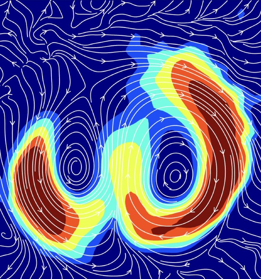
We developed image analysis tools to capture the nematic orientation field of 3D tissue surfaces. Tested on epithelial aggregates, zebrafish hearts, myoblasts on spheres & micro-vessels, we combined soft matter physics with exp. biology.
15.08.2025 12:13 — 👍 91 🔁 38 💬 8 📌 1
Start-Shape-Stop: Cell communication mechanisms controlling organ size
Accurate growth control is critical for the achievement of proportional organs during animal development and repair processes. Either extra or deficie…
Hot off the press!!! Proudly presenting our lab's new review on how do cells communicate to control organ size :) We focus specially on dynamic connections that operate at different timescales to regulate organ growth and morphogenesis. #devbio #SizeandShape www.sciencedirect.com/science/arti...
09.08.2025 08:39 — 👍 63 🔁 19 💬 3 📌 0
Heartiest congratulations and thanks to @rashmi-priya.bsky.social on the first of many studies from the lab, and for supporting me through its morphogenesis from start to finish 🫀
06.08.2025 15:22 — 👍 2 🔁 0 💬 0 📌 0
A huge thanks to all authors for their work in bringing this project to life @jcornwallscoones.bsky.social @mcramel.bsky.social @kirtigupta.bsky.social @jamesbriscoe.bsky.social and our colleagues and facilities @crick.ac.uk 13/n
06.08.2025 15:19 — 👍 1 🔁 0 💬 1 📌 0
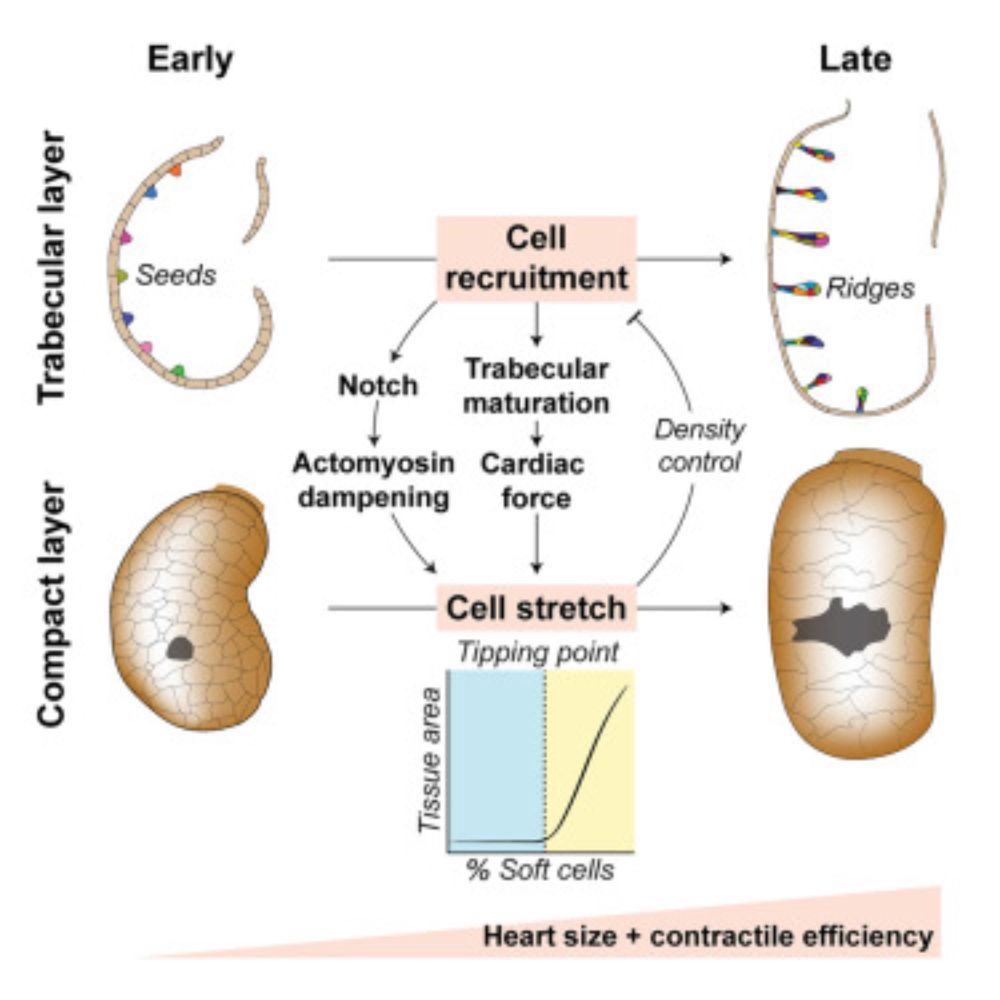
Looking forward - a deeper understanding of these design principles will give us better insight into what makes development robust, and how it can be steered to produce diversity, novelty, and disease 12/n
06.08.2025 15:15 — 👍 1 🔁 0 💬 1 📌 0
Together, we learn that the shape and size of the heart aren’t hardwired, instead they’re worked out through a flow of information across scales, giving rise to self-organising and emergent features 👀 11/n
06.08.2025 15:14 — 👍 5 🔁 0 💬 1 📌 0
Not only this, they were also more functionally efficient, owing to a greater blood filling capacity. 10/n
06.08.2025 15:13 — 👍 3 🔁 0 💬 1 📌 0

To test the model, we came up with a neat genetic approach to disrupt actin turnover in the Notch+ population. Following the model predictions, when activated at sufficient density, this made hearts bigger... 9/n
06.08.2025 15:10 — 👍 6 🔁 0 💬 1 📌 0

To understand how coherent changes in organ shape and size could arise from stochastic signalling @jcornwallscoones.bsky.social developed a 3D vertex model, which predicted the ventricle should grow suddenly, when enough cells soften 8/n
06.08.2025 15:09 — 👍 7 🔁 0 💬 1 📌 0

Looking more closely, we found intrinsic changes in actomyosin tension enable stretched cells to change shape in response to organ scale forces. This is a local response to Notch, activated in a stochastic pattern 7/n
06.08.2025 15:08 — 👍 5 🔁 0 💬 1 📌 0

Using drugs to disrupt the heartbeat, we found cells stretch in response to the force of ventricle contraction. This also traps them in the compact layer, meaning trabecular density stabilises at a threshold of force production 6/n
06.08.2025 15:07 — 👍 5 🔁 0 💬 1 📌 0

meanwhile, by unwrapping the heart, we found that compact layer cells stretch. This allows the heart to grow in size despite losing cells from its outer layer 5/n
06.08.2025 15:05 — 👍 5 🔁 0 💬 1 📌 0

Using single cell tracking, we found trabecular cells don’t just divide to form ridges. Instead, they recruit cells from the surrounding compact layer... 4/n
06.08.2025 15:04 — 👍 5 🔁 0 💬 1 📌 0

As the embryo grows, the heart expands in size and forms two layers – an elastic compact layer, and an inner layer of muscular trabecular ridges that help to power heart contraction 3/n
06.08.2025 15:03 — 👍 4 🔁 0 💬 1 📌 0
The heart is a remarkable organ, where form and function arise in parallel – in this case, the heart wall remodels to form a complex architecture, while the heart beats to support blood flow to the peripheral organs 2/n
06.08.2025 15:02 — 👍 9 🔁 0 💬 1 📌 0
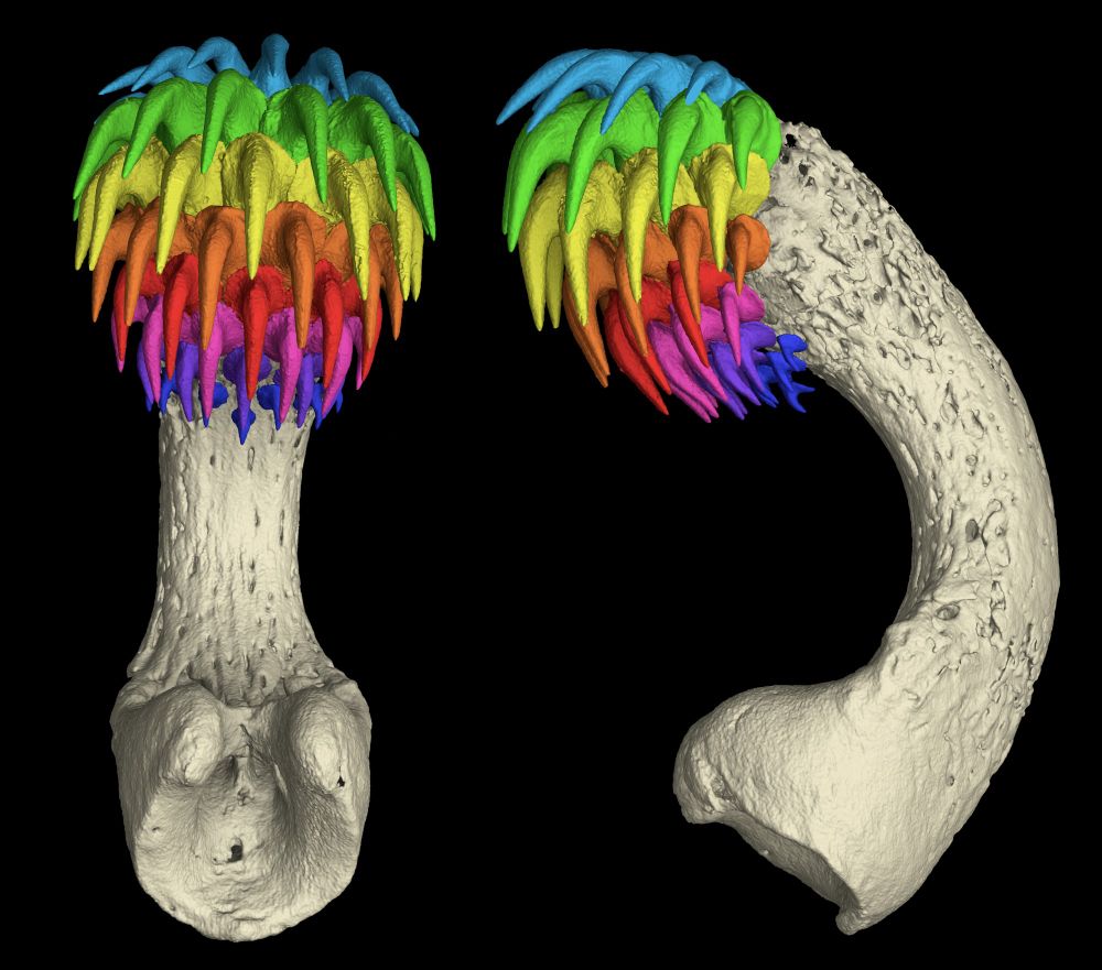
CT scan of the head clasper (tenaculum) from the Spotted Ratfish (Hydrolagus colliei), compete with its rows of shark-like teeth!
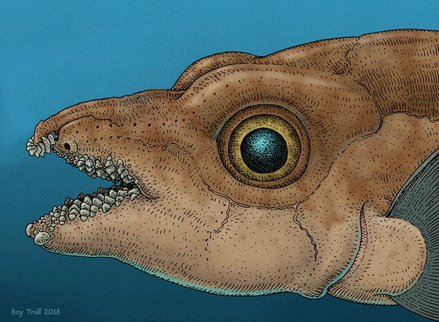
Our paper features fossil reconstruction art (of Helodus simplex) by Ray Troll - https://www.trollart.com/
New Pre-Print Alert! "Teeth Outside the Jaw: Evolution and Development of the Toothed Head Clasper in Chimaeras." We use fossil evidence, development and CT scans through ghost shark ontogeny to describe the emergence of the tenaculum! 👻🦈🦷 @karlycohen.bsky.social
www.biorxiv.org/content/10.1...
08.04.2025 19:08 — 👍 27 🔁 17 💬 0 📌 1
Latest work:
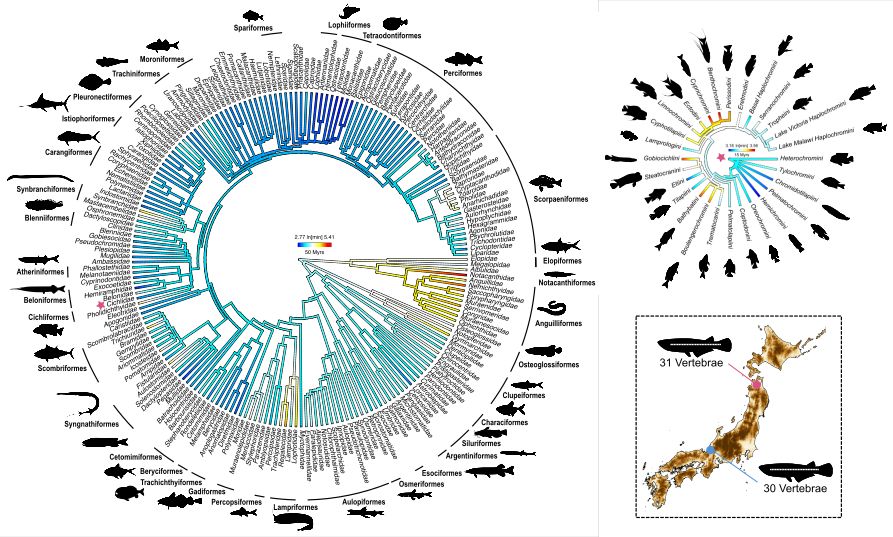
Review on the evolvability of vertebral number, and the developmental processes underpinning it
Written by Callum Bucklow, @bertaverd.bsky.social, and myself
Check it out here: doi.org/10.32942/X2K...
24.03.2025 21:26 — 👍 23 🔁 5 💬 1 📌 3
A fantastic opportunity to take on evolution with experimental embryology
Great lab, mentor, department, and model system 🐠 don't miss out!
05.02.2025 21:00 — 👍 1 🔁 0 💬 0 📌 0
🚨📢 New paper alert! Our work showing that bilateral cellular flows display asymmetry prior to left–right organizer formation in amniote gastrulation is now published in PNAS!! @pnas.org
🥳😃 🐣
Paper link: www.pnas.org/doi/10.1073/...
News article: news.miami.edu/stories/2025...
05.02.2025 17:18 — 👍 47 🔁 9 💬 4 📌 1
An ancient patterning system co-opted to position the chordate forebrain 🧠
Amphioxus spilling more evolutionary secrets - beautiful and rigorous work from @giacomogattoni.bsky.social (but no surprises there!)
31.01.2025 09:30 — 👍 3 🔁 0 💬 1 📌 0
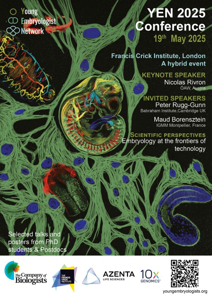
Registration and abstract submission for YEN 2025 is officially open!
We are looking forward to seeing you at the 17th Young Embryologist Network Conference on the 19th May 2025.
Attendence is FREE thanks to our amazing sponsors: @biologists.bsky.social @10xgenomics.bsky.social and Azenta.
30.01.2025 16:36 — 👍 49 🔁 30 💬 1 📌 4

Maximum intensity projection of a live embryonic zebrafish heart at 72 hours post fertilisation, showing myocardial actin (green; Tg(myl7:LifeActGFP)) and endothelial actin (magenta; Tg(fli1a:Ac-TagRFP)). The image is overlaid in the atrium with 3D reconstructions of the myocardial (light blue), endocardial (pink) and extracellular matrix (orange), and in the ventricle with a 3D reconstruction of the myocardium colour-coded to visualise myocardial thickness.
Tools to analyse early heart morphogenesis in detail are limited. @noelresearchlab.bsky.social &co develop computational package called morphoHeart that allows for integrated 3D analysis of both #heart & extracellular matrix morphology in live #zebrafish embryos 🧪 @plosbiology.org plos.io/42DlqtJ
30.01.2025 10:07 — 👍 14 🔁 4 💬 0 📌 0
Spheroids are simple systems with only convex curvature. What about more complex systems?
We teamed up with @tobyandrews.bsky.social & @rashmi-priya.bsky.social, and analyzed the ventricular myocardium of Zebrafish hearts ... and it works! 👇
(Directors: high alignment = red, misaligned = blue)
29.01.2025 23:01 — 👍 3 🔁 1 💬 1 📌 0
How can we accurately measure features on curved 3D tissues? 📐
Normally we rely on lossy 2D projections, but @juliaeckert.bsky.social's new method detects nematic orientation fields on surfaces of arbitrary geometry
We tested it in hearts, and works a charm! 🫀 More in Julia's thread below 👇🏻
30.01.2025 10:10 — 👍 3 🔁 2 💬 0 📌 0
Andrews, T. G. R., & Priya, R. (2024). The Mechanics of Building Functional Organs. Cold Spring Harbor perspectives in biology, a041520. Advance online publication. #EpithelialMechanicsReview
https://cshperspectives.cshlp.org/content/early/2024/06/15/cshperspect.a041520.abstract
30.01.2025 07:00 — 👍 14 🔁 3 💬 0 📌 0
Postdoc at Oxford in neurogenetics🧬Previously PhD at Sanger/Cambridge, ACB at Broad. Harvard cognitive neuro 🧠
Postdoctoral Research Fellow in the Developmental Signalling Lab @The Francis Crick Institute 🐟🔬🧫🎓
Light microscopy at Bruker Fluorescence Microscopy division.
#lightsheet #multiphoton #superresolution #screening
Cell biologist in my previous life. Alumnus @mpicbg.bsky.social
@turkubioscience.bsky.social
Biomedical Picture of the Day: daily intriguing images from global research. IMAGE/RESEARCH EXPLANATIONS AND LINKS TO THE PAPER on bpod.org.uk
Supported by
@leicamicrosystems.bsky.social &
@oxfordbiochemistry.bsky.social, @ox.ac.uk
Developmental biologist, interested in ecosystem interactions - working on the development of coral-algae symbiosis.
Medical faculty of the University of Heidelberg, Aulehla group @EMBL Heidelberg 🎏
Incoming PhD Student with Delas lab at LMCB.
Interested in gene regulation.
Runs.
Physicist having a go at biology, EMBO postdoctoral fellow @UCL in the Mao group, @EMBL alumna
Family & dog - bake, whittle, run, cycle & draw. Nerve repair & biomaterials I teach biology & bioengineering. views=own; he/him. 🇨🇦 🇺🇦 🇬🇧 🇩🇪 🇪🇺 🌍 🌌
It’s way too red!
Blog @ https://bio-mat-sketches-mor.blogspot.com
Developmental biologist. EvoDevo & comparative stem cell dynamics 🐭⌛️👤⏳. Group Leader @BabrahamInst.
@SEBiolDev board member
Post-Doc @katjaroeper.bsky.social lab @pdncambridge.bsky.social
Interested in kidney organoids and lumen formation; Former @honigmann_lab,
@biotec-tud.bsky.social, @cmcb-tud.bsky.social,@mpicbg.bsky.social
and @boulantlab
Scientific writer at the Sanger Institute, Cellular Genetics programme. Formerly at Development journal. Lapsed flypusher (Gurdon Institute, University of Sussex). Creative writing MA at UEA. Likes novels and gardens, lives in Cambridge.
Laboratory Research Scientist in the Organ Morphodynamics lab at the Crick - zebrafish embryology / mechanics in heart development!
Developmental biologist studying endothelial cell differentiation and heterogeneity
Postdoc in the Stone Lab - Institute of Developmental and Regenerative Medicine, Oxford, UK
ELBE Postdoctoral Fellow, CSBD/MPI-PKS/MPI-CBG.
Former PhD, EMBL.
Into cnidarians, morphogenesis and imaging. Seeking minimal rules governing early embryonic development through several interdisciplinary lenses.
Postdoctoral researcher at Cambridge University studying Developmental Metabolism | PhD at Freiburg University | 🇬🇧🇩🇪🇮🇳
Postdoc in the Santos Lab (The Francis Crick Institute)
Investigating how cells navigate change
Trying to appreciate the beauty in nature 🌱