(1/n) DNA-PAINT imaging inside the nucleus at single antibody resolution using TIRF? Ultrathin sectioning makes it happen!
Grateful to share my postdoctoral work introducing “tomographic & kinetically-enhanced DNA-PAINT” or in brief: tkPAINT. Out in @pnas.org!
doi.org/10.1073/pnas...
👇🧵
13.08.2025 14:19 — 👍 68 🔁 22 💬 2 📌 3
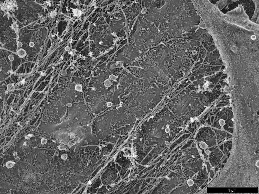
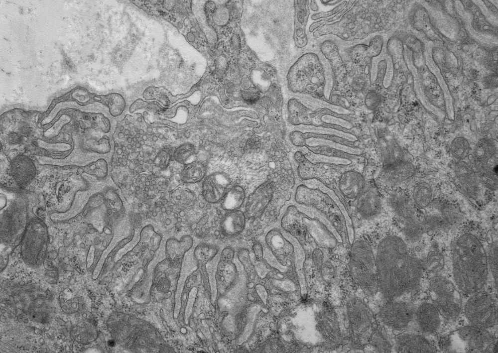
I am excited to share our first Pt replica EM images. It took us a little while, but now we have establish the unroofing, drying and Pt coating workflow 🎉 Great work by our postdoc Luis Wong Dilworth! The image below shows the cytosolic membrane leaflet of a fibroblast 👇
13.02.2025 14:33 — 👍 38 🔁 7 💬 2 📌 0
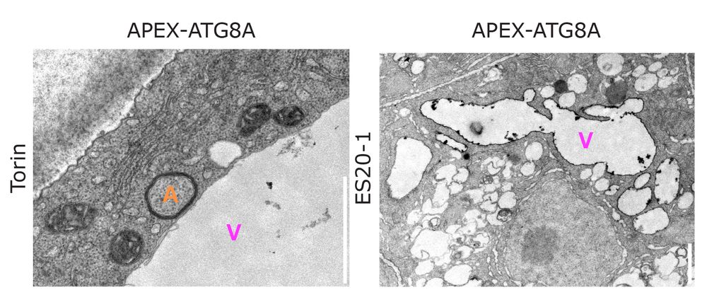
For the first time in plants, we used APEX-based electron microscopy to map the precise localization of ATG8 at the vacuolar membrane after stress! 🚀
Pushing the boundaries of plant cell biology—one EM image at a time. ⚡👀
#PlantScience #ElectronMicroscopy #Autophagy #Vacuole #NaturePlants
07.02.2025 11:11 — 👍 14 🔁 4 💬 1 📌 0
Zooming in on the ultimate connection! 🔬 💥Feast your eyes on this stunning electron microscopy image of a neuromuscular junction #EMFriday #ElectronMicroscopy
10.01.2025 16:04 — 👍 41 🔁 9 💬 0 📌 0
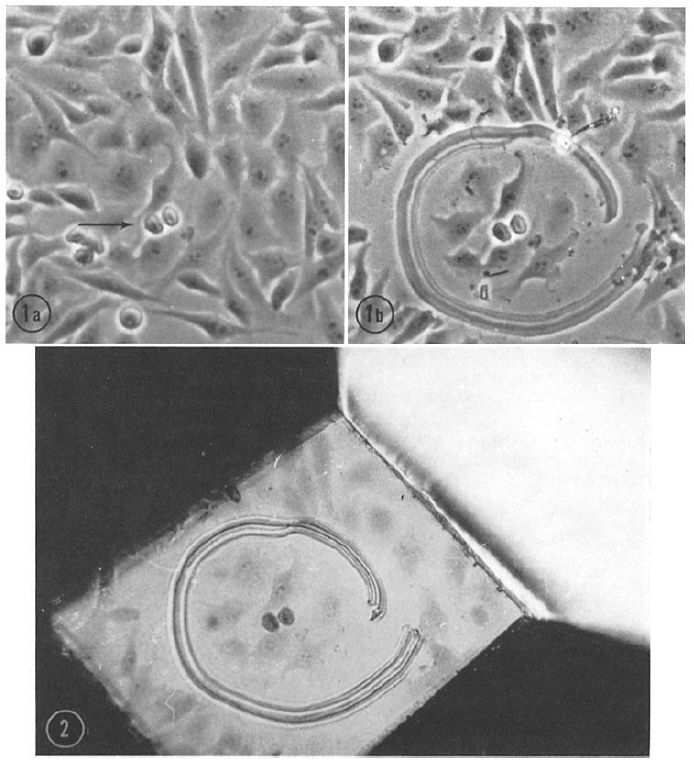
#Correlative Light and Electron Microscopy, #CLEM, is a modern #method combining light and #electronmicroscopy data. Although it is considered “new”, it was already used in the early 1960s, for instance to study the #ultrastructure of mitotic cells in this beautiful example: doi.org/10.1083/jcb....
13.01.2025 07:47 — 👍 6 🔁 1 💬 0 📌 0
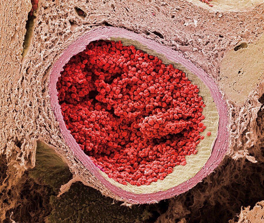
electron microscope image of a section through a foetal aorta filled with red blood cells
12.01.2025 22:09 — 👍 15 🔁 2 💬 0 📌 0

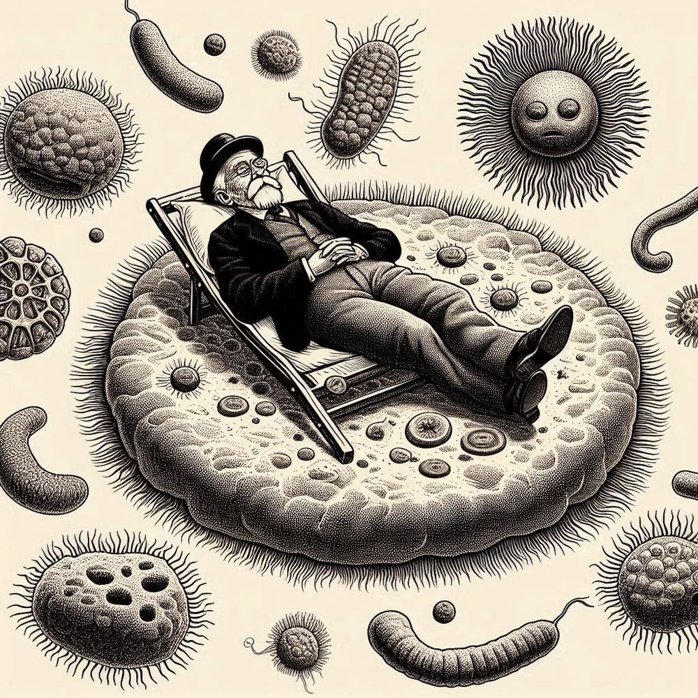
I fear there is deep truth here.
11.01.2025 20:47 — 👍 55359 🔁 13689 💬 1434 📌 841

Beyond misalignment of science in the news and in schools
Almost 40 years ago, the American astronomer, planetary scientist, and science communicator, Carl Sagan, reflected on the role of mass media in science communication. “How much science and technology ...
“One of the challenges in science journalism is the oversimplification of research findings to attract attention. Headlines such as “scientists find cure for cancer” or “ozone layer is healing” are designed to attract readers’ attention but often misrepresent the complexity of scientific research.”
09.01.2025 15:56 — 👍 7 🔁 3 💬 0 📌 0
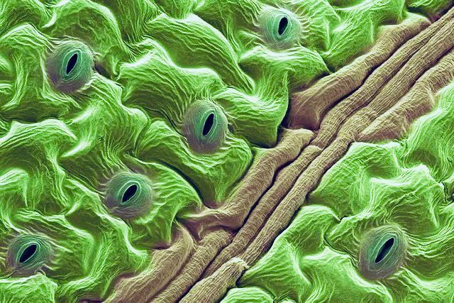
electron microscope image of a coriander leaf
08.01.2025 20:42 — 👍 24 🔁 8 💬 1 📌 2
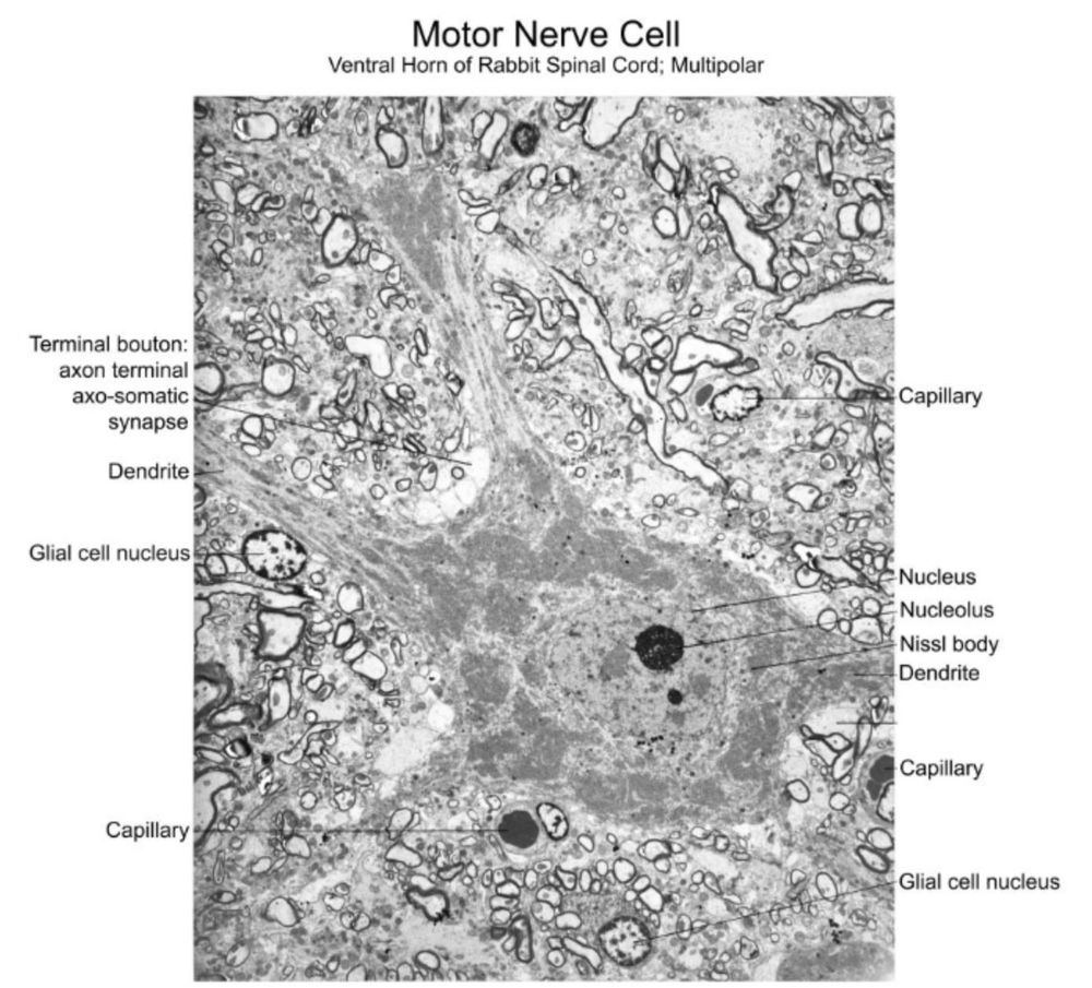
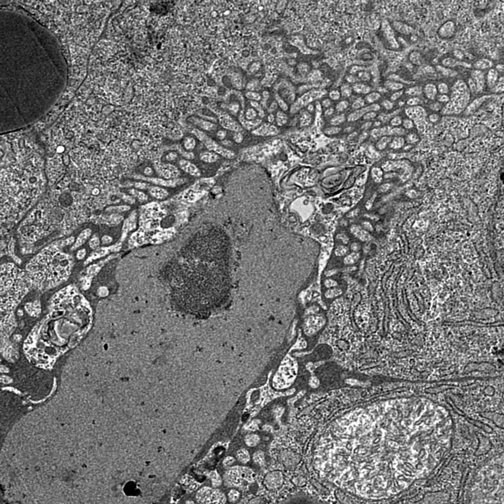
An electron microscopy image of a motor nerve cell (ventral horn of rabbit spinal cord; multipolar). It shows a cell body with a large nucleus. An axo-somatic synapse on that cell body is labeled, as are 2 dendrites. Surrounding the nerve cell are other, labeled features: glial cell nuclei, capillaries.
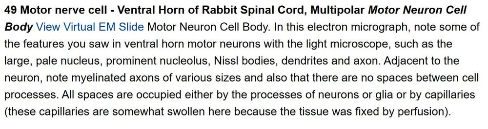
Screenshot of text: 49 Motor nerve cell - Ventral Horn of Rabbit Spinal Cord, Multipolar Motor Neuron Cell Body View Virtual EM Slide Motor Neuron Cell Body. In this electron micrograph, note some of the features you saw in ventral horn motor neurons with the light microscope, such as the large, pale nucleus, prominent nucleolus, Nissl bodies, dendrites and axon. Adjacent to the neuron, note myelinated axons of various sizes and also that there are no spaces between cell processes. All spaces are occupied either by the processes of neurons or glia or by capillaries (these capillaries are somewhat swollen here because the tissue was fixed by perfusion).
Images and instruction span from the area level down to the subcellular. Check out this rabbit motor neuron! Again, the text walks students through what they should be learning and appreciating from the image.
3/n
07.01.2025 11:10 — 👍 2 🔁 1 💬 2 📌 0
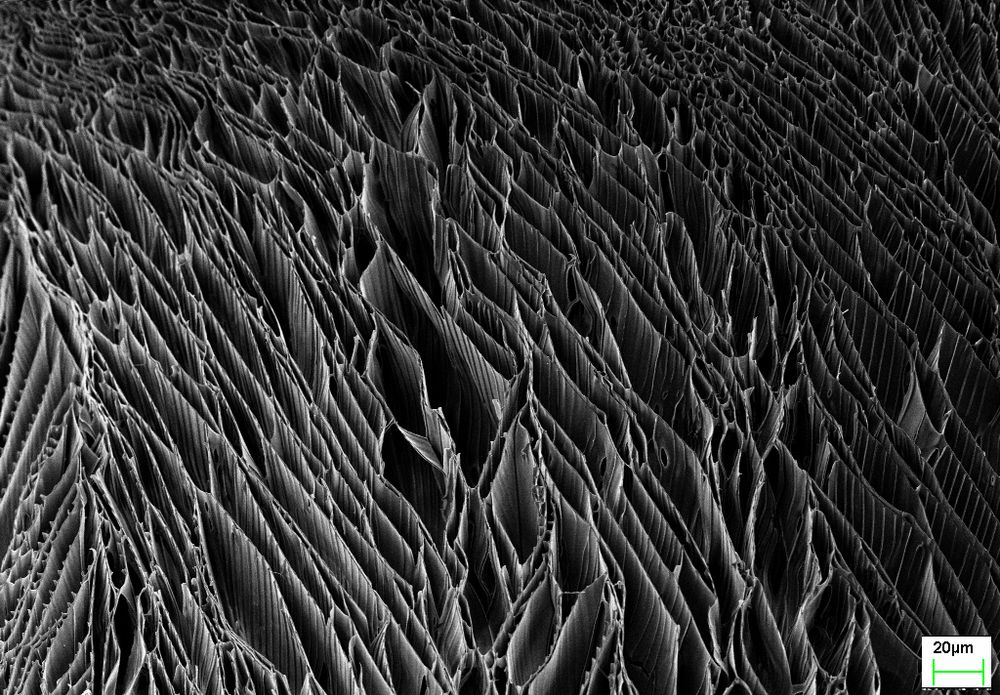
Microscopic image of "sheets" of polymer (plastic) arranged side-by-side
Finding my bearings here on BlueSky. Figuring on dropping more cool "sciencey" stuff on here. I have done quite a bit of electron microscopy... nothing scientifically earth shattering, but I found so many of the images so darned pretty so I'm going to start posting ones I like from time to time.
07.01.2025 19:34 — 👍 3 🔁 1 💬 0 📌 0
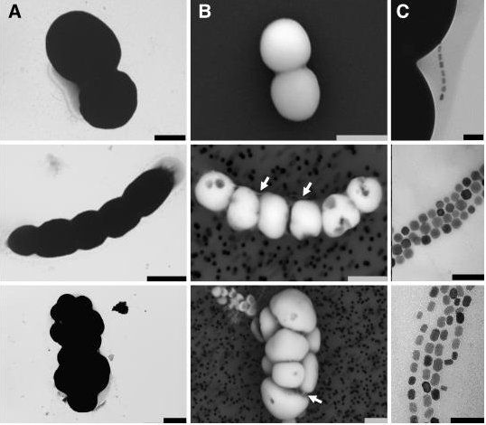
Just published. Check the astonishing diversity of bacteria that can not only form intracellular nanomagnets but also amorphous calcium carbonates that litterally fill the cells. + beautiful EM and cryo-X-ray microscopy images + genomes; follow the link academic.oup.com/ismej/advanc...
08.01.2025 20:27 — 👍 8 🔁 2 💬 0 📌 0
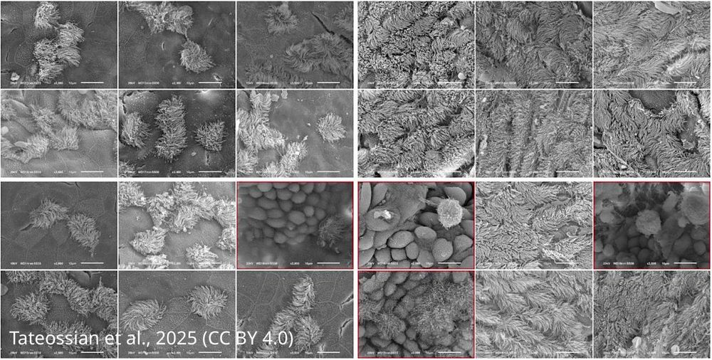
Scanning electron microscopy images from the middle ear of mice showing cilia
Gene discovery sheds light on ‘glue ear’ in people with Down syndrome, paving the way for future targeted therapies.
https://buff.ly/4h6P8eF
08.01.2025 21:01 — 👍 6 🔁 4 💬 0 📌 0
Still our favorite #review! Unfortunately, as relevant today as when it was published, it is an excellent illustration of the #problems arising from the neglect of #ultrastructure and #electronmicroscopy in #cell-biology and the over-reliance on #fluorescence #microscopy.
doi.org/10.1016/0962...
08.01.2025 08:18 — 👍 13 🔁 3 💬 0 📌 0
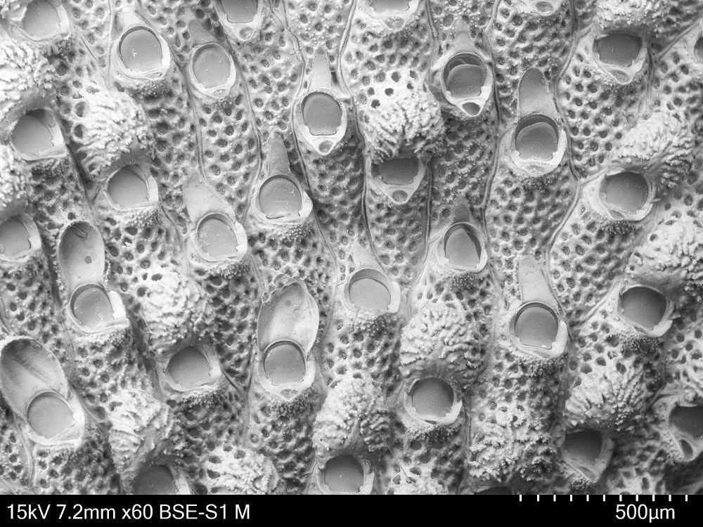
a black and white electron microscopy image of a bryozoan with lots of pores and round, granular ovicells
Potential new invasive species of #bryozoa for Norway alert!
On their way to our #NorDigBryo workshop in Sletvik in October, the people from Bergen did some sampling in some harbors/marinas to look for invasive species and lo and behold:
20.12.2024 14:13 — 👍 7 🔁 3 💬 1 📌 0
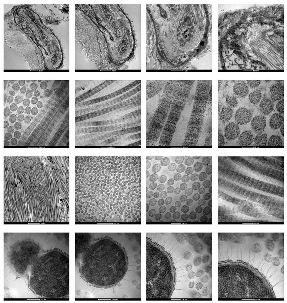
I wanted to end 2024 with these cool Transmission electron microscopy images from my PhD research on raw skin tissue!
Can anyone guess what this structure might be?
#collagen #skinscience
20.12.2024 20:03 — 👍 2 🔁 1 💬 1 📌 0
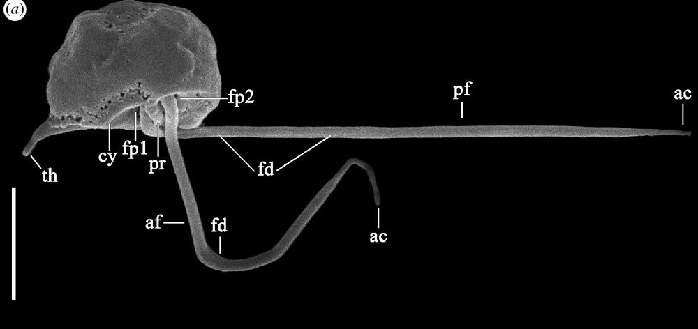
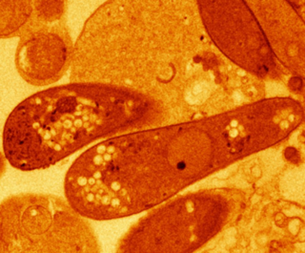
electron microscopy picture of a microbial cell
New #ISEPpapers! The nature of ‘jaws’: a new predatory representative of #Provora and the ultrastructure of nibbling protists royalsocietypublishing.org/doi/10.1098/...
#Protists #Microbes #Biology #TreeOfLife #Microscopy
21.12.2024 18:51 — 👍 22 🔁 4 💬 0 📌 1
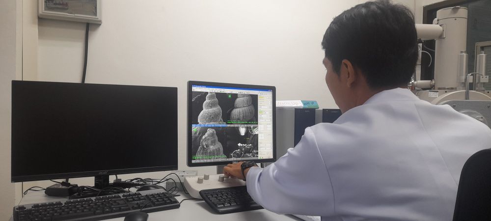
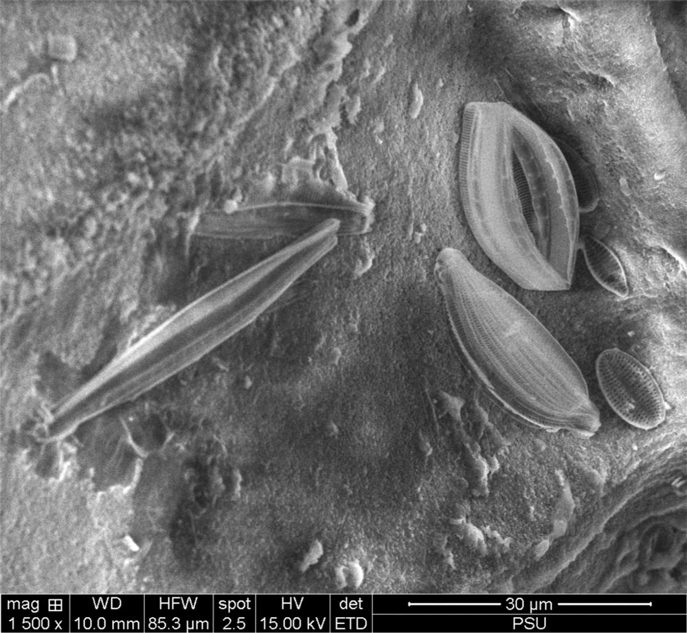
Ending the year with micro-morphology for nerdy taxonomy #1 - Scanning Electron Microscopy never gets old @ Prince of Songkla University, Hat Yai!
ps: Any Diatom people around? We're finding a lot of funky stuff..
24.12.2024 06:36 — 👍 4 🔁 1 💬 0 📌 0
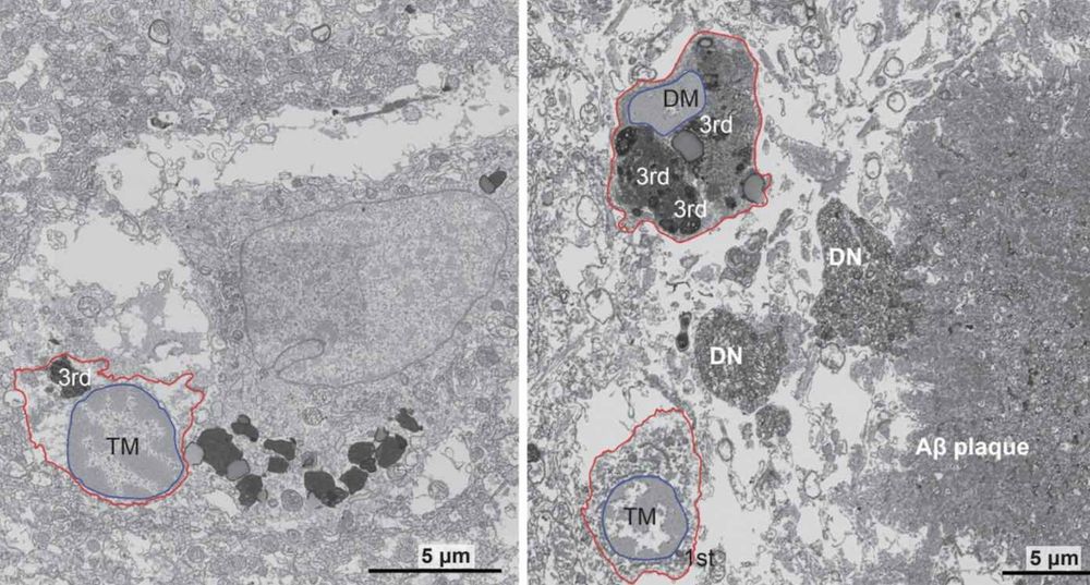
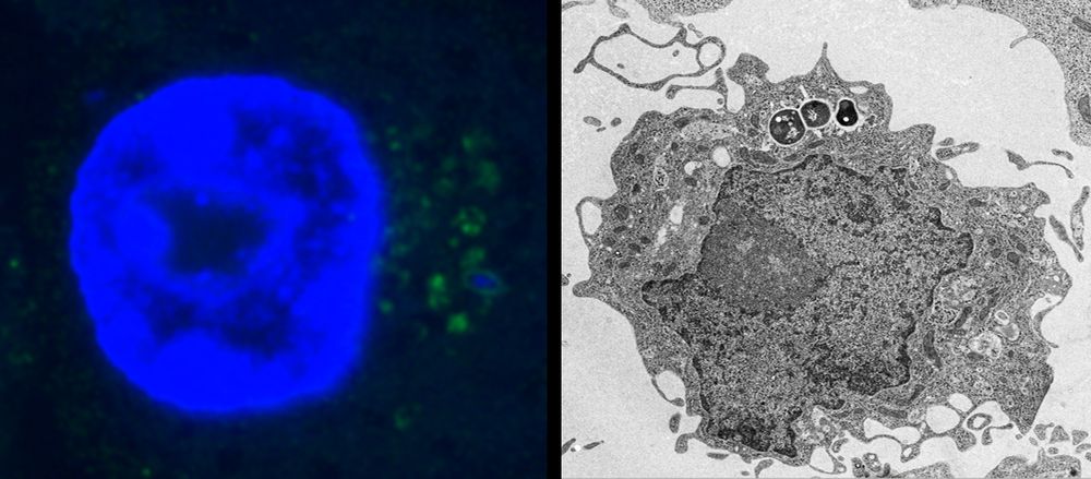
Key Alzheimer’s Breakthrough Identifies Stress-Related Cellular Mechanism Driving the Disease
CUNY scientists made a key Alzheimer's disease breakthrough, identifying a vital mechanism in the brain’s immune cells that drive dementia.
Using electron microscopy, the research team identified an accumulation of “dark microglia”, a subset of microglia associated with cellular stress and neuro-degeneration, in postmortem brain tissues from Alzheimer’s patients. The cells were present at twice the levels seen in healthy-aged people.
31.12.2024 18:11 — 👍 2 🔁 1 💬 0 📌 0
Today my colleague shared with me the most EXQUISITE image of a seed the size of a dust particle, collected from a rare plant called Xylanche in Nepal, and photographed using scanning electron microscopy. Wow.
📷 Renata Piwowarczyk’s work, courtesy of Herbarium LE in St. Petersburg, Russia; scan by Justyna Kasińska from Kielce University of Technology. Thanks for sharing your beautiful work with us x
Today my colleague shared with me the most EXQUISITE image of a seed the size of a dust particle, collected from a rare plant called Xylanche in Nepal, and photographed using scanning electron microscopy. Wow.
03.01.2025 15:22 — 👍 308 🔁 64 💬 7 📌 7
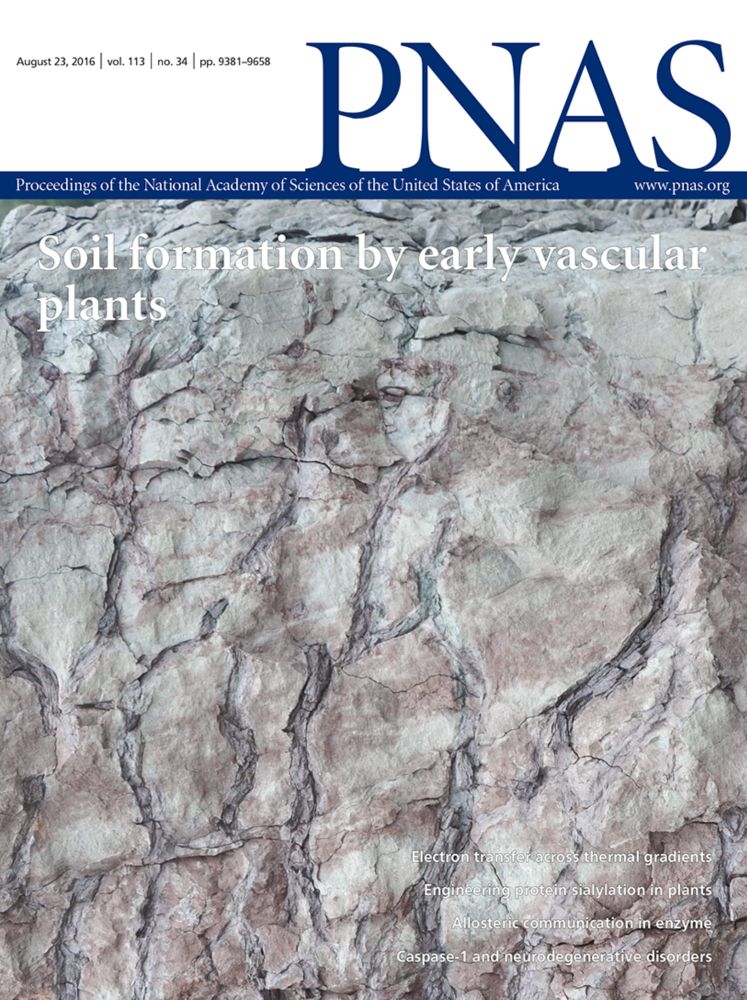
Science in the age of selfies | PNAS
Science in the age of selfies
#Science in the age of #selfies
A short #opinion article that we think hits a very important point!
Albert #Einstein: “an academic career, in which a person is forced to produce #scientific writings in great amounts, creates a #danger of intellectual superficiality”
www.pnas.org/doi/10.1073/...
06.01.2025 07:35 — 👍 4 🔁 2 💬 0 📌 0
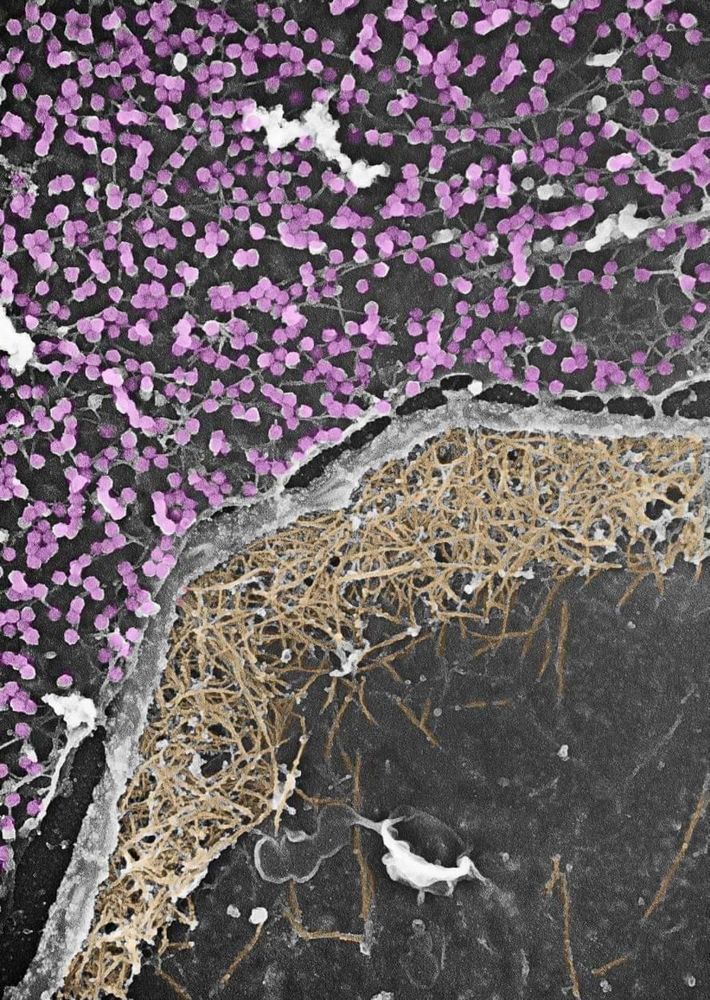
Dynamic duo at the cell's edge: Caveolae and the actin cytoskeleton work together to sense and respond to mechanical stress. Caveolae buffer tension, while actin provides structure and force. Ultimate biomechanics team. #CellBiology #Mechanotransduction
21.12.2024 17:38 — 👍 83 🔁 18 💬 1 📌 1
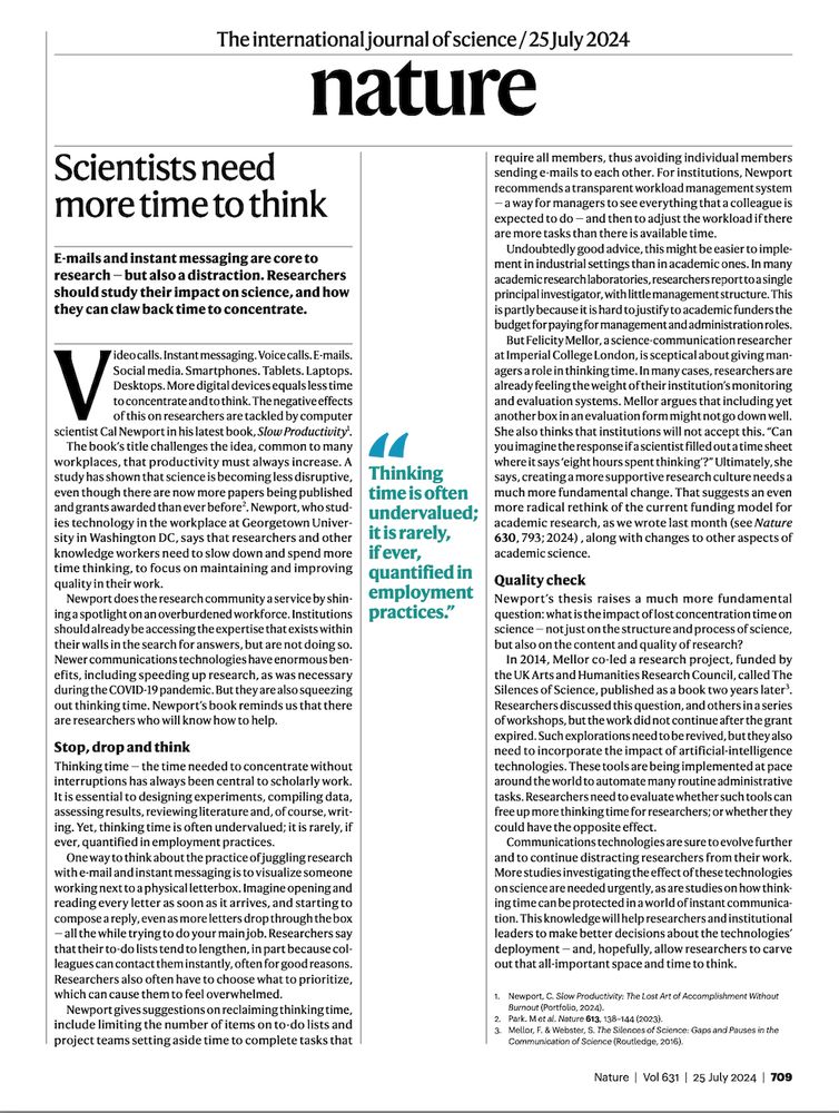
Instead of listing my publications, as the year draws to an end, I want to shine the spotlight on the commonplace assumption that productivity must always increase. Good research is disruptive and thinking time is central to high quality scholarship and necessary for disruptive research.
20.12.2024 11:18 — 👍 1151 🔁 375 💬 21 📌 57
Featured image with the Hughes lab - FocalPlane
Featured image with the Hughes lab - News
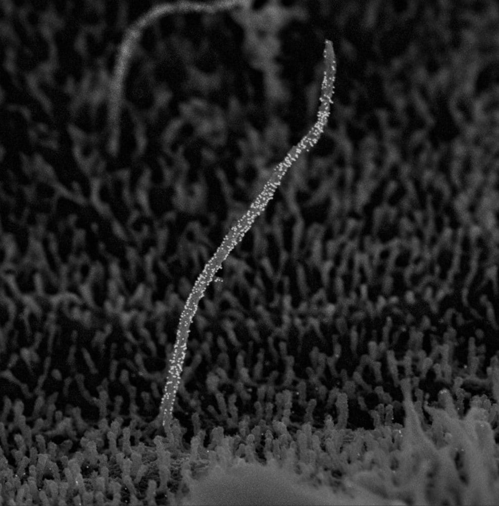
Our final ‘Featured image’ of 2024 is this beautiful immuno-scanning electron micrograph of a primary cilium, acquired by Sanja Sviben. Check out the post to learn more about the image and the research in the Hughes lab: focalplane.biologists.com/2024/12/20/f...
20.12.2024 09:32 — 👍 7 🔁 1 💬 1 📌 0
Is it almost Christmas? Yes!
Is that a reason to stop the @eurobioimaging.bsky.social #VirtualPub? No!
We're closing the year with a big one - a @volumeem1.bsky.social special session from one of our Nodes featuring all their vEM imaging and analysis techniques.
19.12.2024 17:30 — 👍 6 🔁 4 💬 0 📌 0
How dangerous is sodium cacodylate?
Circumstantial evidence suggests that sodium cacodylate may be a far greater health hazard than has been generally assumed. Until further information is available, electron microscopists should see t...
The toxic, #arsenic -containing #Cacodylate buffer is still used in many #electronmicroscopy labs today. Although it is a good buffer, it is no longer needed today as there are better and safer alternatives that perform just as well - #PHEM buffer (Schliwa, 1981) for example.
doi.org/10.1111/j.13...
16.12.2024 07:09 — 👍 4 🔁 1 💬 0 📌 0
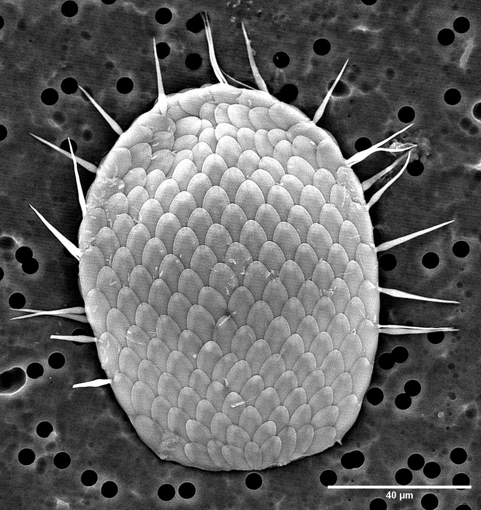
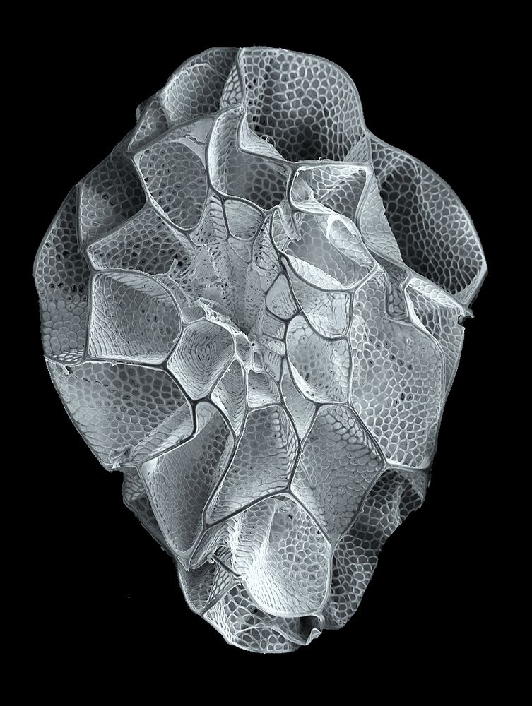
SEM image of a rhizarian testate amoeba (euglyphid) showing its glass scales.
Not a fish or a dragon scale, but a single cell of a #protistsonsky. This rhizarian testate amoeba creates perfect tiny glass scales to build it's shell. From a peat bog. #protistaday.
15.12.2024 13:55 — 👍 168 🔁 31 💬 4 📌 2
Research Associate at the Canadian Museum of Nature, curator of eensy creatures at iNaturalist. Strangely preoccupied with bog ciliates and arcellinid amoebae. Blogger at www.itcamefromthepond.com. Ancestrally biflagellate.
Neuroscience/Electrophysiology/Synapse
Assistant Professor at Akita University
Submicron morphogenesis
Postdoc at University of Vienna @univie.ac.at
A platform for life sciences. Publications, research protocols, news, events, jobs and more. Sign up at https://www.lifescience.net.
Lab Manager @universityofutah.bsky.social @UUNeurobiology | Molecular Mechanisms of Memory | wife and mom of 4 kiddos and a mad dog | views expressed are my own | Lo que escribo es mi opinion.
Ph.D student in Hirabayashi lab (Laboratory of Neurobiology), UTokyo, DC2
Group leader @uio.no - Spatial Immunology & Nanomedicines.
#ERCStG & NFR FRIPRO Young talent
🐟Passionate about fish and their immune system🐟
Biophysicist | Microscopist | Grad Student
Imaging Ageing Endothelium at the
nanoscale | Marie Skłodowska-Curie Actions | Doctoral Networks | Researching fenestrations in bone marrow, brain, and liver
The BIU is a joint NHS and University of Southampton facility providing 2D and 3D light, electron and X-ray imaging for research, diagnostic and commercial users.
Research fellow at Aston Institute of Membrane Excellence. Previously folding proteins at The Crick and membrane proteins at King's College London.
Chromatin Biochemistry and Structural Biology @Center for Molecular Medicine Cologne (CMMC), University Clinic Cologne, Germany
PhD student researching migrant biology at the University of Exeter Cornwall 🪰🔬💪🧬 https://experts.exeter.ac.uk/40094-oliver-poole
Long-distance runner 🏃♂️⛰️
We are at the forefront of #mentalhealth & #neuroscience #research. We collaborate across industries & disciplines to find answers to global #health challenges.
🇪🇸 Group Leader | Julian Lab @ BOKU University 🇦🇹
🔬 Vacuolar defense & plant stress responses
🧬 ATG8ylation | Non-canonical autophagy | VQC
https://boku.ac.at/en/btlw/impb/julian-lab
Geoscientist | Trail runner | Electron microscopist at the Uni of Glasgow | Latvian-born serial expat
https://www.gla.ac.uk/schools/ges/research/researchfacilities/gems/