The latest preprint from the Hansel lab at UChicago. My excellent grad colleague Abby Silbaugh shows that activating a supervisory signal local to the cerebellum can, via thalamus, influence whisker stim-induced plasticity in mouse S1. A fantastic demo that the brain + its plasticity is distributed!
22.04.2025 17:04 — 👍 1 🔁 1 💬 0 📌 0
Thanks Cedric!
18.04.2025 21:24 — 👍 1 🔁 0 💬 0 📌 0
Huge thanks goes to my mentor Christian Hansel, the UChicago neuro institute, and to many for their ideas and feedback along the way: @tingfenglin.bsky.social, Abby Silbaugh, Aurora Ferrell, Donald Huang, Ruth Anne Eatock, Wei Wei, Mark Sheffield, and Peggy Mason
18.04.2025 15:04 — 👍 1 🔁 0 💬 1 📌 0
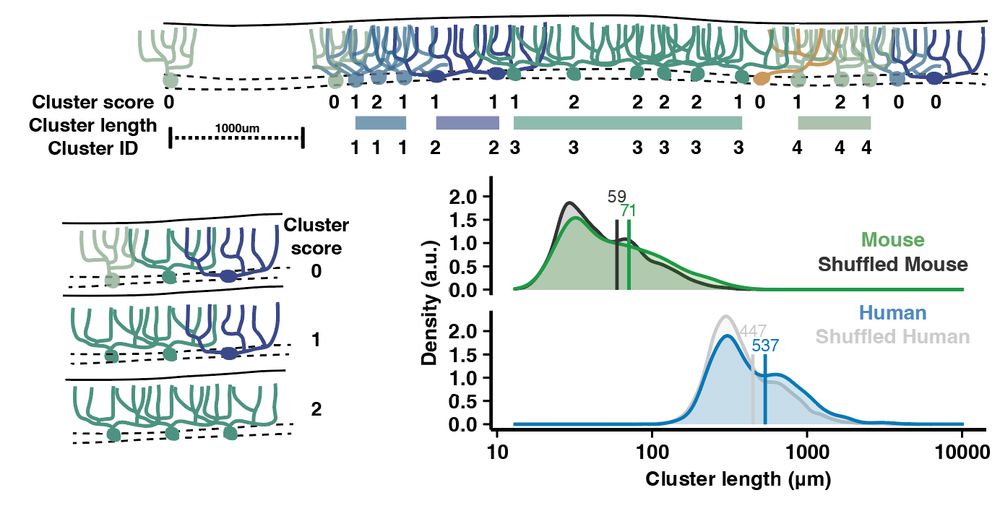
Purkinje cells of similar morphologies also tended to cluster near each other. If morphology dictates some features of PC function, then this cell-cell clustering may generate patches of functional similarity in the cortex
18.04.2025 15:04 — 👍 2 🔁 0 💬 1 📌 0
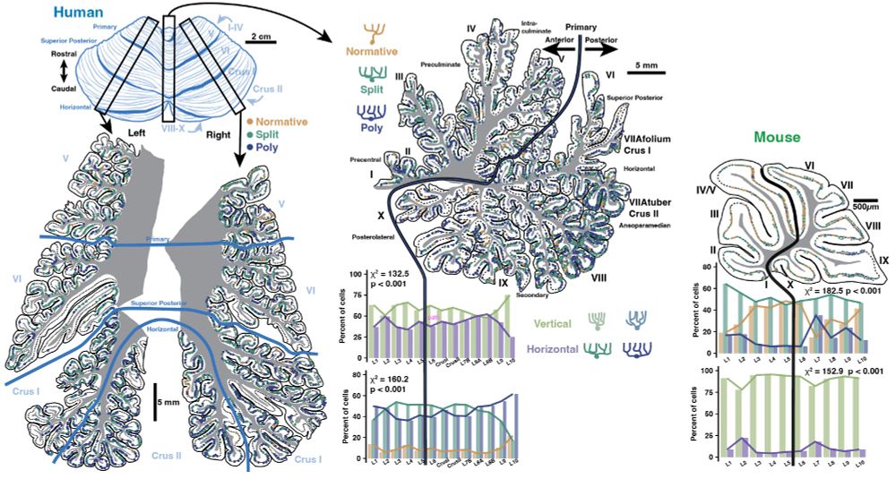
Building on our 2023 analysis of PC morphology by region, the anterior-posterior gradient in the hemispheres is bilateral, distinct from the pattern in vermis (in both species), and inter-hemisphere similarity covaries with functional symmetry
18.04.2025 15:04 — 👍 1 🔁 0 💬 1 📌 0
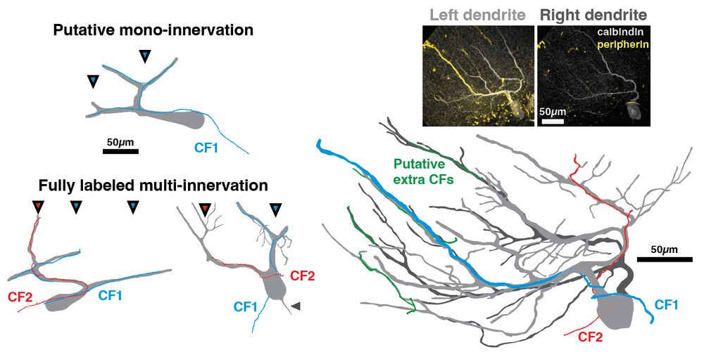
Using peripherin as a putative label for climbing fibers (CFs), we show that human PCs can indeed receive multiple CFs (and on separate dendritic compartments), confirming a hypothesis from our previous work
18.04.2025 15:04 — 👍 1 🔁 0 💬 1 📌 0
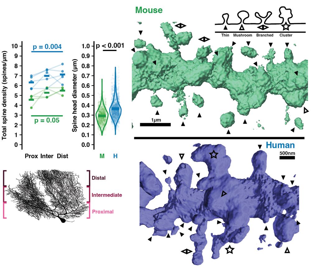
Outward growth of dendrites (w conserved diameter but 3x longer individual branches) may influence how human cells sample and integrate input. Human dendritic spines are also denser, larger, and show more complex morphologies including a ‘spine cluster’ motif
18.04.2025 15:04 — 👍 1 🔁 0 💬 1 📌 0
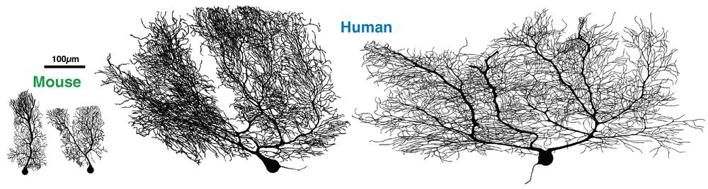
Despite having only 2x thicker cerebellar cortex, human Purkinje cell (PC) dendrites spread laterally to a total length 11x longer than mouse. This far exceeds our previous measure, revealing they are >2x the total length of human pyramidal neurons
18.04.2025 15:04 — 👍 3 🔁 0 💬 1 📌 0
Human neurons are incredibly complex and Purkinje cells are, by far, the largest in our brain. We give a detailed characterization of their dendrite and spine morphology and distribution across cerebellar regions compared with mouse. Check it out!
11.09.2024 13:29 — 👍 0 🔁 0 💬 0 📌 0
The aliens are within.....
31.10.2023 18:13 — 👍 1 🔁 0 💬 0 📌 0
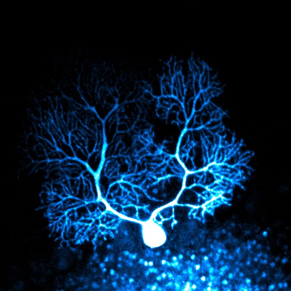
For my first blue post, a blue Purkinje cell!
This cell was loaded with dye during whole cell patch clamp in sliced mouse tissue, and oops, some dye spilled out on the granule cells below during the approach...
19.10.2023 12:54 — 👍 10 🔁 1 💬 2 📌 0
into brain evolution & development, open science, art & science https://www.youtube.com/watch?v=6hMNZHsrNHw, music, making, javascript, contemporary dance https://www.youtube.com/watch?v=OZfHj7F2FzQ
website: katjaq.github.io
Brains, Data and Science
Prof in Computational Neuroscience at Western University
Cerebellum and Motor Control
Neurobiology PhD student at UChicago.
Mechanisms of learning and memory in distributed brain circuits, and other things.
Neuroscience Professor at Harvard University. Personal account and posts here. Research group website: https://vnmurthylab.org.
Postdoc @ the Leslie Vosshall lab, Rockefeller University.
Molecular-neuro-evolution + mosquitoes
Random thoughts & interesting papers
neurobiologist, ornithologist, scruffy looking nerfherder, social distancing since 1974, currently at U Lethbridge, Treaty 7 lands
🇨🇦 🇦🇺 stuff
open access link for our book: https://direct.mit.edu/books/oa-monograph/6000/Bird-Brains-and-BehaviorA-Synthesis
Assistant Professor Stanford. Neuroscientist and psychiatrist. Cellular and molecular mechanisms of human brain development and disease.
Evo-Devo neurobiologist | Associate Prof. at Institute of Genomics, University of Tartu | SFARI Fellow | EMBO Installation Grantee | Cerebellum development: how it evolved and how it goes wrong
Biomedical Picture of the Day: daily intriguing images from global research. IMAGE/RESEARCH EXPLANATIONS AND LINKS TO THE PAPER on bpod.org.uk
Supported by
@leicamicrosystems.bsky.social &
Dept of Biochemistry, @ox.ac.uk
Neurophysiologist working on motor control and neural population dynamics. Assistant Prof. at the Case Western Reserve University School of Medicine.
https://sauerbreilab.org/
Professor of Neuroscience. Studying neural development, regeneration, and control of innate behaviors at Johns Hopkins.
@sethblackshaw at Twitter.
PhD Student @ Brandeis University
Vision Neuroscience and Tennis Fanatic
A veterinary ophthalmologist @ucdavis, 👁 scientist, & mom looking to make the 🌎 clearer for people & animals. Co-director of CMSTP T32 for DVMs getting their PhD. Views are my own. 🐴🦜🥰
https://covsl.vetmed.ucdavis.edu/
Neuroscientist+Child Neurologist. McNair Scholar #RareDisease, #neurogenetics, inhibition/circuits, #autism, #epilepsy, & brain disorders. RT≠endorse.
Cutting-edge research, news, commentary, and visuals from the Science family of journals. https://www.science.org
PI @Rockefeller University. Investigator @HHMI. Instigator of clonal raider ant project #CRAP. 🐜 Evolution, Behavior & Neuroscience. Posts science and photography. 🧠 📸
https://www.rockefeller.edu/research/2280-kronauer-laboratory/
VP/CSO @HHMI.bsky.social & Head of #VosshallLab @rockefelleruniv Toward Excellent, Inclusive, & Open Science
The Rockefeller University is a world-renowned center for research and graduate education in the biomedical sciences, chemistry, bioinformatics and physics.
Postdoc at @JustusKebschull lab at @JohnsHopkins. I am interested in evolution of cerebellum-ish, spinal cord and everything in between. @cimec_unitrento, @oist_edu PhD.