We're excited to welcome Jeff Chen and JinYoung Lee to the team! 🎉
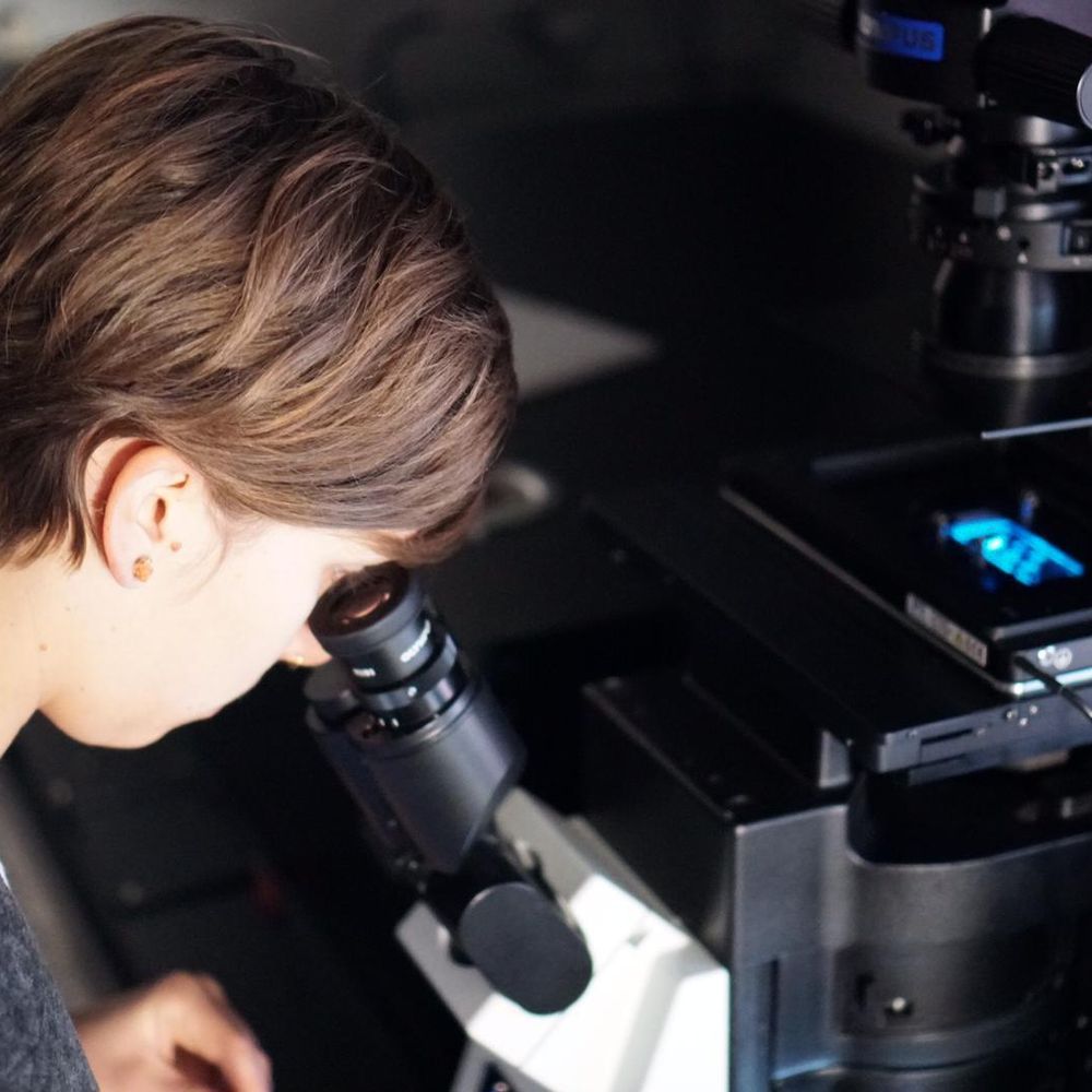
Both are University of Toronto undergrads joining us for a gap year. Jeff will work on barcode design and viral payloads, while JinYoung will focus on expansion microscopy and imaging.
10.06.2025 16:49 — 👍 1 🔁 1 💬 0 📌 0
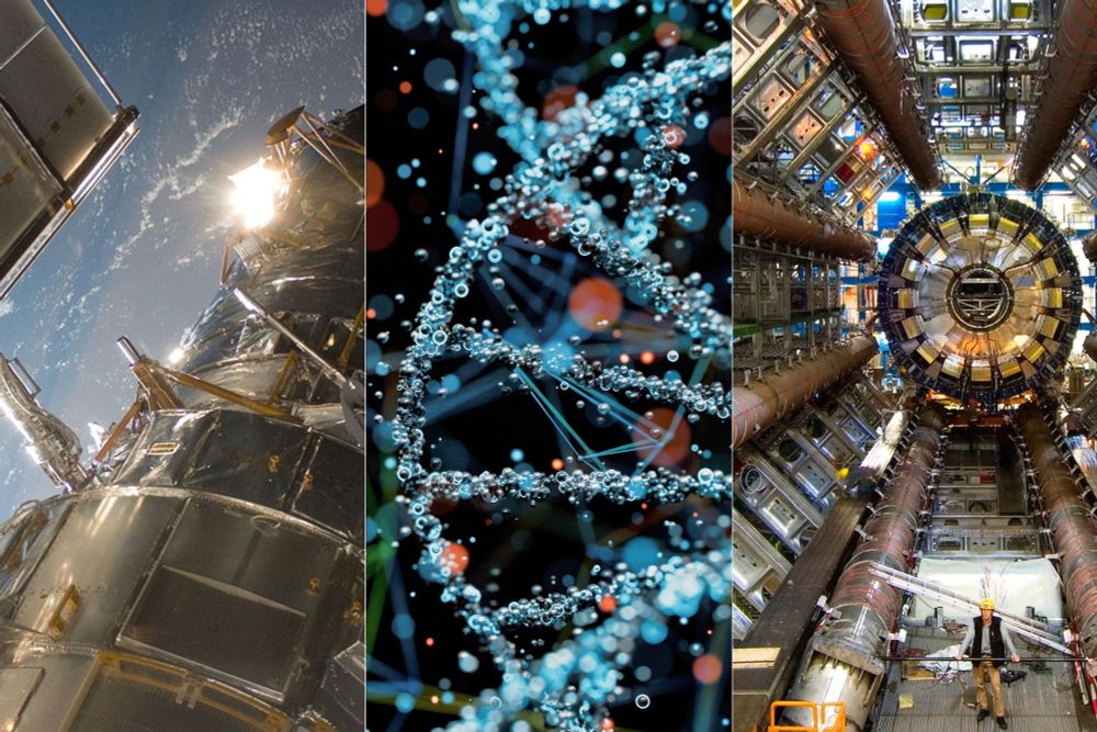
Former MIT researchers advance a new model for innovation
Developed by former MIT researchers, focused research organizations (FROs) undertake large research efforts and have begun to contribute to scientific advances.
"Academic research groups and startups are essential drivers of scientific progress. But some projects, like the Hubble Space Telescope or the Human Genome Project, are too big for any one academic lab or loose consortium..."
09.06.2025 19:45 — 👍 7 🔁 4 💬 2 📌 1
E11 Bio is launching Volara: an open-source Python library for block-wise processing of large volumetric microscopy datasets! 🧪🔬
Volara features block-wise task abstractions, making scalable image processing more accessible, robust, and repeatable.
Read more: e11.bio/blog/volara
29.05.2025 15:29 — 👍 35 🔁 18 💬 2 📌 2
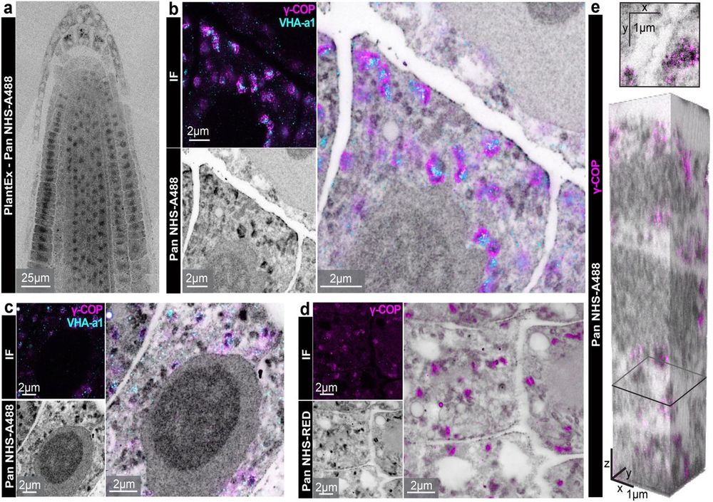
Pan-protein labeling reveals ultrastructure in expanded Arabidopsis thaliana roots.
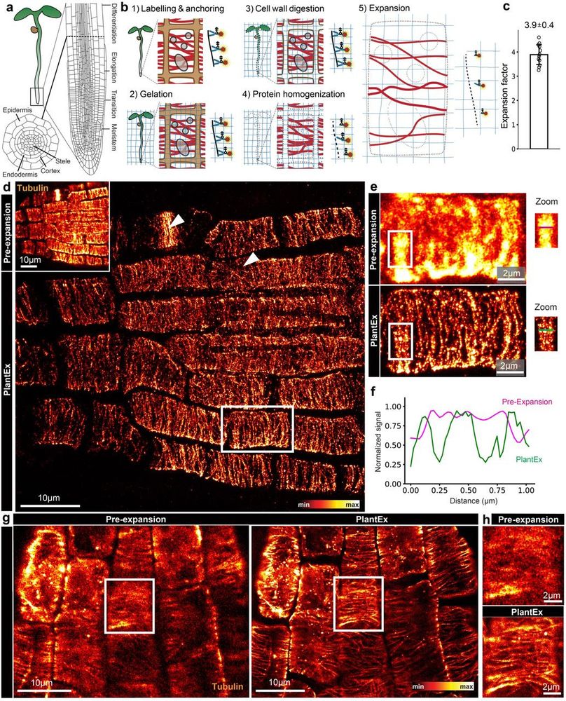
PlantEx provides sufficient resolution to visualize individual microtubules in expanded Arabidopsis thaliana roots.
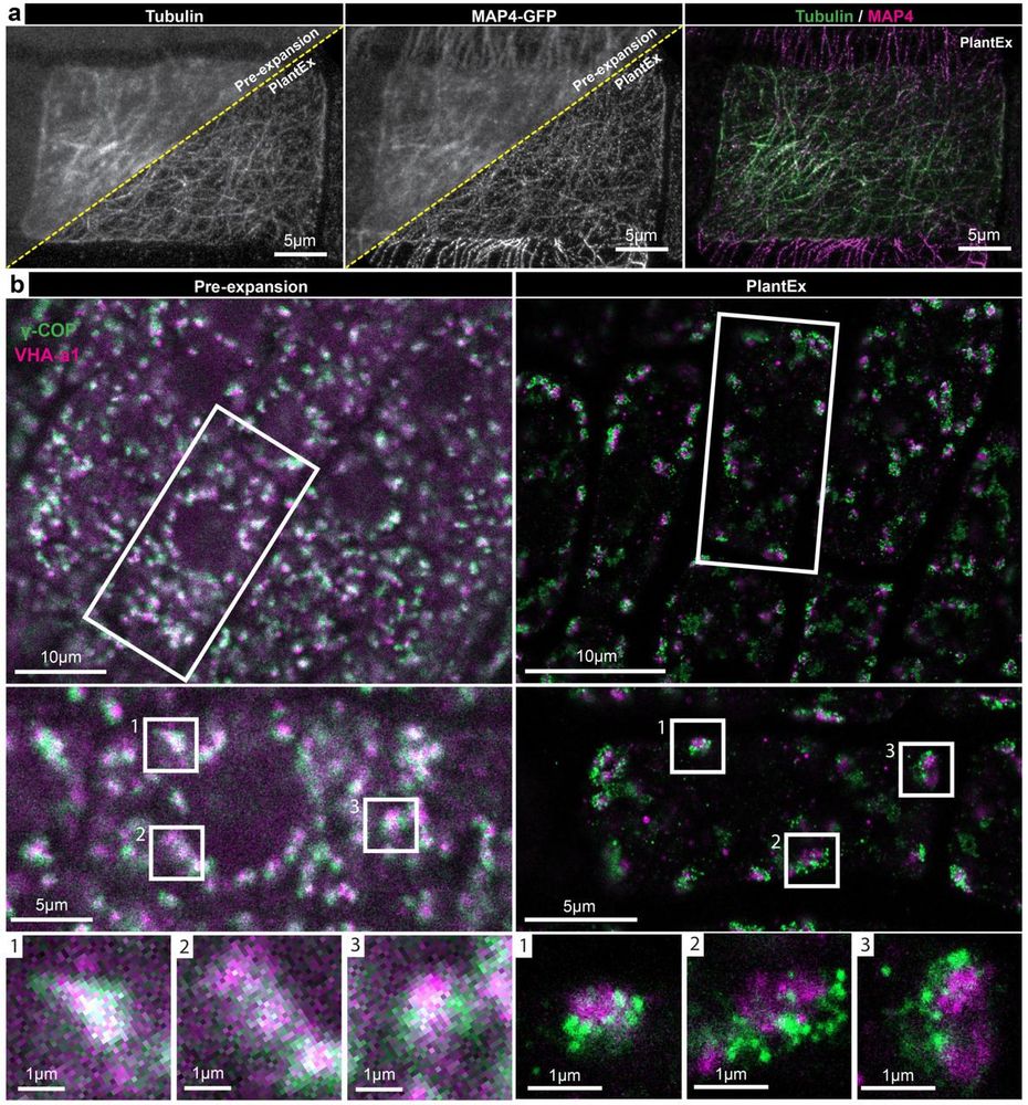
Golgi vesicles can be visualized at super-resolution in Arabidopsis thaliana roots via PlantEx expansion microscopy.
I'm really hoping to see some applications of PlantEx this year — tell your plant friends! 🍀
If you are a plant biologist yourself and interested, skeptical, or struggling implementing expansion microscopy — just write me; it's easy & I'd love to be of assistance! 🌺
www.biorxiv.org/content/10.1...
16.02.2025 02:26 — 👍 1 🔁 1 💬 0 📌 0
Our work at @e11bio.bsky.social in connectomics is featured in @asimovpress.bsky.social ! We’re building new tools to map the brain with greater precision and scale.
10.02.2025 18:16 — 👍 2 🔁 0 💬 0 📌 0
GitHub - wiebkejahr/slm_control
Contribute to wiebkejahr/slm_control development by creating an account on GitHub.
Do you work with confocal, STED or MINFLUX microscopes, commercial or homebuilt? Do you struggle with control of your spatial light modulator or with incorporating adaptive optics, or do you just need more flexibility? Have a look at my SLM control code:
github.com/wiebkejahr/s...
20.01.2025 20:38 — 👍 11 🔁 2 💬 1 📌 1
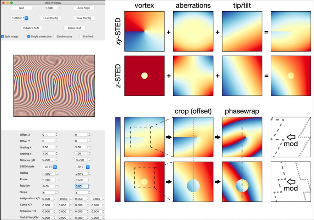
Image showing a GUI with many numeric inputs on the left side and color coded intensity maps, illustrating a workflow, on the right side.
🔬peeps!
I am delighted to finally publish my code for controlling a spatial light modulator (SLM) to perform adaptive optics & to sculpt the vortex beams for STED & MINFLUX microscopy - complete w graphical user interface for easy use!
github.com/wiebkejahr/s...
Short user manual🧵
15.01.2025 15:31 — 👍 45 🔁 10 💬 4 📌 2
Here at E11BIO we have been working hard to make optical #connectomics accessible for the scientific community. Check out our journey!
06.12.2024 18:57 — 👍 3 🔁 0 💬 0 📌 0
Nature Reviews Neuroscience features reviews, perspective articles and the latest research news.
https://www.nature.com/nrn/
We report on the latest news in all fields of science. See also @snexplores.bsky.social
Cutting-edge research, news, commentary, and visuals from the Science family of journals. https://www.science.org
Science lover. Waterpolo player. Sport & concert-addicted. Queer 🏳️🌈🙂 She/her
A journal dedicated to publishing the latest advances across all areas of cell biology. Part of @natureportfolio.nature.com
nature.com/ncb/index.html
Nature Communications is an open access journal publishing high-quality research in all areas of the biological, physical, chemical, clinical, social, and Earth sciences.
www.nature.com/ncomms/
The funder-researcher collaboration and open-access publisher for research in the life and biomedical sciences.
Follow @eLifeCommunity.bsky.social
Sharing what’s new in #microscopy for science & education. 🔬
Carl-Zeiss-Promenade 10, Jena 07745
zeiss.ly/i_bio
Journal of Cell Biology publishes peer-reviewed research on all aspects of cellular structure and function. Published by Rockefeller University Press @rupress.org
🌐 https://rupress.org/jcb
PLOS is a non-profit organization on a mission to drive open science forward with measurable, meaningful change in research publishing, policy and practice.
Nature Reviews Neurology is a clinical review journal from Springer Nature. Follow us for the latest updates in neurology from the journal and beyond. www.nature.com/nrneurol
Research, Education and Business. Vienna BioCenter is a life science cluster that includes IMP, IMBA, GMI, Max Perutz Labs, as well as the Faculty of Life Science & CeMESS (both University of Vienna) and 35+ biotechs.
Engineer in optics and image processing
The IMP is a leading life science centre in Europe with 220 researchers from 40 countries. Part of the ViennaBioCenter.
🧬 Join our world-class courses, conferences, and workshops at the forefront of molecular life science and its applications. 👩🏻🔬 www.embl.org/events/ 🔬
Molecular Microscopy and Spectroscopy Lab (https://vicidominilab.github.io) at the @iitalk.bsky.social.
"A #SPADarray detector in any laser-scanning microscope"