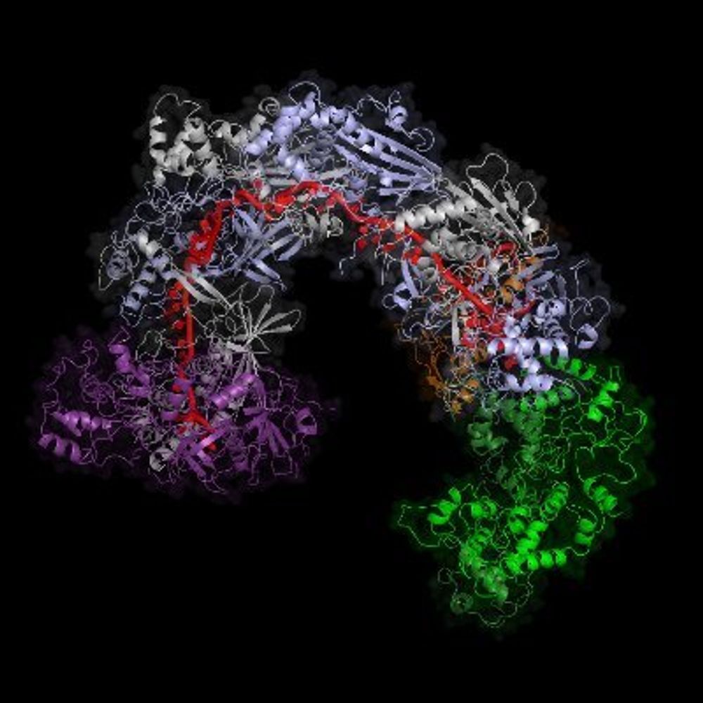
Much needed addition to the AlphaFold Database! 💻 Large potential for use in #StructuralBiology #Phage research.
19.05.2025 11:23 — 👍 11 🔁 0 💬 0 📌 0
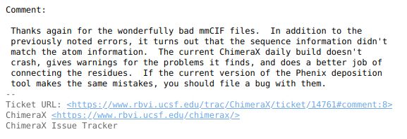
Always happy to provide "wonderfully bad mmCIF files" to make #chimeraX better!
28.04.2025 08:01 — 👍 7 🔁 0 💬 0 📌 0
Nice work and awesome #scicomm thread!
18.04.2025 16:46 — 👍 2 🔁 0 💬 0 📌 0
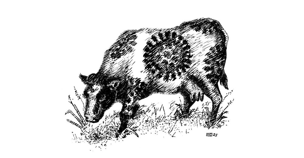
A picture of a Fresian cow whose markings resemble influenza viruses
Our study on pasteurising influenza viruses in milk is now out! It's a nice simple story: influenza viruses (including H5N1) are killed really effectively by pasteurisation, but in raw milk they stay infectious - obvious public health implications of both points... (1/2)
rdcu.be/d73te
30.01.2025 17:12 — 👍 157 🔁 84 💬 8 📌 5
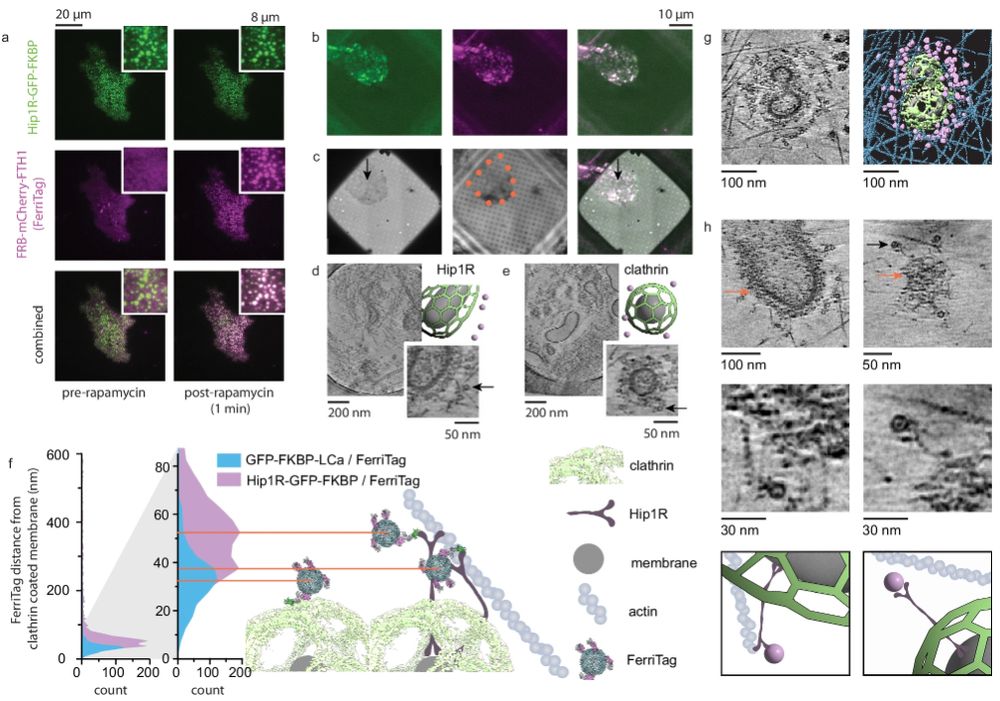
This is a scientific figure from the paper mentioned in the post. It shows fluorescence and electron microscopy images and associated schematics for using the ferritag to image clathrin coated pit proteins.
I'd like to draw your attention to this truly excellent paper from the Taraska lab on cryoET of plasma membrane associated proteins. Everyone who is thinking about probes in the cryoET space should also see what they could do with ferritag (fig 6) www.nature.com/articles/s41...
22.01.2025 16:40 — 👍 404 🔁 58 💬 11 📌 7
I appreciate the stats! Thanks, @pdbeurope.bsky.social for a summary!
13.01.2025 11:45 — 👍 4 🔁 1 💬 0 📌 0
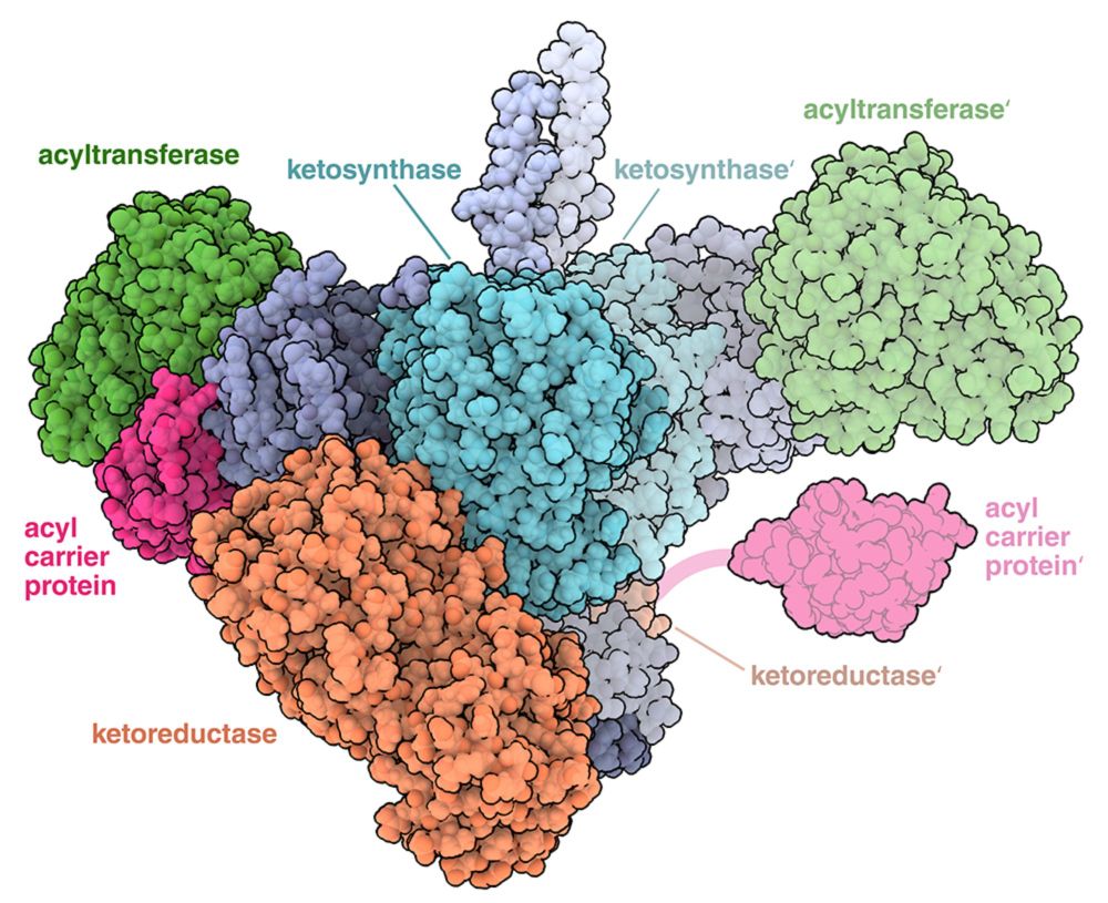
PDB101: Molecule of the Month: Assembly Line Polyketide Synthases
Large multienzyme complexes that synthesize diverse small molecules in a stepwise manner.
I'm happy and humbled to take on a new role with @rcsbpdb.bsky.social. I'll be continuing David Goodsell's work on the Molecule of the Month, starting with my first article on Assembly Line Polyketide Synthases! Check it out (animation included!)
pdb101.rcsb.org/motm/301
07.01.2025 18:32 — 👍 165 🔁 39 💬 5 📌 1
Amazing work and resource! 😍
06.01.2025 13:35 — 👍 2 🔁 0 💬 0 📌 0
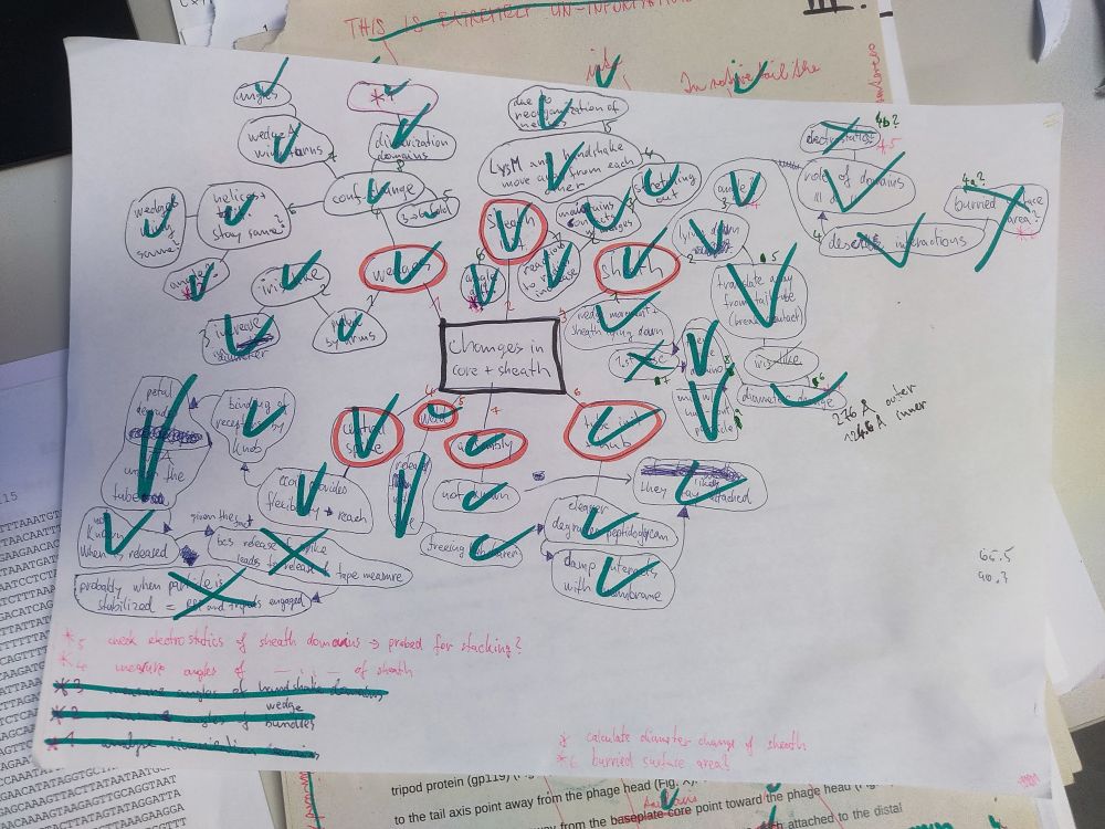
The image is a mind map drawn on white paper, used for organizing thoughts before writing a paragraph. It contains various interconnected words and phrases, with many items circled, underlined, or checked off using green and red pens. Some words and phrases are marked with red circles, possibly indicating key points, while others have green checkmarks denoting completion. Additional notes and arrows are scattered across the sheet, suggesting a workflow for developing ideas. The lower section includes handwritten notes in red and green ink, outlining tasks like measuring angles, verifying structures, and calculating specific parameters.
🎄Xmas office cleaning 🗑️
...and how do you organize your thoughts? 🫵
#PhDlife #PhDSky #mindmap
17.12.2024 09:40 — 👍 3 🔁 0 💬 0 📌 0
I knew it wouldn't be long before we see #cryoEM structures of a #phage cousin infecting #Archea 🦠 🧪
Beautiful structures from @daumlab.bsky.social, congrats!
11.12.2024 12:51 — 👍 19 🔁 1 💬 0 📌 0
impressive structures! Congrats!
10.12.2024 13:01 — 👍 1 🔁 0 💬 0 📌 0
Thanks, I am glad you like it!
26.11.2024 19:36 — 👍 1 🔁 0 💬 0 📌 0
can I join the list? ✌️🙂
26.11.2024 10:55 — 👍 0 🔁 0 💬 1 📌 0
Dear #StructuralBiology #PhD #PostDoc peers, join in!
22.11.2024 08:12 — 👍 4 🔁 0 💬 0 📌 0
Thanks for doing this! Can you add me to the list? I am a PhD candidate in Structural Biology program at CEITEC in Brno, CZ ✌️
22.11.2024 08:02 — 👍 1 🔁 0 💬 1 📌 1
are #CryoEMemes back?😀
15.11.2024 21:14 — 👍 0 🔁 0 💬 1 📌 0
Hi Daniel, great idea! Please add me there🤙
15.11.2024 21:12 — 👍 1 🔁 0 💬 1 📌 0
Another useful starter pack 👇
15.11.2024 21:11 — 👍 1 🔁 1 💬 0 📌 0
In case someone is looking for this...
14.11.2024 21:48 — 👍 8 🔁 4 💬 0 📌 0
Hi, can you add me to the list, please? I'd be happy to connect with other structural biologists!
14.11.2024 21:45 — 👍 0 🔁 0 💬 0 📌 0
I would like to be added to the list🙌
14.11.2024 19:06 — 👍 0 🔁 0 💬 1 📌 0
Just uploaded a revised version of the manuscript on bioRxiv 🙌
We made some changes to the text and figures, mainly to clarify the naming of proteins.
Yeah, it's a challenge to describe things within two different 10MDa structures 🐘
08.11.2024 13:00 — 👍 4 🔁 2 💬 1 📌 0
thank you! 🙂
24.09.2024 19:42 — 👍 0 🔁 0 💬 0 📌 0
Thanks for reading! If you found this thread interesting, pass it along!
bsky.app/profile/jbin...
Hopefully, the peer-reviewed version will follow!
Also, if you find any mistakes or have suggestions, drop me a ✉️
11/11
23.09.2024 16:40 — 👍 3 🔁 0 💬 0 📌 0
Thanks to my amazing co-first author Marta Šiborová and people from the Plevka Lab for their incredible support 🙌
Special shoutout to all collaborators for their contributions 📈💡
10/11
23.09.2024 16:38 — 👍 3 🔁 0 💬 1 📌 0
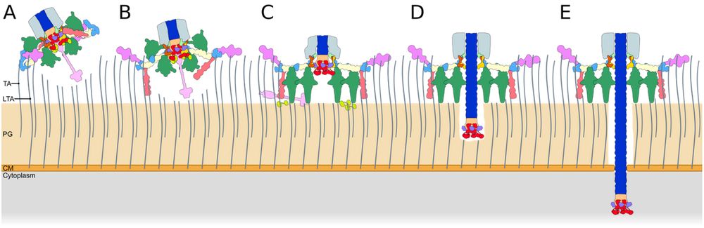
Schematic illustration of phage attachment and penetration through a bacterial cell wall. The diagram shows multiple stages of a baseplate interacting with the bacterial envelope, progressing from initial attachment to full penetration. The bacterial layers, including peptidoglycan (PG) and cytoplasmic membrane (CM), are labeled, with viral components depicted in different colors, illustrating the stepwise mechanism of infection.
The tail sheath contraction pushes the tail tube's tip into the host cytoplasm, where the genome is ejected 🧬 and infection occurs.
Our structural study of phi812 cell wall binding opens up the possibility of designing phi812 to target specific #SAureus strains 🎯
9/11
23.09.2024 16:36 — 👍 5 🔁 0 💬 1 📌 0
Changes in the baseplate arms' positions are relayed to the wedge modules and tail sheath initiator proteins, causing their iris-like expansion 👁️ and triggering a gradual tail sheath contraction. This 🌊-like process drives the tail tube through the host cell wall 💉.
8/11
23.09.2024 16:32 — 👍 3 🔁 0 💬 1 📌 0
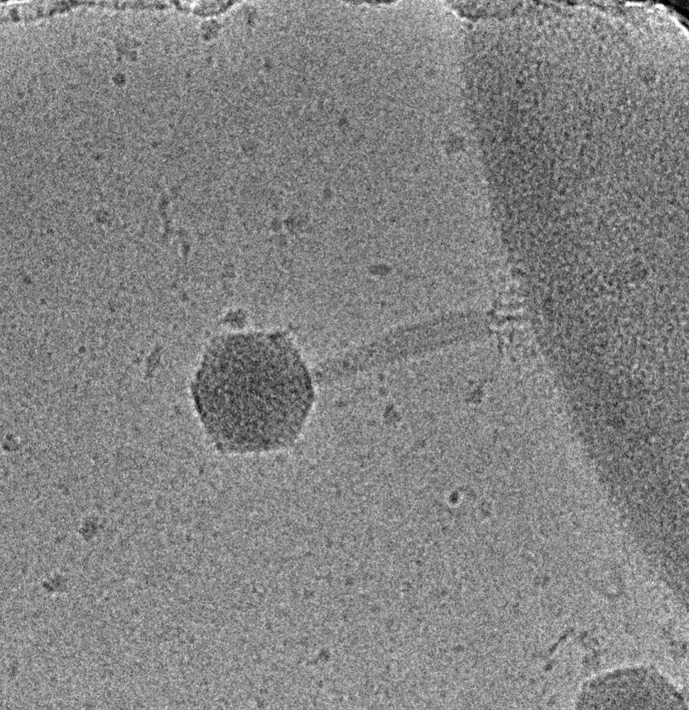
Cryo-electron micrograph of phage phi812 with a semi-contracted tail sheath. Phage is attached to a cell wall of S. aureus.
The transformation of tripod complexes frees the hub protein domains to degrade peptidoglycan ✂️ and facilitate penetration of the tail tube through the cell membrane.
But how does the tail tube move towards the cell❓
7/11
23.09.2024 16:30 — 👍 4 🔁 0 💬 1 📌 0
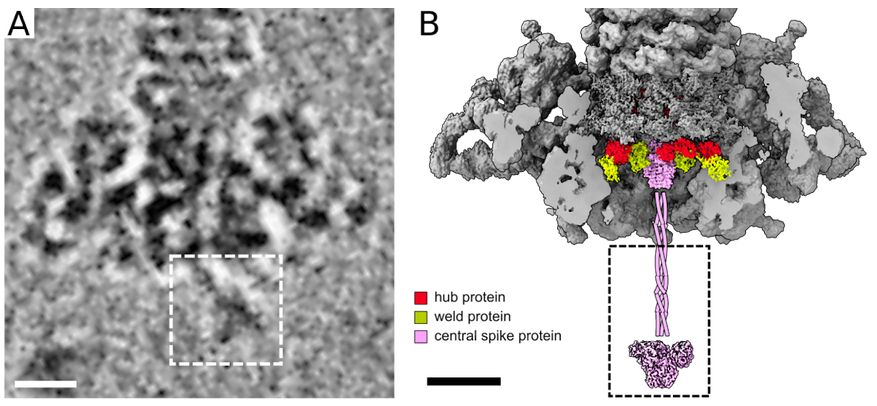
Panel A: Cryo-electron micrograph of the phi812 baseplate. The white dashed rectangle contains the coiled-coil, knob, and petal domains of the trimer of central spike proteins. Scale bar 100 Å. Panel B: The surface representation of the cryo-EM reconstruction of the native phi812 baseplate is colored in gray except for central spike, hub, and weld proteins. The front part of the baseplate has been removed. Scale bar 100 Å.
The central spike protein of phi812 protrudes 22 nm from the baseplate and degrades teichoic acids✂️, enabling the tripod complexes to anchor the phage on the cell wall ⚓️.
6/11
23.09.2024 16:28 — 👍 4 🔁 0 💬 1 📌 0
News from the Sternberg lab at Columbia University, Howard Hughes Medical Institute.
Posts are from lab members and not Samuel Sternberg unless signed SHS. Posts represent personal views only.
Visit us at www.sternberglab.org
Structural Bioinformatics👨💻🧬
PhD student at Atkinson Lab, Lund University
Archaea, Eukaryotes, DNA replication & repair, Evolution enthusiast.
Biochemist, Structural biologist.
Postdoc #UniKiel
Unfocused wildlife 📸, Smasher of🏸. Opinions are my own
Crime writer based in Vienna, bringing the city's dark corners and quirky tales to life.
https://a.co/d/0ye5EL5
#slavaukraini🇺🇦
#Pro-Democracy
#Resist
Krimiautor aus Wien, der die dunklen Ecken und skurrilen Geschichten der Stadt zum Leben erweckt.
Engineering Human Resources. A job board and career portal for engineers, researchers and developers. Our website: https://www.engineering.hr
M.D. | Physicist | Dr. rer. nat. | Investigating Tissue and Neurodegeneration by #CryoET, PostDoc @uniGoettingen, previous MPI Biochemistry
The VIGILANT project is funded by #HorizonEurope and develops broad spectrum antivirals for pandemic preparedness. Find more information at https://linktr.ee/VIGILANT_EU
Industry Scientist in SD | Sequencing + Cells
🦠 Postdoc @jcvi.org
🧠 PhD @utah.edu
I enjoy playing with family, reading science fiction and history, gardening, board games, and jazzercise. I'm a father, husband, Navy veteran, and graduate student studying computer science at George Mason University.
Mastodon: https://scicomm.xyz/@David
Science Events helps you organise a scientific conference or workshop. Use our platform to organise your next event! Visit our platform at https://www.sci.events
PhD student at Trevor Lithgow's group Monash BDI. Facultative bioinformatician who in love with phages and their dark matters.
I write dark Victorian fiction and specialize in the long 19th century.
She/Her 🏳️🌈🇪🇸✡️ (I'm always kind)
#writer #historian #Lesbian #Hispanic #feminist #goth #vampires
NO DMs (mutuals) | TerfsDNI
Phage biologist focused on virus-host interactions and bacterial pathogenesis.
Official Account for the Lorne Protein Meeting, Australia.
lorneproteins.org
We provide a cloud-native platform for cryo-EM data analysis which integrates cloud storage, scalable compute and an intuitive web-app for fast and easy end-to-end cryo-EM data analysis.
Scientist @ Thermo Fisher Scientific
Structural biologist by day 🧠 manga artist by night 🎨 PhD loading ~99% 👨👩👧 mom 📍Wien, AT
linkedin.com/in/anna-santa-molbio
researchgate.net/profile/Anna-Santa-2?ev=hdr_xprf