Please comment on this! The complexity of living systems cannot be fully recapitulated by in vitro or computational approaches alone. While advances in non-animal model technologies offer complementary tools, they lack the full physiological and developmental context of a living organism.
10.07.2025 22:41 — 👍 47 🔁 31 💬 0 📌 1
Thank you Mark! Yes, I will probably talk about this story!
15.01.2025 11:15 — 👍 0 🔁 0 💬 0 📌 0
This work was a collaboration with Toshihiko Fujimori, Noriyuki Kinoshita, and a former PhD student Thanh Phuong Nguyen, who supported the imaging and laser ablation experiments. And special thanks to Mikio who gave me the freedom to pursue this issue in his lab! (13/n)
14.01.2025 10:26 — 👍 3 🔁 0 💬 0 📌 0
Many important questions remain. How do their neighbors recognize junction-deficient cells? How does cell compression lead to apoptosis? What are the physiological functions of this process in vivo? These issues will keep us busy in the future! (12/n)
14.01.2025 10:26 — 👍 2 🔁 0 💬 1 📌 0
Further exploring the underlying molecular mechanisms, we found that mechanosensing in the ‘winner’ cells via the Hippo signaling and an adherens junction component afadin was required to eliminate ZO-1/ZO-2 double KO cells. (10/n)
14.01.2025 10:26 — 👍 2 🔁 0 💬 2 📌 0
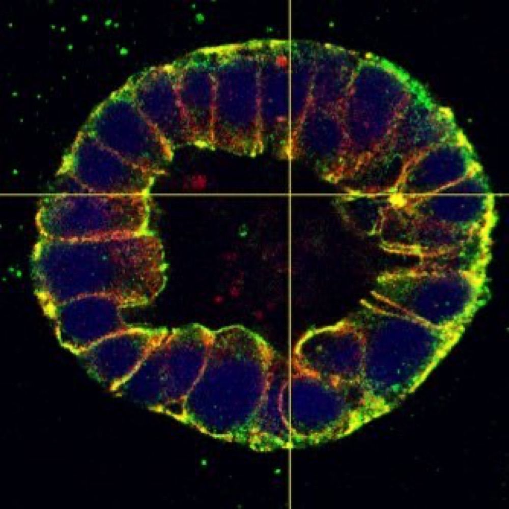
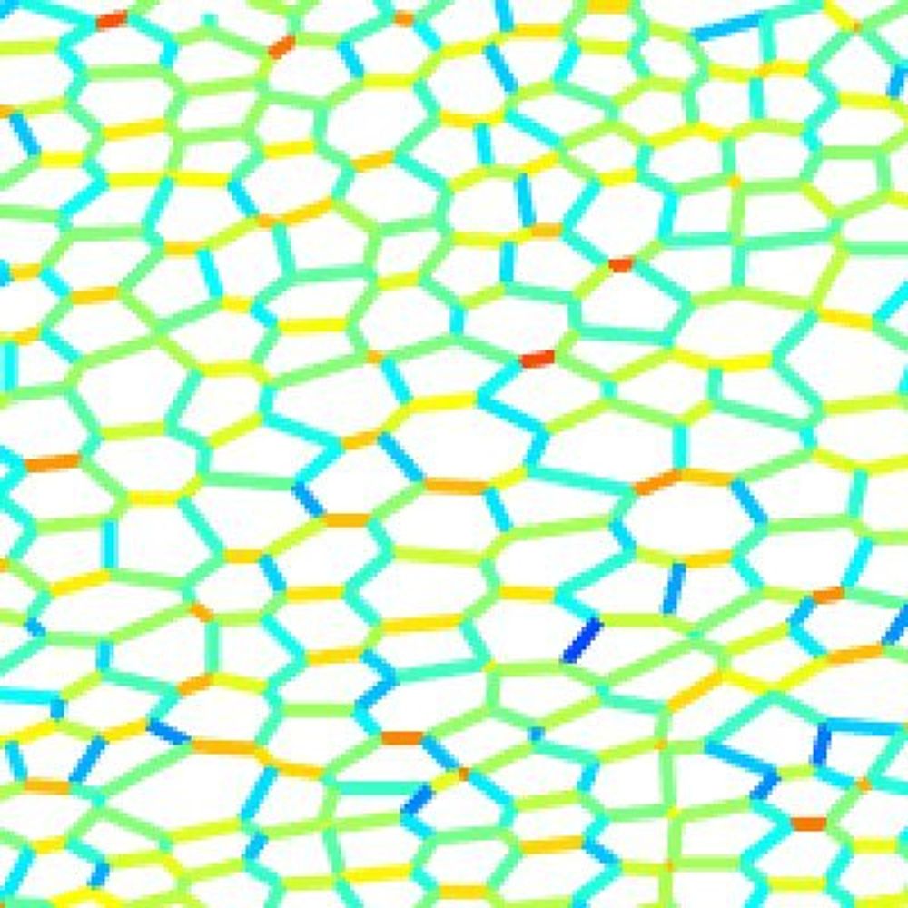
These results suggested that the ‘loser’ cells are eliminated by mechanical compression. Indeed, when we modified the coculture ratio, we found that ZO-1/ZO-2 double KO cells (magenta) needed to be surrounded by normal cells (green) to be eliminated. (9/n)
14.01.2025 10:26 — 👍 1 🔁 0 💬 1 📌 0
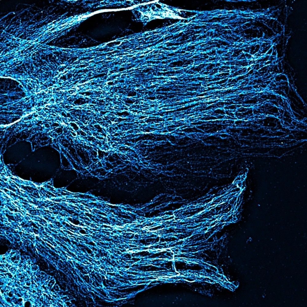
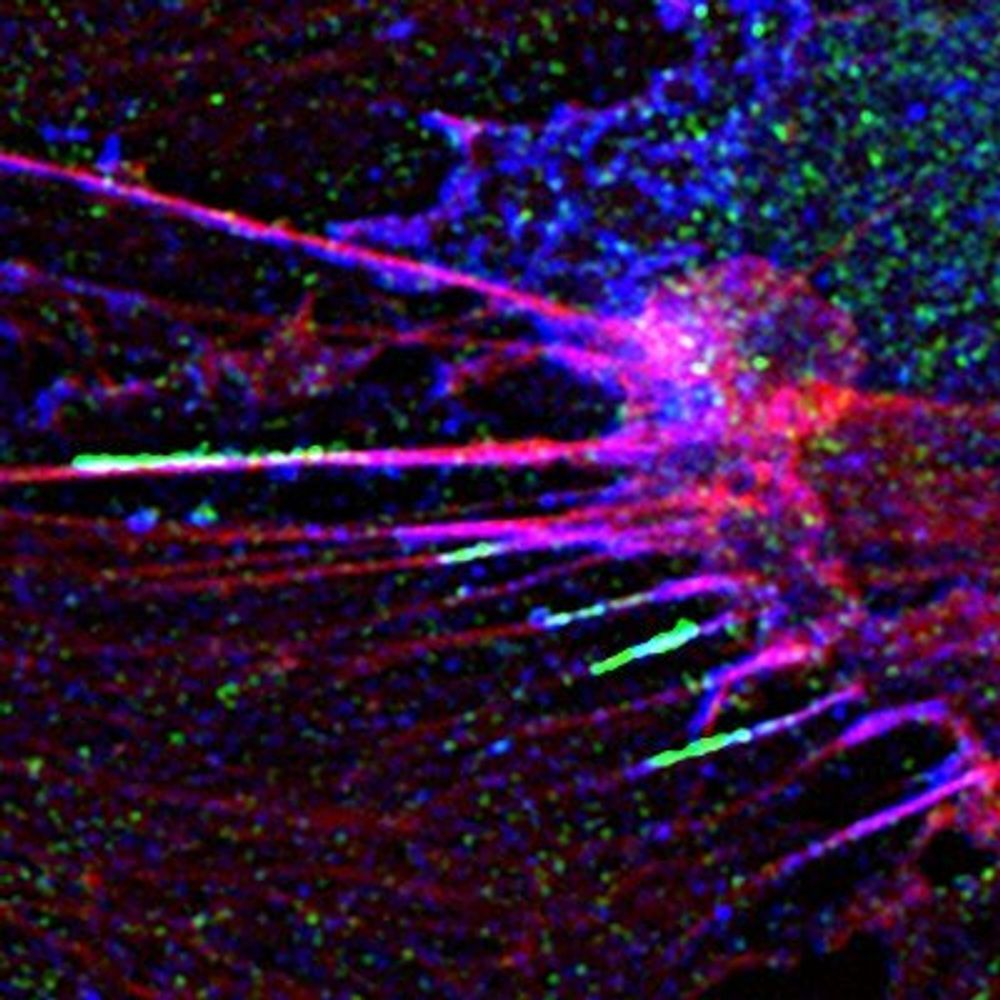
ROCK was required to form supracellular actomyosin cables formed in the normal cells at the clone boundary. The actomyosin cables constricted in a purse-string-like manner to eliminate ZO-1/ZO-2 double KO cells. (8/n)
14.01.2025 10:26 — 👍 1 🔁 0 💬 1 📌 0

We performed a chemical compound library screening to elucidate the underlying molecular mechanisms and found that ROCK plays a key role in eliminating ZO-1/ZO-2 double KO cells. (7/n)
14.01.2025 10:26 — 👍 1 🔁 1 💬 1 📌 0

Importantly, epithelial barrier function measured by transepithelial electric resistance progressively recovered as the elimination proceeded. These results suggested that cell-cell junction-deficient cells are eliminated from epithelia to maintain epithelial barrier homeostasis. (6/n)
14.01.2025 10:26 — 👍 1 🔁 0 💬 1 📌 0

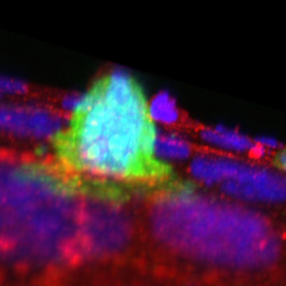
Indeed, by carefully observing the time course of the coculture, we found that ZO-1/ZO-2 double KO cells are eliminated by apoptosis when surrounded by normal cells! (5/n)
14.01.2025 10:26 — 👍 1 🔁 0 💬 1 📌 0

At first, we thought it was a simple mistake, but finally, we realized that some KO cells remained at the edge of the colonies. This observation reminded us of the cell competition phenomena initially characterized in Drosophila. (4/n)
14.01.2025 10:26 — 👍 7 🔁 1 💬 1 📌 0
Having a background in Drosophila genetics, I decided to mix the KO cells with normal cells, akin to the mosaic analysis in flies, and compare the phenotype. However, we repeatedly encountered a problem – a failure to recover ZO-1/ZO-2 double KO cells in the coculture. (3/n)
14.01.2025 10:26 — 👍 3 🔁 0 💬 1 📌 0
This project started when I joined Mikio Furuse’s lab in National Institute for Physiological Sciences. ZO-1/ZO-2 double KO cells were just established in the lab then, and I decided to look at their phenotypes. (2/n)
14.01.2025 10:26 — 👍 1 🔁 0 💬 1 📌 0
Please add me!
18.11.2024 11:47 — 👍 0 🔁 0 💬 0 📌 0
Assistant Professor at the University of Michigan
Watching cells do cool things during development🐣🐁🧫🔬
huyckelab.org
Cancer Cell Biologist studying how epithelial barriers get corrupted during tumor invasion and metastasis. Assistant Professor @MCDB @UMich. thewestlab.com
zookeeper to two tiny humans, professor, cell herder, bioelectrician, 'professional' storyteller, and waterbears just because. see us at:
cohenlab.princeton.edu
When I am not trying to understand embryos, I enjoy mountains, good music and gardening
PhD student @yap-lab.bsky.social at IMB,UQ🔬Studying mechanobiology of apoptotic extrusion in epithelia
Postdoc at UCSF in Weiner Lab (@oweinerlab.bsky.social)
PhD UC Berkeley in Park Lab
(she/her)
Molecular logic of complex cell behaviors.
Orion Weiner's lab at UCSF. Account run by lab members.
#CellMigration #CellPolarity #Mechanics #OptoTools
weinerlab.com
Where Cell-Cell adhesion meets RNAi - and other intriguing encounters. Molecular and Cellular Biology Lab at the Medical University of South Carolina in Charleston.
https://medicine.musc.edu/departments/regenerative-medicine/research/kourtidis-lab
Cell Polarity, Migration and Cancer lab, Institut Pasteur Paris & CNRS
Cell biology group with interest in glial cell migration and glioblastoma invasion, cytoskeleton, mechanobiology, RhoGTPases.
We explore how cells stick together and fall apart..focussing on how cell-cell adhesion, apoptotic extrusion and tissue mechanics regulate epithelial homeostasis 🧫🔬
@AlphaYap’s lab at IMB, Brisbane
Sugimura lab at the Department of Bioinformatics and Systems Biology, Faculty of Science, The University of Tokyo.
Associate Professor at Amsterdam UMC, University of Amsterdam / Vascular Biology / Mechanotransduction / Junctions / Adhesion / Cell migration / Angiogenesis / Atherosclerosis / Capillary malformations
/ https://www.medicalbiochemistry.nl/stephan-huveneers
Postdoc @GeorgiaTech. Previously @SyracuseU and @Penn. PhD @RiceUniversity. Soft matter and biophysics theory
cell & tissue biology, & sometimes frisbee | she/her
assistant professor @ UW Biochemistry
Biologist interested in Skin development & regeneration, ECM, Stem cell @RIKEN Kobe, Japan.
Developmental biology fascinated about the mane (sur)faces/phases of epithelial morphogenesis: from mechanics to evolution.
https://www.bdr.riken.jp/en/research/labs/wang-yc/index.html
Biomechanics, Developmental biology, Cell biology
PhD student at Nano-LSI, Kanazawa Univ. (D1)
#Mechanobiology #DevelopmentalBiology #Biophysics
Cell biologist interested in cancer, in love with the archipelago and good books