📢 Learn #QuPath across 🇵🇹!
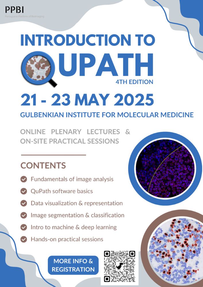
The 4th edition of our Intro to QuPath course is happening:
🗓️ May 21–23, 2025
📍 PPBI Nodes across Portugal
🔹 Register by May 11
📝 Info & registration: see comments
@ljimicrocore.bsky.social
📢 Learn #QuPath across 🇵🇹!
The 4th edition of our Intro to QuPath course is happening:
🗓️ May 21–23, 2025
📍 PPBI Nodes across Portugal
🔹 Register by May 11
📝 Info & registration: see comments
Join us! Science Homecoming helps scientists reconnect with communities by writing about the importance of science funding in their hometown newspapers. We’ve mapped every small newspaper in the U.S. and provide resources to get you started. Help science get back home 🧪🔬🧬 🏠
sciencehomecoming.com
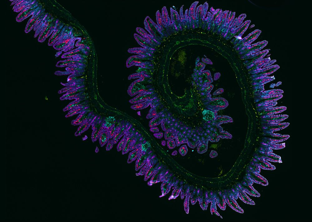
Orion 15 channel imaging of a normal murine ileum spiral. Courtesy of Simon Goldstein, La Jolla Institute for Immunology. The image shows the wavy lining of the ileum, with finger-like villa reaching out. Cells are highlighted with green, purple, pink, and blue florescent markers.
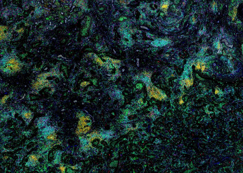
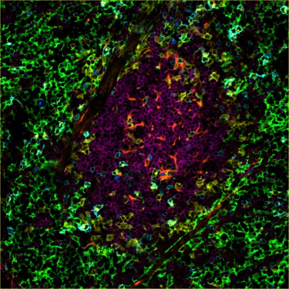
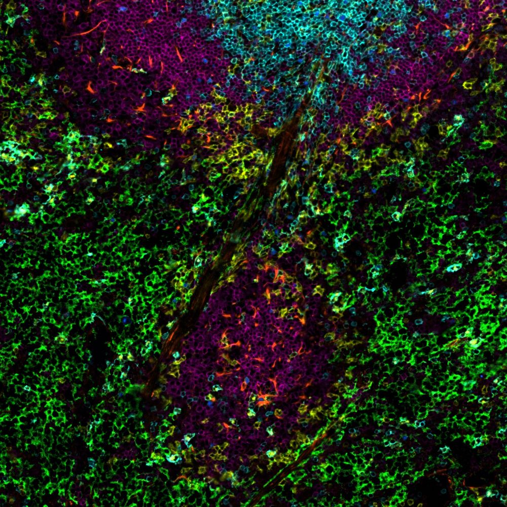
Orion 18 channel imaging of a human lung tumor with dense immune aggregates. Courtesy of Simon Goldstein, La Jolla Institute for Immunology. Image shows cells against a background. Cells are highlighted with green, yellow, blue, and pink florescent markers. The image appears abstract, with clumps of cells forming loose swirls of color.
#Microscopy gives us an absolutely breathtaking view of the immune system 😍
A huge congratulations to LJI's Simon Goldstein and the LJI Microscopy and Histology Core for having TWO stunning images featured in the 2025 @rarecyte.bsky.social Calendar rarecyte.com/imagecontest/ #immunology #biology
What do you see ?
07.02.2025 13:05 — 👍 16 🔁 2 💬 0 📌 0We are with you! Stay strong! #myindirect
08.02.2025 07:03 — 👍 2 🔁 0 💬 0 📌 0The NIH overhead cut doesn't just hurt universities.
It's deadly to the US economy.
The US is a world leader in tech due to the ecosystem that NIH and NSF propel. It drives innovation for tech transfer, creates a highly-skilled sci/tech workforce, and fosters academic/industry crossfertilization.
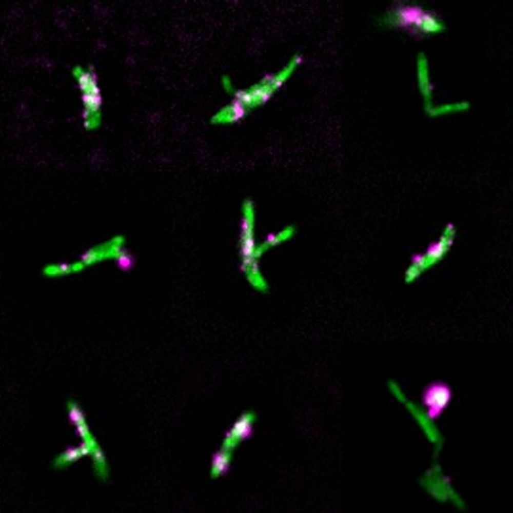
Join us! In two days we will teach you how to analyze complex immunofluorescence multiplex data. It will take you weeks to months to learn this on your own!
23.01.2025 09:24 — 👍 3 🔁 1 💬 0 📌 0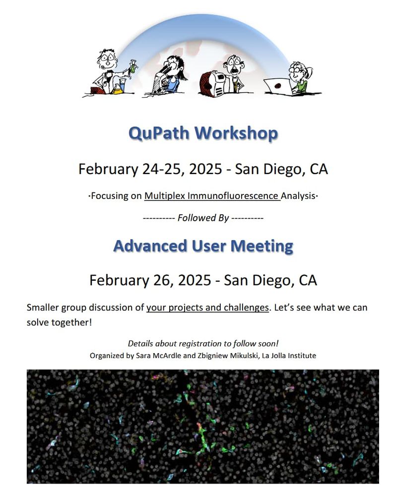
Announcement for a QuPath Workshop in San Diego, CA, on Feb 24-25, 2-25. The following day there will be an Advanced User Meeting.
📢Announcing a QuPath Workshop 📢
February 24-25, 2025, in San Diego, CA, at the La Jolla Institute for Immunology.
This year’s event will be all about Multiplex Immunofluorescence Analysis.
The workshop will be followed by an 🎉 Advanced QuPath User’s Meeting 🎉- February 26, 2025.
Hello, World!
24.11.2024 07:36 — 👍 8 🔁 2 💬 2 📌 0