#RésultatScientifique 🔎 | Comment les filaments d'actine s'organisent à l'intérieur des cellules et des tissus vivants
🔬 @institutfresnel.bsky.social 🤝 @univ-amu.fr @centralemed.bsky.social @cnrs.fr
📍 @cnrs-dr12.bsky.social
@aurelienvilledieu.bsky.social
Postdoc in Jérôme Gros's team | Former PhD student in Yohanns Bellaïche's team | Multi-scale study of tissue morphogenesis | Live-imaging and quantitative biology
#RésultatScientifique 🔎 | Comment les filaments d'actine s'organisent à l'intérieur des cellules et des tissus vivants
🔬 @institutfresnel.bsky.social 🤝 @univ-amu.fr @centralemed.bsky.social @cnrs.fr
📍 @cnrs-dr12.bsky.social

Out today. 🙏 again to everyone for this wonderful piece of work, in particular to Aurelie @aurhin.bsky.social Chase @chasebolt.bsky.social and Brent @homeobox.bsky.social. 🙏 also to the Harris lab @fish4walking.bsky.social and @neilshubin.bsky.social @biology-unige.bsky.social @college-de-france.fr
17.09.2025 17:30 — 👍 95 🔁 41 💬 2 📌 1
We are all super happy and proud to see our work on the function and evolution of the #cephalic #furrow published in @nature.com. Let me say a few things about the background and history of this work on the #Evolution_of_Morphogenesis (1/12)
04.09.2025 08:21 — 👍 347 🔁 119 💬 16 📌 8
Our latest: We developed a chemo-optogenetic system for precise spatiotemporal control of morphogen production. Using dual light + small molecule control of Sonic Hedgehog production, we recapitulated neural tube patterning in vitro & measured spread of Shh
🧵
www.sciencedirect.com/science/arti...
Latest paper elifesciences.org/articles/107... closes an important cycle in our efforts to study regeneration: week-long recordings allow us to observe the behaviour of cells during the entire course of regeneration in a crustacean leg – bright objects in movie are fluorescent nuclei of cells. 1/6
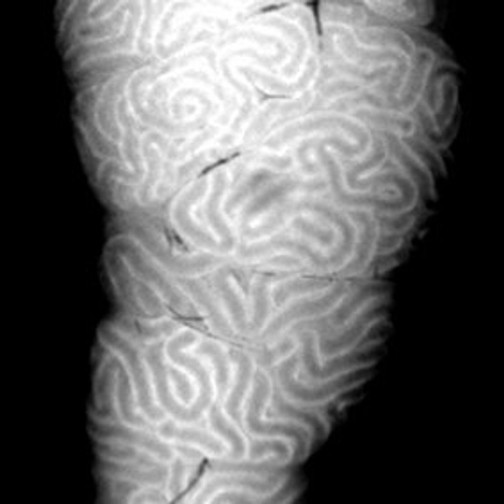
08.08.2025 17:39 — 👍 142 🔁 50 💬 2 📌 3Very happy that the first article from my postdoc work in the Tomancak lab is now published @PNAS! www.pnas.org/doi/10.1073/.... We studied the self-organization of actin in aggregates made from Hydra cells. Thread below (1/9)
06.08.2025 13:09 — 👍 144 🔁 41 💬 3 📌 4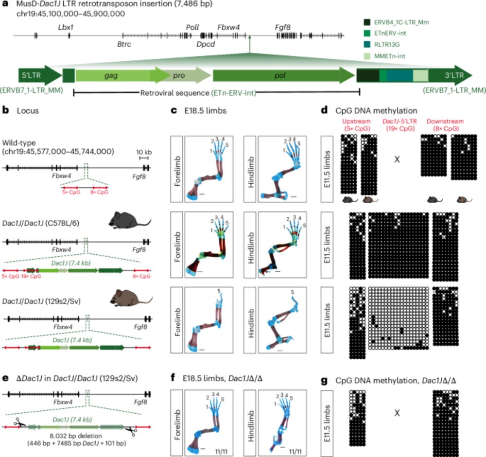
Finally out! 🥳 Our paper showing how a transposable element (TE) insertion can cause developmental phenotypes is now published @natgenet.nature.com 🧬🦠🐁
Below is a brief description of the major findings. Check the full version of the paper for more details: www.nature.com/articles/s41588-025-02248-5
If you’ve been following this account closely, you might already know methods to probe mechanics in vitro, but what about in live embryos?
I’m @amichaut.bsky.social, and I’m going to share a few great papers on this aspect of #EpithelialMechanics.
Here is what I have been up to in the lab of @stephangrill.bsky.social together with @neipel.bsky.social and many others.
Intrigued by avian left right symmetry breaking we found that a tissue-scale active torque drives chiral flow and requires mechanical coupling to the underlying tissue.

🏳️🌈Our editor @slefkopoulos.bsky.social discussed with @jzylicz.bsky.social about the importance of celebrating Pride, representation in science, and more! Happy Pride!
Read here: rdcu.be/ewfba
bit.ly/3Uc72TO
Many thanks to Ruoheng Li and @prelights.bsky.social for featuring our preprint!
10.07.2025 17:08 — 👍 6 🔁 3 💬 0 📌 0🚨 A very interesting conference on gastrulation coming up soon 🚨 Don't miss the chance to interact with these renowned speakers! Early bird deadline: July 15th
03.07.2025 13:25 — 👍 2 🔁 1 💬 0 📌 0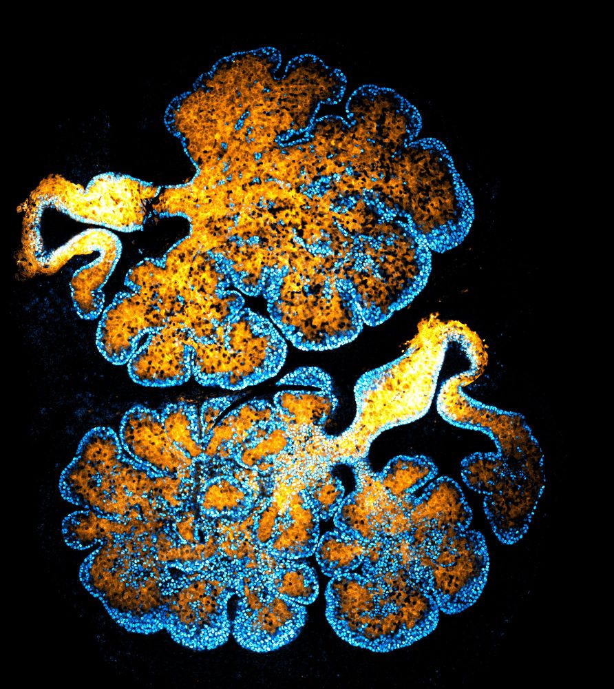
🧵Just out !
We reveal how 4 branched epithelia—mammary, lacrimal, salivary & prostate—use a conserved YAP–Notch–p63 circuit to self-organize during development & regeneration.
Here’s the story👇
authors.elsevier.com/c/1lM285Sx5g...
Work done in @frelab.bsky.social at
@institutcurie.bsky.social
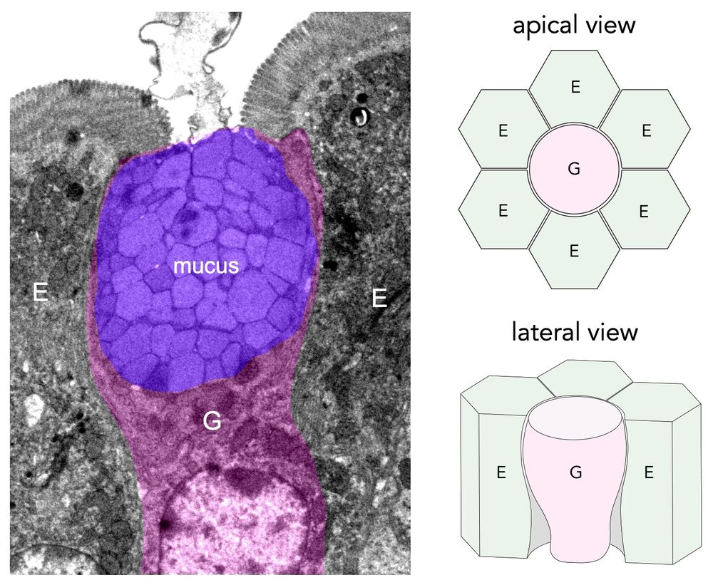
🥳 Thrilled to share our lab's first preprint, led by our talented postdoc Justine Creff! 👩🏻🔬 We tackled a fundamental question: how the epithelium withstands mechanical stress at the interface of cells with distinct geometries and mechanics, such as enterocytes (E) and goblet cells (G) (1/9)
06.04.2025 22:24 — 👍 65 🔁 17 💬 2 📌 3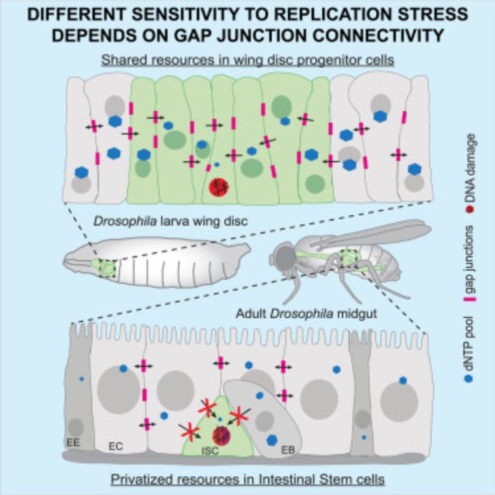
Very happy to share our latest lab publication!
The hard work of @bboumard.bsky.social, Gwenn Le Meur and collaborators!
Cell-type-specific nucleotide sharing through gap junctions impacts sensitivity to replication stress in Drosophila: Developmental Cell
authors.elsevier.com/a/1lDFi_Yv6z...
Thanks a lot Diana !!
24.05.2025 13:22 — 👍 0 🔁 0 💬 0 📌 0The effector caspases drive cell death, but their activation can often be survived, so how do cells make this decision? Our new preprint from @levayerr.bsky.social shows that instantaneous caspase activity is important, but past activation has a key role to play!
www.biorxiv.org/content/10.1...

Our results thus show that primitive streak formation is induced by NODAL activation in the posterior hypoblast. The movement of the hypoblast (which is a counter-rotating movement) is a consequence of primitive streak formation – and not a cause of it, as previously thought.
19.05.2025 09:16 — 👍 3 🔁 0 💬 0 📌 0
Finally, we investigated whether the anterior hypoblast could inhibit primitive streak formation, as previously thought. By grafting the anterior hypoblast onto the forming primitive streak, we could show that it does not. On the contrary, the grafted hypoblast starts to express NODAL.
19.05.2025 09:13 — 👍 1 🔁 0 💬 1 📌 0In this movie, you can for example see that the full ablation of the hypoblast completely inhibits primitive streak formation, while a partial ablation that leaves a piece of posterior hypoblast on each side leads to the formation of two embryonic axes (forming conjoined twins).
19.05.2025 09:11 — 👍 1 🔁 0 💬 1 📌 0Using functional analyses and tissue-recombination experiments, we confirmed that the posterior hypoblast is indeed the inducer of primitive streak formation through NODAL signaling activation.
19.05.2025 09:10 — 👍 1 🔁 0 💬 1 📌 0
Using HCR-RNA-FISH, we observed that the expression of NODAL, a master activator of primitive streak formation, initiates in the posterior hypoblast and then spreads to the entire posterior blastoderm.
19.05.2025 09:10 — 👍 1 🔁 0 💬 1 📌 0The hypoblast thus remains in constant contact with the epiblast, which is incompatible with the idea that its movement controls primitive streak formation by locally lifting NODAL inhibition. This contradiction led us to reinvestigate the molecular mechanisms underlying primitive streak formation.
19.05.2025 09:09 — 👍 1 🔁 0 💬 1 📌 0By intercalating a porous filter between the epiblast and the hypoblast, we further showed that the hypoblast requires physical contact with the epiblast to achieve counter-rotating flows. Thus, forces generated in the epiblast propagate by mechanical coupling to the hypoblast, setting it in motion.
19.05.2025 09:08 — 👍 2 🔁 0 💬 1 📌 0By generating a two-color chimera, we observed that the counter-rotating flows of the epiblast and hypoblast are highly synchronized. As a result, the hypoblast and epiblast move very little in relation to each other.
19.05.2025 09:07 — 👍 1 🔁 0 💬 1 📌 0We have adapted live-imaging techniques previously used to characterize tissue flows in the avian epiblast to describe, this time, tissue flows in the hypoblast. This allowed us to discover that, like the epiblast, the hypoblast exhibits counter-rotating flows (also known as Polonaise movements).
19.05.2025 09:05 — 👍 1 🔁 0 💬 1 📌 0
In birds and mammals, primitive streak formation is thought to occur when an anterior movement of the hypoblast (visceral endoderm in mice) lifts its inhibition on the posterior epiblast, allowing NODAL signaling activation. However, in birds, a precise description of this movement was lacking.
19.05.2025 09:01 — 👍 1 🔁 0 💬 1 📌 0
🚨New preprint
Glad to share my postdoc work on the role of the hypoblast in primitive streak induction! Discover how live imaging observations led us to revisit the molecular mechanisms governing embryonic axis induction
Thanks to @jeromegros.bsky.social and all co-authors!
doi.org/10.1101/2025...

🚨📣 New preprint alert!
Excited to share my new postdoc work on direct force and mechanical properties measurements in live embryos!
A great team effort with A. Chamolly (theory), under the supervision of F. Corson and @jeromegros.bsky.social
📄 www.biorxiv.org/content/10.1...
#devbio #mechanics
ICYMI: Commentary on "mechanism" in dev bio
Ozpolat et al argue for diverse approaches to mechanism - from molecular to systems level
journals.biologists.com/dev/article/...