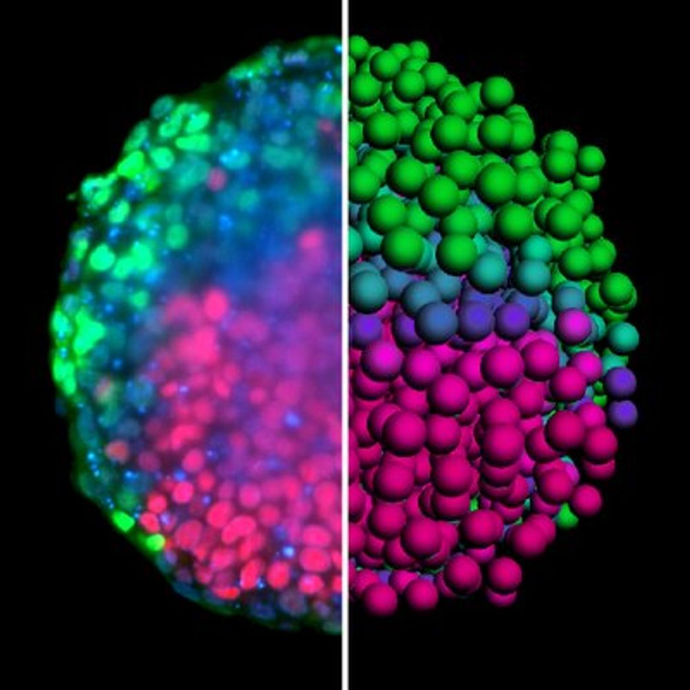
Pierre-François Lenne's Avatar

Pierre-François Lenne

@pflenne.bsky.social

Biophysicist interested in cell dynamics and tissue morphogenesis at IBDM and Turing Center for Living Systems Group: https://www.morphotiss.org/ https://www.ibdm.univ-mrs.fr/physical-approaches-to-cell-dynamics/ https://centuri-livingsystems.org

892 Followers  |  214 Following  |  43 Posts  |  Joined: 16.11.2024  |  1.8587

Latest posts by pflenne.bsky.social on Bluesky

Katia took the initiative to start a Xenopus project in the lab a few years ago —and now her work is highlighted by Development! Huge congrats, Katia!

31.07.2025 10:39 — 👍 28    🔁 7    💬 0    📌 0
Video thumbnail

Feat. @guignardlab.bsky.social @pflenne.bsky.social @shamtlili.bsky.social @philipperoudot.bsky.social
The Tapenade pipeline is fully open-source with user-friendly notebooks and Napari plugins: github.com/GuignardLab/... 🫒

15.07.2025 11:48 — 👍 6    🔁 2    💬 1    📌 0
Preview
Sham Tlili, winner of the CNRS 2025 bronze medal - IBDM | Institut de Biologie du Développement de Marseille A distinction that rewards a promising and already fruitful scientific career.

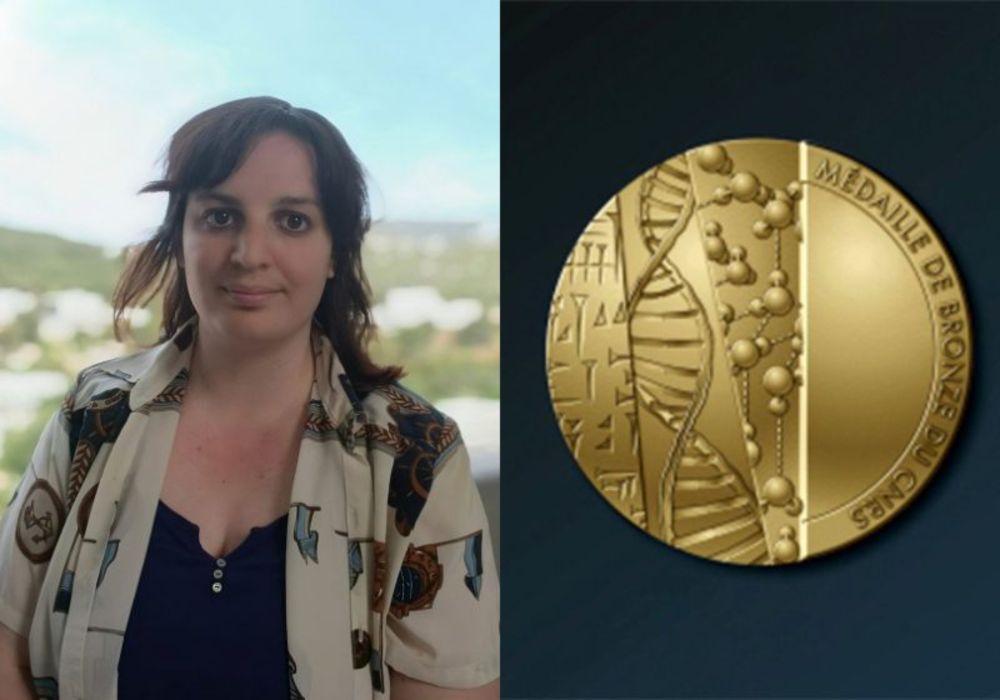
🥇Sham Tlili has been awarded the CNRS 2025 Bronze Medal! She studies how physical forces shape living tissues by using mouse stem cell models known as gastruloids. A unique blend of physics & biology.

🔗 Read more: www.ibdm.univ-amu.fr/sham-tlili-w...

27.06.2025 09:00 — 👍 44    🔁 14    💬 2    📌 0

Thank you so much for sharing your thoughts on our article!

10.04.2025 18:33 — 👍 10    🔁 0    💬 1    📌 0
Post image Post image

Excited to share that IBDM (Institut de Biologie du Développement de Marseille) is expanding its research groups this year! A great place for developmental biology and interdisciplinary research located on the beautiful Marseille-Luminy campus!
Deadline for applications: March 30th, 2025

17.01.2025 07:11 — 👍 57    🔁 51    💬 2    📌 1
Bayesian Force Inference package for Matlab This package allows the user to perform 2D Bayesian Force Inference in Matlab Reference: Experimental validation of force inference in epithelia from cell to tissue scale W. Kong, O. Loison, P. Shiva...

Hi Romain, Thanks for noticing it. I transferred some old Twitter posts to BlueSky, but it didn't work that well.
Code: data.mendeley.com/datasets/78n...
Ref: www-nature-com.insb.bib.cnrs.fr/articles/s41...

16.01.2025 12:49 — 👍 6    🔁 1    💬 0    📌 1

Congrats on this beautiful work!

15.12.2024 14:21 — 👍 1    🔁 0    💬 1    📌 0

Thrilled that our team clinched both 1st and 2nd place in the image contest! 🥇🥈 Huge applause for everyone’s creativity and excitement in front of the microscope. Let’s keep inspiring each other! 💪✨ #Teamwork #Science

15.12.2024 14:17 — 👍 9    🔁 1    💬 0    📌 0

And usually, the sky is blue in Cassis, near Marseille!

19.11.2024 07:35 — 👍 5    🔁 0    💬 0    📌 0
Post image

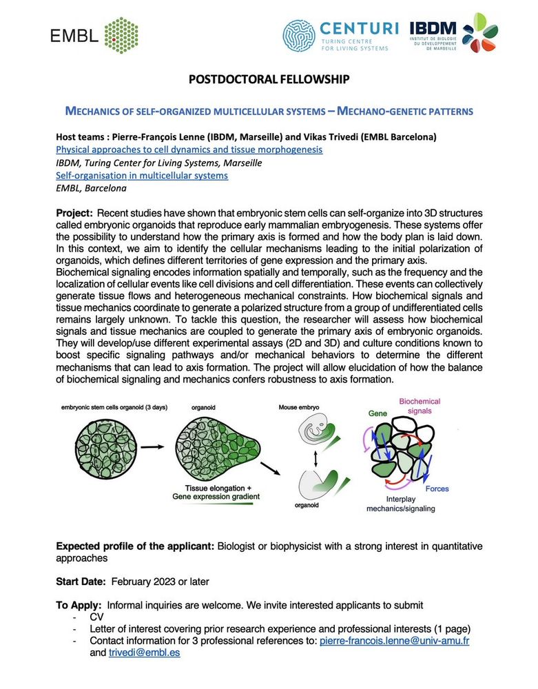
Vikas Trivedi's @LabTrivedi @EMBLBarcelona @the_prbb and my group @Equipe_lenne @IBDMmarseille @centuri_ls are recruiting for a Postdoc project on the emergence of mechano-genetic patterns using embryonic organoids!
Start Date: Feb 2023 or later
Informal inquiries are welcome!

20.10.2022 11:49 — 👍 0    🔁 0    💬 0    📌 0
Two-point optical manipulation reveals mechanosensitive remodeling of cell-cell contacts in vivo Biological tissues acquire reproducible shapes during development through dynamic cell behaviors. These events involve the remodeling of cell contacts driven by active cytoskeletal contractile forces. However how cell-cell contacts remodel remains poorly understood because of lack of tools to directly apply forces at cell-cell contacts to produce their remodeling. Here we develop a dual-optical trap manipulation method to impose different force patterns on cell-cell contacts in the early epithelium of the Drosophila embryo. Through different push and pull manipulations at the edges of junctions, the technique allows us to produce junction extension and junction shrinkage. We use these observations to constrain and specify vertex-based models of tissue mechanics, incorporating negative and positive mechanosensitive feedback depending on the type of remodeling. We show that Myosin-II activity responds to junction strain rate and facilitates full junction shrinkage. Altogether our work provides insight into how stress produces efficient deformation of cell-cell contacts in vivo and identifies unanticipated mechanosensitive features of their remodeling. Significance statement The highly organized tissues and organs that form our body emerge from internal dynamic activities at the cellular level. Among such activities, cell shape changes and cell rearrangement, cell extrusion and cell division sculpt epithelial tissues into elongated sheets, tubes and spherical cavities. Remodeling of cell-cell contacts, powered by actomyosin contractility, is key to all these transformations. Although much is known about the molecular machinery and biochemical signals that regulate remodeling of cell contacts, there is a lack of approaches to directly probe the mechanics of cell contacts and therefore assess their ability to resist or deform in response to mechanical loads. We developed an experimental technique to manipulate and exert contractile and extensile forces to cell-cell junctions. Our results lead to a specific physical model of junctional mechanics, with implications in the modeling of collective cell behavior in epithelial tissues. ### Competing Interest Statement The authors have declared no competing interest.

How do cell-cell contacts remodel in vivo? We address this question here:

biorxiv.org/content/10.110…

19.07.2022 14:39 — 👍 0    🔁 0    💬 0    📌 0
Post image

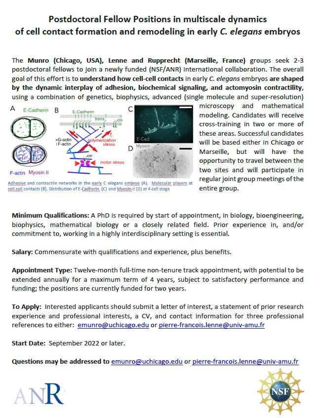
The Munro (Chicago), Lenne and Rupprecht (Marseille) groups seek 2-3 postdoctoral fellows to join a newly funded (NSF/ANR) international collaboration on the multiscale dynamics of cell contact formation and remodeling.

10.05.2022 12:59 — 👍 15    🔁 21    💬 1    📌 0
Cell-state transitions and collective cell movement generate an endoderm-like region in gastruloids A cellular and biophysical study on embryonic stem cell aggregates reveals that the endoderm can form by a three-step mechanism involving a Wnt/beta-catenin-mediated epiblast fragmentation, tissue flow and cell segregation.

Our work on endoderm formation in gastruloids is out!@eLife

doi.org/10.7554/eLife.…

12.04.2022 11:28 — 👍 0    🔁 0    💬 0    📌 0
Post image

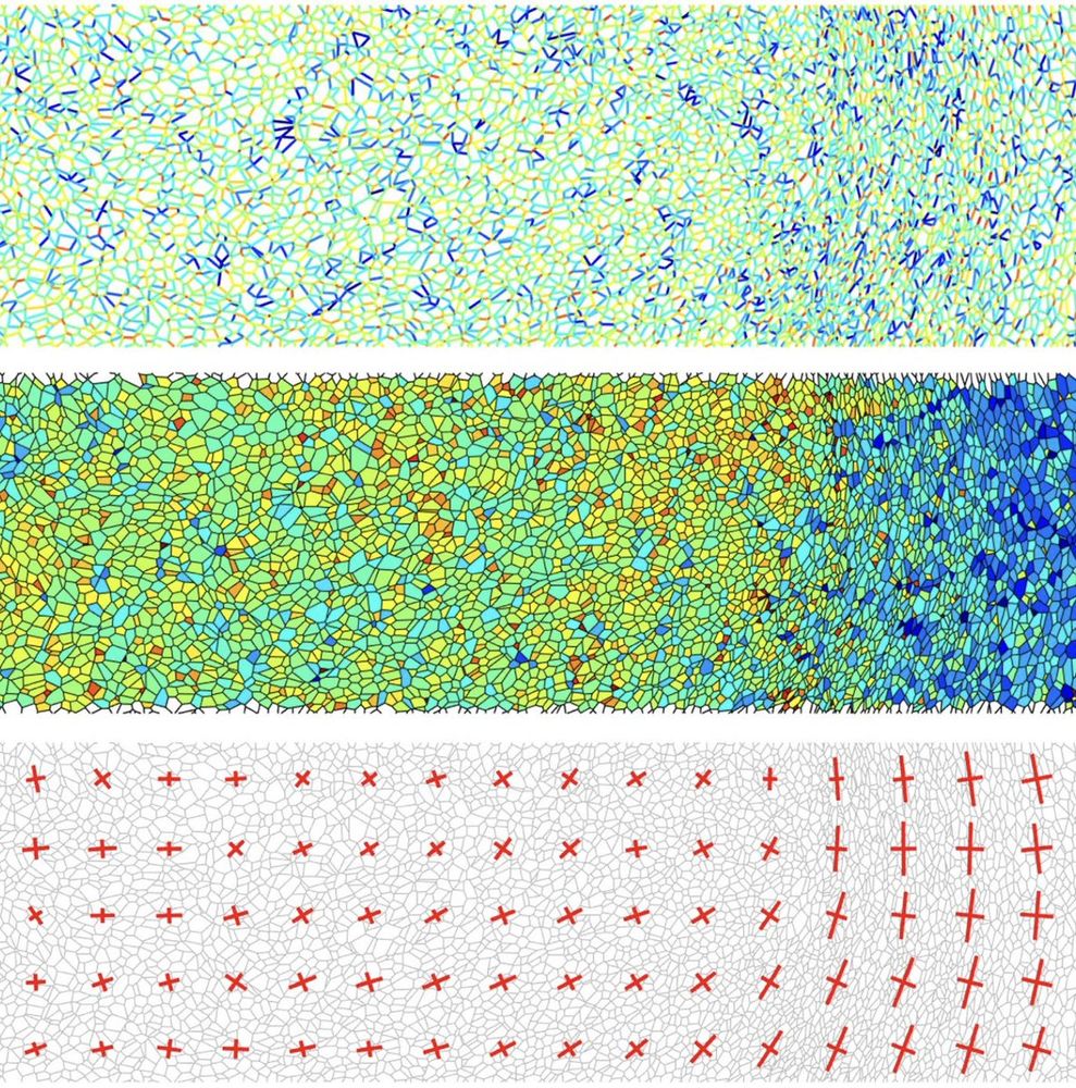
Our code for force inference in epithelial tissues is available online!
Feel free to contact us if you're interested in using it or if you want to collaborate.
Code :Ref :

nature.com/articles/s4159… data.mendeley.com/datasets/78ng4…

08.02.2022 10:33 — 👍 22    🔁 5    💬 1    📌 1

Very happy to welcome in our group Alice Gros @_AliceGros as a new Centuri @centuri_ls PhD student on a joint project with Léo Guignard @GuignardLab on morphogenesis of self-organized multicellular systems #Gastruloids

04.10.2021 13:29 — 👍 0    🔁 0    💬 0    📌 0
Roadmap for the multiscale coupling of biochemical and mechanical signals during development - IOPscience Roadmap for the multiscale coupling of biochemical and mechanical signals during development, Pierre-François Lenne, Edwin Munro, Idse Heemskerk, Aryeh Warmflash, Laura Bocanegra-Moreno, Kasumi Kishi, Anna Kicheva, Yuchen Long, Antoine Fruleux, Arezki Boudaoud, Timothy E Saunders, Paolo Caldarelli, Arthur Michaut, Jerome Gros, Yonit Maroudas-Sacks, Kinneret Keren, Edouard Hannezo, Zev J Gartner, Benjamin Stormo, Amy Gladfelter, Alan Rodrigues, Amy Shyer, Nicolas Minc, Jean-Léon Maître, Stefano Di Talia, Bassma Khamaisi, David Sprinzak, Sham Tlili

Very grateful to great colleagues and pleased that our roadmap on multi-scale coupling of biochemical and mechanical signals during development is now onlinein IOPPBio @IOPPBio @WarmflashLab
@TimESaunders
@JgrosL
@a_michaut
@EdouardHannezo
@ZevGartner

ow.ly/eRun50EodEL

14.04.2021 16:16 — 👍 0    🔁 0    💬 0    📌 0

Very fortunate to welcome today a new postdoc in the group, Valentin Dunsing @DunsingValentin, who will explore/exploit "fluctuations" in morphogenesis.
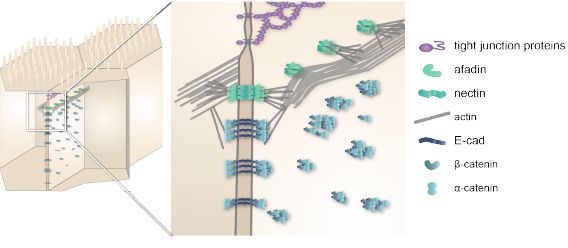
01.04.2021 06:49 — 👍 1    🔁 0    💬 0    📌 0
Cell Junction Mechanics beyond the Bounds of Adhesion and Tension Cell-cell adhesion is more than the sum of its molecular parts. In this perspective, Lenne, Rupprecht, and Viasnoff aim at bridging scales from adhesion molecules and actomyosin networks to junctional integrity and mechanical resistance of tissues and organs.

See our new perspective on cell junction mechanics written together with @Viasnofflab and @JFRupprecht_OM

zpr.io/HW2PN

18.01.2021 07:44 — 👍 0    🔁 0    💬 0    📌 0
Super-resolution imaging uncovers the nanoscopic segregation of polarity proteins in epithelia Epithelial tissues acquire their integrity and function through the apico-basal polarization of their constituent cells. Proteins of the PAR and Crumbs complexes are pivotal to epithelial polarization, but the mechanistic understanding of polarization is challenging to reach, largely because numerous potential interactions between these proteins and others have been found, without clear hierarchy in importance. We identify the regionalized and segregated organization of members of the PAR and Crumbs complexes at epithelial apical junctions by imaging endogenous proteins using STED microscopy on Caco-2 cells, human and murine intestinal samples. Proteins organize in submicrometric clusters, with PAR3 overlapping with the tight junction (TJ) while PALS1-PATJ and aPKC-PAR6β form segregated clusters that are apical of the TJ and present in an alternated pattern related to actin organization. CRB3A is also apical of the TJ and weakly overlaps with other polarity proteins. This organization at the nanoscale level significantly simplifies our view on how polarity proteins could cooperate to drive and maintain cell polarity. ### Competing Interest Statement The authors have declared no competing interest.

Check our latest manuscript on the nanoscopic segregation of polarity proteins in epithelia using superresolution. A project led by @PierreMangeol and a great collaboration with Le Bivic team.

biorxiv.org/content/10.110…

13.08.2020 07:19 — 👍 0    🔁 0    💬 0    📌 0

Honoured and thrilled to become a EMBO member! A big thank you to all the great members of my team, past and present, and to my inspiring colleagues.

07.07.2020 15:33 — 👍 0    🔁 0    💬 0    📌 0
Cell-state transitions and collective cell movement generate an endoderm-like region in gastruloids Shaping the animal body plan is a complex process that involves the spatial organization and patterning of different layers of cells. Recent advances in live imaging have started to unravel the cellular choreography underlying this process in mammals, however, the sequence of events transforming an unpatterned group of cells into structured territories is largely unknown. Here, using 3D aggregates of mouse embryonic stem cells, we study the formation of one of the three germ layers, the endoderm. We show that the endoderm can be generated from an epiblast-like state by a three-tier mechanism: a release of islands of E-cadherin expressing cells, their flow toward the tip of the 3D aggregate, and their segregation. In contrast to the prevailing view, this mechanism does not require epithelial to mesenchymal transitions and vice-versa but rather a fragmentation, which is mediated by Wnt/β- catenin, and a sorting process. Our data emphasize the role of signaling and cell flows in the establishment of the body plan. ### Competing Interest Statement The authors have declared no competing interest.

Our first paper on endoderm formation using #gastruloidswith @AMA_Lab

biorxiv.org/content/10.110…

24.05.2020 19:22 — 👍 0    🔁 0    💬 0    📌 0
Preview
Signez la pétition La recherche scientifique a besoin d’un plan d’urgence

Signez la pétition : La recherche scientifique a besoin d’un plan d’urgencevia @ChangeFrance

chng.it/DjnW7knV

13.05.2020 13:00 — 👍 0    🔁 0    💬 0    📌 0
Post image

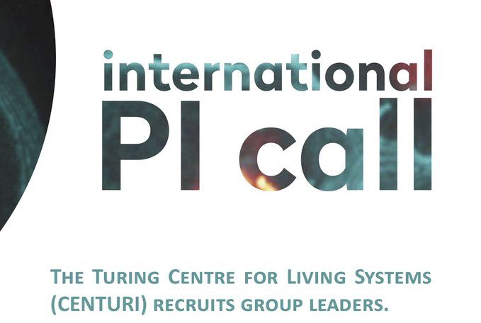
We seek to attract new PIs, computer scientists, physicists, or mathematicians with a theoretical and/or computational biology project, in the Turing Center for Living Systems (CenTuri) in Marseille.

View the offer here


centuri-livingsystems.org/wp-content/upl…

14.01.2020 10:09 — 👍 0    🔁 0    💬 0    📌 0
Post image

Check our latest manuscript on supramolecular architecture of epithelia:

biorxiv.org/content/10.110…

18.10.2019 08:14 — 👍 0    🔁 0    💬 0    📌 0
Post image

Last days to register/submit an abstract to our meeting on "Self-organization in multicellular systems" in Cargèse, Corsica, Sept 30 - Oct 4.

centuri-livingsystems.org/csm2019/

24.06.2019 15:06 — 👍 0    🔁 0    💬 0    📌 0
Assembly of a persistent apical actin network by the formin Frl/Fmnl tunes epithelial cell deformability Tissue remodeling during embryogenesis is driven by the apical contractility of the epithelial cell cortex. This behavior arises notably from Rho1/Rok induced transient accumulation of non-muscle myosin II (MyoII pulses) pulling on actin filaments (F-Actin) of the medio-apical cortex. While recent studies begin to highlight the mechanisms governing the emergence of Rho1/Rok/MyoII pulsatility in different organisms, little is known about how the F-Actin organization influences this process. Focusing on Drosophila ectodermal cells during germband extension and amnioserosa cells during dorsal closure, we show that the medio-apical actomyosin cortex consists of two entangled F-Actin subpopulations. One exhibits pulsatile dynamics of actin polymerization in a Rho1 dependent manner. The other forms a persistent and homogeneous network independent of Rho1. We identify the Frl/Fmnl formin as a critical nucleator of the persistent network since modulating its level, in mutants or by overexpression, decreases or increases the network density. Absence of this network yields sparse connectivity affecting the homogeneous force transmission to the cell boundaries. This reduces the propagation range of contractile forces and results in tissue scale morphogenetic defects. Our work sheds new lights on how the F-Actin cortex offers multiple levels of regulation to affect epithelial cells dynamics.

Assembly of a persistent apical actin network by the formin Frl/Fmnl tunes epithelial cell deformability

biorxiv.org/content/10.110…

24.06.2019 14:56 — 👍 0    🔁 0    💬 0    📌 0
Spatiotemporal dynamics of calcium transients during embryogenesis of Drosophila melanogaster Calcium signaling plays a crucial role in the physiology of the organs but also in various aspects of the organogenesis of the embryo. High versatility of calcium signaling is encoded by the dynamic variation of intracellular calcium concentration. While the dynamics of calcium is important, little is known about it throughout the embryogenesis of the largest class of animals, insects. Here, we visualize calcium dynamics throughout embryogenesis of Drosophila using a fluorescent protein-based calcium indicator, GCaMP3, and report calcium transients in epithelium and neuronal tissues. Local calcium transients of varying duration were detected in the outer epithelium, trachea and neural cells. In addition, gap-junction-dependent calcium waves were identified at stage 16 in the outer epithelium and in the trachea at stage 17. Calcium transient waveform analysis revealed different characteristics as a function of the duration, location and frequency. Detailed characterization of calcium transients during embryogenesis of Drosophila will help us better understand the role of calcium signaling in embryogenesis and organogenesis of insects.

Olga's paper on calcium transients in the Drosophila embryo is in BiorXiv:

disq.us/t/3b8ulq0

06.02.2019 13:30 — 👍 0    🔁 0    💬 0    📌 0

Girish's paper on how mechanical forces affect E-cadherin adhesion and junction dynamics is now onlineTowards a distinction between shear and tensile stresses.

rdcu.be/bb2Hp

27.11.2018 10:29 — 👍 0    🔁 0    💬 0    📌 0
Post image

This week, I participated in #Declics2018 with @CercleFSER. Half a day in a nearby high school to explain to students how knowledge progresses. A unique opportunity to share our experience as a researcher and answer students' questions.

25.11.2018 19:04 — 👍 0    🔁 0    💬 0    📌 0
Force inference predicts local and tissue-scale stress patterns in epithelia Morphogenesis relies on the active generation of forces, and the transmission of these forces to surrounding cells and tissues. Hence measuring forces directly in developing embryos is an essential task to study the mechanics of development. Among the experimental techniques that have emerged to measure forces in epithelial tissues, force inference is particularly appealing. Indeed it only requires a snapshot of the tissue, as it relies on the topology and geometry of cell contacts, assuming that forces are balanced at each vertex. However, establishing force inference as a reliable technique requires thorough validation in multiple conditions. Here we performed systematic comparisons of force inference with laser ablation experiments in three distinct Drosophila epithelia. We show that force inference accurately predicts single junction tensions, tension patterns in stereotyped groups of cells, and tissue-scale stress patterns, in wild type and mutant conditions. We emphasize its ability to capture the distribution of forces at different scales from a single image, which gives it a critical advantage over perturbative techniques such as laser ablation. Our results demonstrate that force inference is a reliable and efficient method to quantify the mechanics of epithelial tissues during morphogenesis.

Wei's work on Biorxiv "Force inference predicts local and tissue-scale stress patterns in epithelia

biorxiv.org/content/early/…

23.11.2018 16:22 — 👍 0    🔁 0    💬 0    📌 0

@pflenne is following 19 prominent accounts