ORCID
Hi! Can I be added to the feed please?
orcid.org/0000-0003-14...
02.10.2025 01:04 — 👍 0 🔁 0 💬 0 📌 0
I’m grateful to everyone who worked on this project with me–Aidan, Richard, @yoitsjasmine.bsky.social , @molbiolgv.bsky.social , and Chantal. As always, huge thanks to @kranzuschlab.bsky.social and Amy for making it possible for me to work on these kinds of questions!
02.10.2025 00:32 — 👍 2 🔁 0 💬 0 📌 0

A model of the function of viral IF4F.
This unique viral replication strategy shows that you can build sophisticated translation regulation through a very simple cap-binding complex. Perhaps there are contexts in which cellular organisms also make use of similar strategies?
02.10.2025 00:32 — 👍 0 🔁 0 💬 1 📌 0
But why do these viruses not just rely on the host cap-binding complex? We found that mimivirus replication is unusually resistant to abiotic stresses in a way that depends on viral translation factors. Could it be an adaptation to the unusual stresses faced by the amoeba host?
02.10.2025 00:32 — 👍 0 🔁 0 💬 1 📌 0

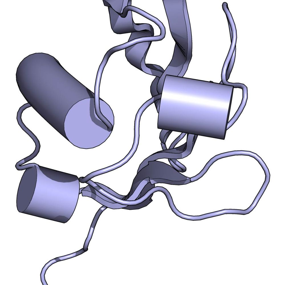
Crystal structure of viral IF4E bound to a viral cap structure.
How does the viral cap-binding complex specifically promote viral translation? Viral mRNAs carry a unique 5′ UTR motif: a conserved +1A followed by AU-rich sequences. A crystal structure of vIF4E shows exactly how this mRNA cap is recognized!
02.10.2025 00:32 — 👍 0 🔁 0 💬 1 📌 0

Transmission electron micrograph showing viral factories in amoebae infected with mimivirus.
This effect becomes obvious when looking at viral factories by TEM. Early in infection these large structures form independent of the viral cap-binding complex, but when this complex is disrupted viral particles cannot assemble.
02.10.2025 00:32 — 👍 0 🔁 0 💬 1 📌 0
We call these proteins viral IF4A, IF4E, and IF4G. They 1) form a complex, 2) are essential for viral replication, and 3) act as bona fide translation factors, promoting synthesis of viral structural proteins late in infection.
02.10.2025 00:32 — 👍 0 🔁 0 💬 1 📌 0
In amoeba infected by mimivirus, the prototypical giant DNA virus, we found dozens of viral proteins bound to ribosomes—including three that are homologous to the eukaryotic mRNA cap-binding complex (eIF4A, eIF4E, eIF4G).
02.10.2025 00:32 — 👍 0 🔁 0 💬 1 📌 0

Transmission electron micrograph of a mimivirus particle.
Giant DNA viruses encode a stunning number of proteins that were long thought unique to living organisms. Among them: translation factors, the master regulators of protein synthesis. But are these viral proteins functional?
02.10.2025 00:32 — 👍 1 🔁 0 💬 1 📌 0

>18,000 new genomes of giant DNA viruses! An incredible trove of new genes and insights into evolution of host-virus interactions from @fmschu.bsky.social and @jgi.doe.gov
www.biorxiv.org/content/10.1...
29.09.2025 19:10 — 👍 18 🔁 6 💬 0 📌 0
Congrats! Beautiful work
22.07.2025 17:54 — 👍 2 🔁 0 💬 1 📌 0
Shameful. Sorry Jason!
25.03.2025 20:39 — 👍 1 🔁 0 💬 1 📌 0
Cool work! Will you add phages or viruses of the rest of eukaryotes?
21.12.2024 21:00 — 👍 1 🔁 0 💬 1 📌 0
The Ramsey philosophy of biology lab at KU Leuven, Belgium.
https://www.theramseylab.org • #HPbio #philsci #philsky #evosky #paleosky #cogsci
NYC; Lab of Environmental Microbiology @rockefelleruniv.bsky.social
Assistant Professor at the University of Waterloo. Head of the Environmental Virology and Ecology Research Group (ENVERG) Into algae, viruses, environmental microbiology, oceanography. Views are my own.
Assistant Prof and Group Leader at Max Perutz Labs, University of Vienna
Associate Professor of Microbiology & Immunology
UNC Chapel Hill
Viral pathogenesis, arboviruses, virus-host interactions, antiviral immunity
www.lazearlab.org
Scientist, mom, & dog mom. She/they. Trans rights are human rights. Long covid survivor. Pissed about the climate crisis. Tell your dog I said hi
Professor and Chair of Cell Biology at harvardcellbio , all things ubiquitin, autophagy, protein/organelle quality control & proteomics (website:harper.hms.harvard.edu). Mostly posting from @harvardcellbio.bsky.social
Assistant Professor @ ISTA
Using cryo-EM to understand how bacteria defend themselves
https://bravo-lab.org/
Science integrity consultant and crowdfunded volunteer, PhD.
Ex-Stanford University. Maddox Prize/Einstein F Award winner
NL/USA/SFO.
#ImageForensics
@MicrobiomDigest on X.
Blog: ScienceIntegrityDigest.com
Support me: https://www.patreon.com/elisabethbik
Phage Biology, Bacterial defense systems, CRISPR-Cas, Microbial communities, Mucus interactions
http://www.fnobregalab.org
http://www.klebphacol.org
http://www.phage-collection.org
protein biochemistry and evolution | viruses and antibodies | faculty @uofubiochem.bsky.social
starr.biochem.utah.edu
Albert Einstein College of Medicine: Educating students to become caring, skilled physicians and fostering biomedical and translational research.
Assistant Professor of Emerging Infectious Diseases,
Duke-NUS Medical School, Singapore.
Adj. Investigator, A*STAR Infectious Diseases Labs
#CellBiologyofVirusInfection
#FunctionalGenomics
https://sites.google.com/view/ysolab
Molecular virologist with a background in poxviruses 🦠 Views my own.
Associate Professor @ UCL, London. Computational Biology, Structural Bioinformatics, machine Learning, Proteins, Viruses. https://profiles.ucl.ac.uk/103340-gorka-lasso-cabrera
Virologist at LSU Health, Shreveport
Assistant Professor at UCIrvine. Biochemistry and structural biology enthusiast. Dabbling in innate immunity and microbiology. Phage defense, cyclic nucleotides, and cool enzymes. (he/him/his) https://faculty.sites.uci.edu/morehouselab/
Assistant professor of Biological Sciences at Pitt, studying evolution, genes, and microbes. Same as @tera_levin in the other place. #NewPI She/her
PhD student @soreklab.bsky.social. Researching the arms race between bacteria and phages 🦠🛡⚔️ A proud father of 3 cats. 🏳️🌈
Immunologist x cell biologist x microbiologist at MRC LMB in Cambridge. Occasionally birdwatching. Less nerdy than my bio sounds. Views my own.