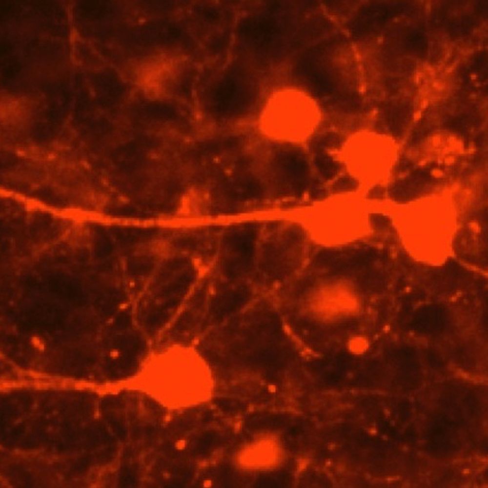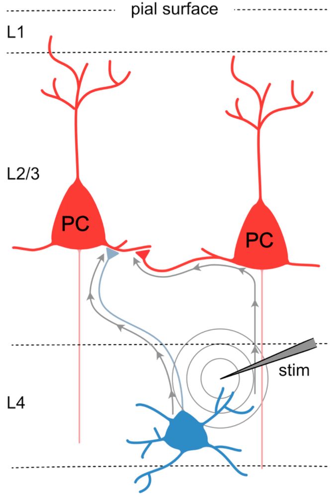
🧪 We recently published our first micro-publication! This paper highlights the pitfalls of using extracellular stimulation to recruit synapses in neocortical circuits, as it lacks specificity. www.micropublication.org/journals/bio...
19.06.2025 18:04 — 👍 28 🔁 2 💬 2 📌 0
I'd like to make a list of PhD programs that (1) are in Europe (which does include the UK); (2) offer opportunities in systems neuroscience; and obviously (3) pay a stipend. Please share and/or respond to add your suggestions?
17.06.2025 17:25 — 👍 21 🔁 6 💬 3 📌 0
We are super excited about this news!!! 🤩 #ERCAdG
17.06.2025 13:09 — 👍 33 🔁 4 💬 3 📌 0
Oops the original post was deleted! The news is here: bsky.app/profile/cham...
17.06.2025 19:02 — 👍 23 🔁 1 💬 2 📌 0
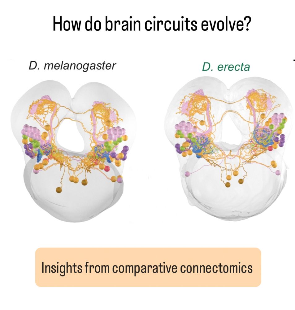
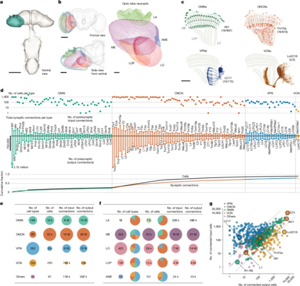
How do brain circuits evolve? We started looking for some answers by using synapse-resolution cross-species comparative connectomics on an entire olfactory circuit 👇
bit.ly/44aVm9E
12.06.2025 11:04 — 👍 145 🔁 60 💬 5 📌 6
Posting this after some recent conversations with potential international applicants - still time to apply to our Masters courses and International PhD Academy for 2025 entry - join the diverse and vibrant Neuroscience community on our beautiful campus next to Brighton
13.05.2025 16:12 — 👍 6 🔁 6 💬 0 📌 0
I am very happy and thankful for having been part of this amazing journey. Enjoy exploring this incredibly rich dataset!
09.04.2025 18:42 — 👍 3 🔁 1 💬 0 📌 0
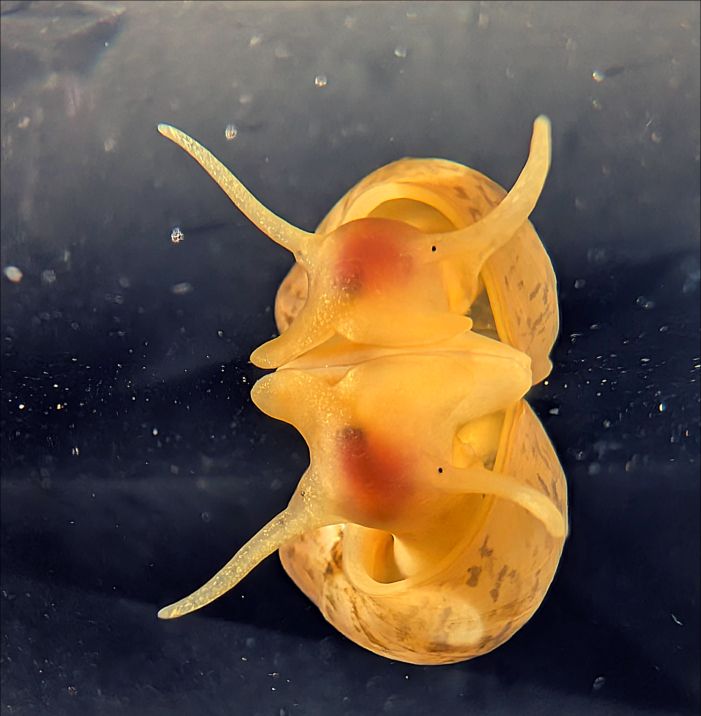
Aw, we got the cover for our new paper on X-ray imaging and atlas building in the snail brain. www.pnas.org/doi/10.1073/... Thanks @pnas.org .. and to @sussexneuro.bsky.social @leverhulme.bsky.social @ukri.org for funding support #invertebrate #brain #neuroscience THREAD: bsky.app/profile/kevi...
05.03.2025 10:24 — 👍 27 🔁 9 💬 1 📌 2
Michael Crossley led the experimental work, supported by Anna Simon, @arndroth.bsky.social and
@enzomarra.bsky.social Thanks to @sussexneuro.bsky.social @leverhulme.bsky.social @ukri.org and @diamondlightsource.bsky.social for funding support. Thanks for reading! 10/10
28.02.2025 09:40 — 👍 2 🔁 0 💬 1 📌 0
Our approach should readily generalize to other model systems with comparable brain sizes (e.g. other molluscs, crustacea, annelids, insects). On its own, it won’t yield a full wiring diagram, but it does rapidly provide a detailed overview map for atlas building and comparative studies. 9/10
28.02.2025 09:38 — 👍 4 🔁 1 💬 1 📌 0

This provides the locations of principal feeding-circuit cell types, including motoneurons, CPG neurons and modulatory cells, alongside a detailed summary of their main functional properties. 8/10
28.02.2025 09:37 — 👍 3 🔁 1 💬 1 📌 0
We also brought together the anatomical mapping and functional information to establish the beginnings of a fully scalable functional cell atlas of the brain of Lymnaea stagnalis: sites.google.com/view/snailbr... 7/10
28.02.2025 09:36 — 👍 2 🔁 0 💬 3 📌 0
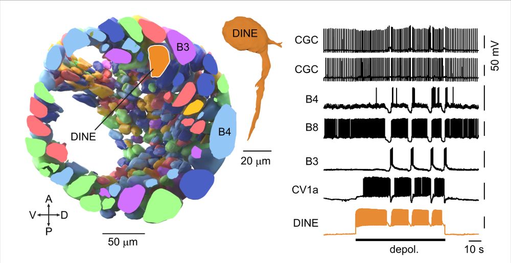
The consistent positioning of neurons across Lymnaea brains means the atlas can guide follow-up functional experiments. Targeting a non-superficial region led to the discovery of DINE (“Diamond Neuron”), an apt name 😜 because it activates the food ingestion circuitry. 6/10
28.02.2025 09:35 — 👍 3 🔁 0 💬 1 📌 0
The 3D reconstruction revealed the organization of neurons beneath the surface layer for the first time. It turns out around half the neurons (coloured orange) are non-superficial - a hidden world of circuit components that can now be studied. 5/10
28.02.2025 09:35 — 👍 2 🔁 0 💬 1 📌 0
We then used the excellent volume image-sharing, annotation, and reconstruction platform
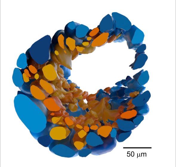
@webknossos.org to fully reconstruct the buccal ganglia (one side is shown here) housing the main feeding circuitry, yielding the first accurate estimate of the total number of neurons: ~1100. 4/10
28.02.2025 09:34 — 👍 3 🔁 0 💬 1 📌 0
Michael Crossley led the experimental work, supported by Anna Simon, @arndroth.bsky.social and @enzomarra.bsky.social Thanks to @sussexneuro.bsky.social @leverhulme.bsky.social @ukri.org and @diamondlightsource.bsky.social for funding support. Thanks for reading! 10/10
28.02.2025 08:49 — 👍 1 🔁 2 💬 0 📌 0
Our approach should readily generalize to other model systems with comparable brain sizes (e.g. other molluscs, crustacea, annelids, insects). On its own, it won’t yield a full wiring diagram, but it does rapidly provide a detailed overview map for atlas building and comparative studies. 9/10
28.02.2025 08:47 — 👍 1 🔁 1 💬 1 📌 0
Sample image showing detailed information on different neuron types in the Lymnaea brain.
This provides the locations of principal feeding-circuit cell types, including motoneurons, CPG neurons and modulatory cells, alongside a detailed summary of their main functional properties. 8/10
28.02.2025 08:45 — 👍 1 🔁 0 💬 1 📌 0
We also brought together the anatomical mapping and functional information to establish the beginnings of a fully scalable functional cell atlas of the brain of Lymnaea stagnalis: sites.google.com/view/snailbr... 7/10
28.02.2025 08:44 — 👍 1 🔁 0 💬 1 📌 0
Figure shows a cross section view of the ganglia with an orange neuron, DINE, highlighted. It also shows electrophysiological traces with DINE driving a robust fictive feeding rhythm.
The consistent positioning of neurons across Lymnaea brains means the atlas can guide follow-up functional experiments. Targeting a non-superficial region led to the discovery of DINE (“Diamond Neuron”), an apt name 😜 because it activates the food ingestion circuitry. 6/10
28.02.2025 08:44 — 👍 1 🔁 0 💬 1 📌 0
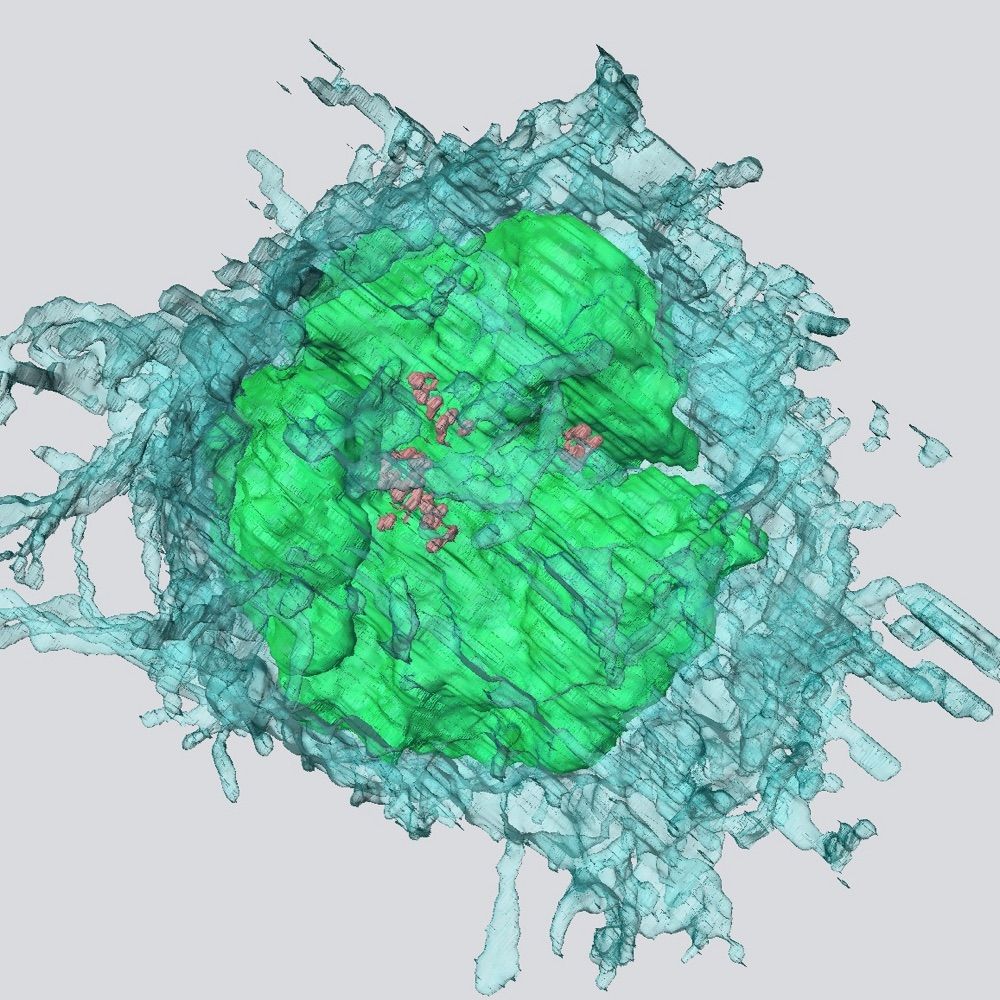
Image shows a cross section through the snail ganglia with superficial neurons shown in blue and internalized neurons in orange. There are similar numbers of each.
The 3D reconstruction revealed the organization of neurons beneath the surface layer for the first time. It turns out around half the neurons (coloured orange) are non-superficial - a hidden world of circuit components that can now be studied. 5/10
28.02.2025 08:42 — 👍 1 🔁 0 💬 1 📌 0
We then used the excellent volume image-sharing, annotation, and reconstruction platform @webknossos.org to fully reconstruct the buccal ganglia (one side is shown here) housing the main feeding circuitry, yielding the first accurate estimate of the total number of neurons: ~1100. 4/10
28.02.2025 08:41 — 👍 1 🔁 0 💬 1 📌 0
Lymnaea_Buccal_Ganglia_Synchrotron-X-ray_Tomography | WEBKNOSSOS
View this dataset in WEBKNOSSOS
A full CNS scan took ~3 mins, and higher-res stacks (voxel size: 0.325 µm) <20 mins. Browse a sample here: wklink.org/2643 3.5/10
28.02.2025 08:40 — 👍 3 🔁 1 💬 1 📌 0
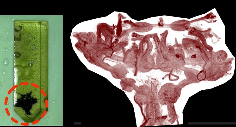
Image shows embedded snail brain and X-ray overview of structure
We plastic-embedded the 3x3x2 mm3 brain and performed X-ray tomography imaging at the Diamond Synchrotron Facility @diamondlightsource.bsky.social
28.02.2025 08:38 — 👍 2 🔁 1 💬 2 📌 0
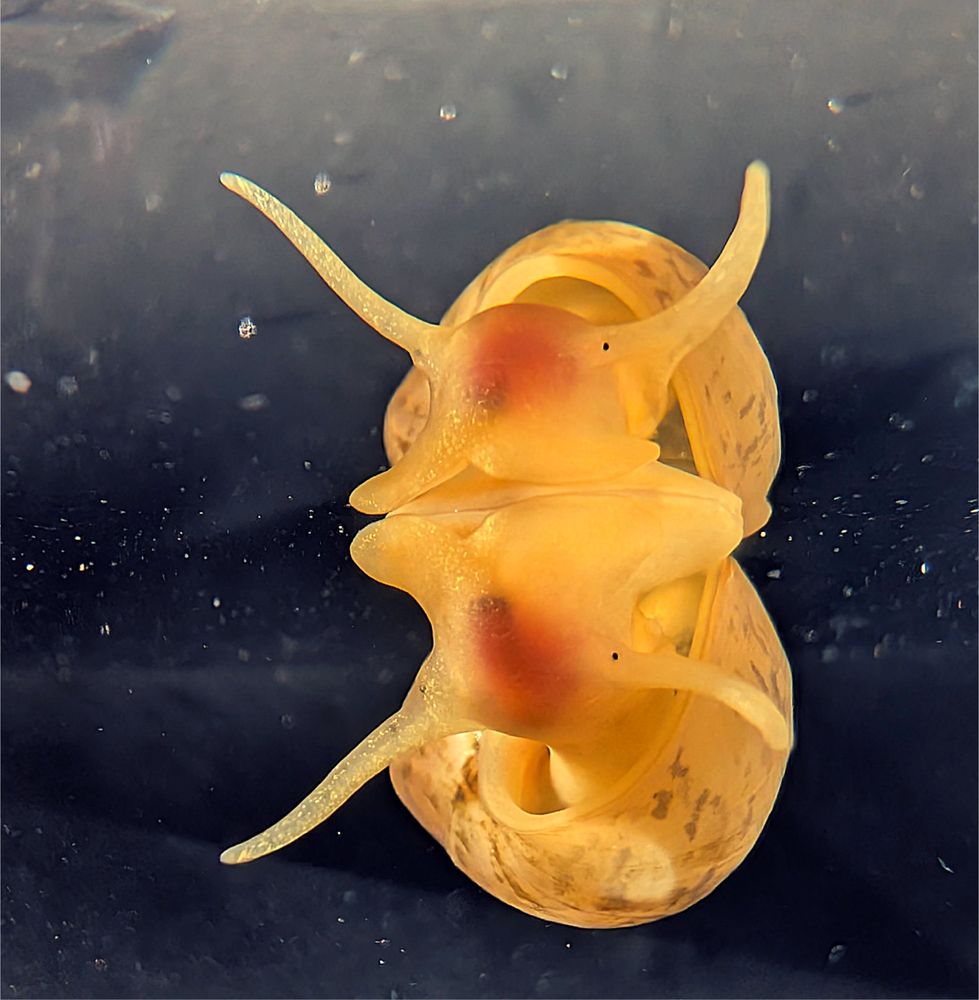
The mollusc Lymnaea is a classical system for neural circuit studies. However, we lack a cell-level atlas of its multi-mm scale brain to guide functional investigations. The solution? 2/10
28.02.2025 08:37 — 👍 2 🔁 1 💬 1 📌 0
Neuroscience Group Leader at Champalimaud Centre for the Unknown. Zebrafish behavior and brain-wide imaging. Also some Danionella and Devario. Might post about music and Rubik's cubes.
At the Sussex Researcher School we support our emerging researchers: Junior Research Associates (JRAs), Postgraduate Researchers (PGRs) and Early Career Researchers (ECRs).
PGRs: @SussexPGRs.bsky.social
ECRs: @SussexECRs.bsky.social
University of Sussex
At @bsmsmedschool.bsky.social I am integrating medicine & conservation in Papua New Guinea + improving scabies control in Britain & Ethiopia. Chair @wildhealthcic.bsky.social Views own. #PublicHealth #MedicalParasitology
https://linktr.ee/DrJMiddleton
We aim to reveal the neuronal ensemble mechanisms of motivated behaviours guided by food-associated cues. We are located in weird and wonderful Brighton on the southern English coast!
(lab homepage: http://tinyurl.com/y44g9a7u)
Executive Editor/Team Leader Open Access Science Journals Sage Publishing
Opinions = mine
http://linkedin.com/in/jlovick-editor
#oncology #cancerresearch #medicine #biology #cardiology #neurology #microbiology #publichealth #healthcare #technology
Neuroscientist fascinated by sensory physiology and how internal state modulates neural circuits
https://johnstonlab.org/
Science journalist & biology nerd. ❤️🧪 Lover of brains & microbes & weird animals. Opinions mine obviously.
At the Electron Microscopy Science Technology Platform at the Francis Crick Institute. Imaging life at the nanoscale with innovative new tech and citizen science.
Welcome to our official account 👋
Nestled in the South Downs, we're a UK university located just 10 mins from beachy Brighton and an hour from London.
www.sussex.ac.uk
Seaslug neuroethologist at UMass Amherst
Post-doc researcher at the University of Sussex 👨🔬 researching insect neuroscience and behaviour 🐝🧠🦗🔩 | First Gen | He/him 🏳️🌈🏳️⚧️ | European, immigrant 🇪🇺🇮🇹🇬🇧🇬🇧
Biologist. PhD Student in Neuroscience at the University of Santiago de Compostela (NEURODEVO group).
Focusing on neurogenesis in the shark retina 🦈👁🧠
Views my own. He/him.
Wellcome CDA fellow at University of Manchester | Postdoc at Sheffield University 🏢, interested in neurovascular coupling in health, ageing and neurodegeneration 🧠🔬
Manchester based neuroscientist. Researching circadian rhythms and vision but with other interests too!
Professor of Marine Ecology at GEOMAR / Kiel University, trained ecoevophysiologist, interested in coastal invertebrates, algae & climate change.
We study presynaptic function and dysfunction, specifically how neurotransmission is sustained during neuronal activity.
Evolutionary biologist (invertebrates, vision, brains), Junior Group Leader @multipleye-lab.bsky.social @MfNBerlin.bsky.social. Gradual learner of 🇩🇪 She/her. Views mine, all mine!
Neuroscientist and engineer. Studying spatial navigation in the miniature translucent fish Danionella cerebrum. Postdoc in Briggman Lab at MPINB.